组织工程与重建外科杂志 ›› 2020, Vol. 16 ›› Issue (3): 237-240.doi: 10.3969/j.issn.1673-0364.2020.03.016
崔基浩1, 李创1( ), 马文斌2, 李春1, 徐万宏1
收稿日期:2020-04-01
修回日期:2020-04-28
出版日期:2020-07-27
发布日期:2020-06-26
基金资助:
Jihao CUI1, Chuang LI1(), Wenbin MA2, Chun LI1, Wanhong XU1
Received:2020-04-01
Revised:2020-04-28
Online:2020-07-27
Published:2020-06-26
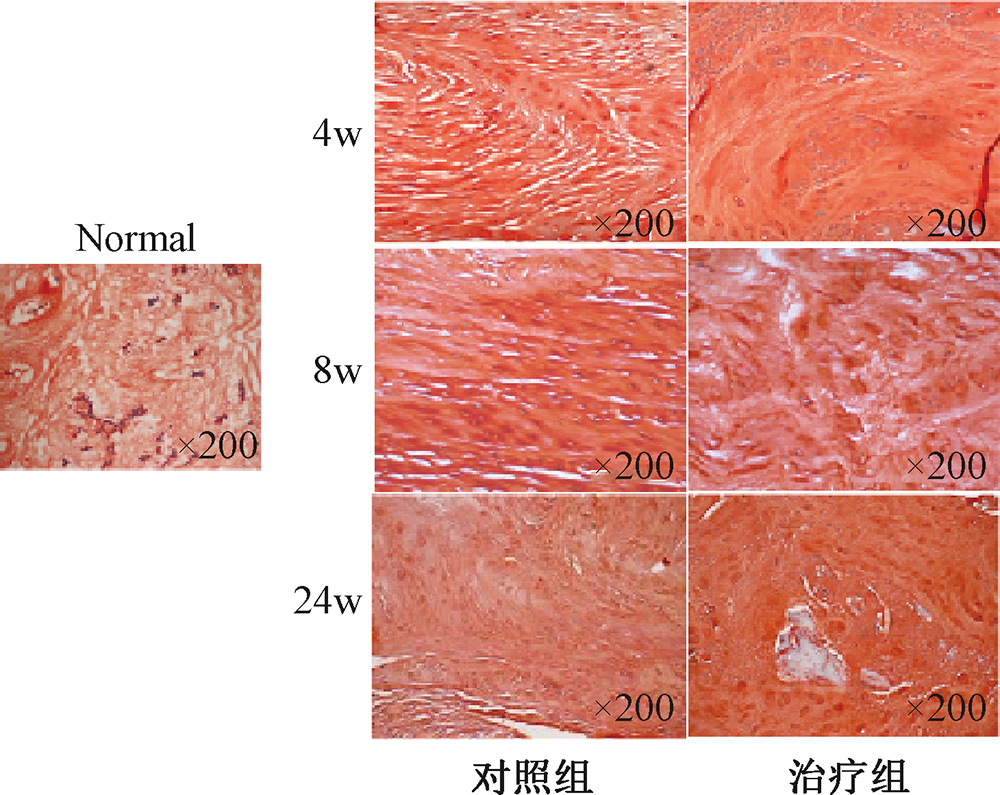
摘要: 目的 研究自体骨髓单核细胞移植对兔退行性病变后椎间盘的修复作用。 方法 选取新西兰大白兔12只,随机分为对照组(n=6)与治疗组(n=6)。取新西兰大白兔髂后上棘骨髓,密度梯度离心法提取骨髓单核细胞。两组实验兔麻醉后使用16 G穿刺针进行L4/5、L5/6、L6/7椎间盘穿刺造模。治疗组将骨髓单核细胞注入到穿刺造模的椎间盘中,对照组不予治疗。第4、8、24周分别进行椎间盘的影像学(X线和MR)与组织学观察。 结果 在椎间隙高度维持方面,随着周数的增加,治疗组优于对照组;在MR T2W1加权像的信号强度及组织形态方面,治疗组明显优于对照组。组织学观察显示,与治疗组相比,对照组中的髓核细胞数量减少,胞外基质收缩、不均。 结论 自体骨髓单核细胞移植对兔退行性病变后的椎间盘具有一定的修复再生作用。
中图分类号:
崔基浩, 李创, 马文斌, 李春, 徐万宏. 自体骨髓单核细胞移植对兔退行性病变后椎间盘的修复作用[J]. 组织工程与重建外科杂志, 2020, 16(3): 237-240.
Jihao CUI, Chuang LI, Wenbin MA, Chun LI, Wanhong XU. Regeneration of Degenerated Intervertebral Disc with Autologous Bone Marrow Mononuclear Cells in Rabbit Models[J]. Journal of Tissue Engineering and Reconstructive Surgery, 2020, 16(3): 237-240.
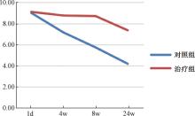
图1
各组各时间点椎间盘高度指数(DHI)

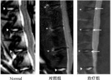
图2
术后第8周各组椎间盘MR T2W1加权像

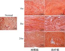
图3
各组各时间点椎间盘组织学观察
| [1] | Hartvigsen J, Hancock MJ, Kongsted A , et al. What low back pain is and why we need to pay attention[J]. Lancet, 2018,391(10137):2356-2367. |
| [2] | Smith LJ, Silverman L, Sakai D , et al. Advancing cell therapies for intervertebral disc regeneration from the lab to the clinic: Recommendations of the ORS spine section[J]. JOR Spine, 2018,1(4):e1036. |
| [3] | 施长城, 姚海 . 退变性椎间盘疾病诊断与治疗技术的研究进展[J]. 中国医疗设备, 2015,30(3):1-7. |
| [4] | Soltan M, Smiler D, Choi JH , et al. Bone marrow: orchestrated cells, cytokines, and growth factors for bone regeneration[J]. Implant Dent, 2009,18(2):132-141. |
| [5] | Song F, Tang J, Geng R , et al. Comparison of the efficacy of bone marrow mononuclear cells and bone mesenchymal stem cells in the treatment of osteoarthritis in a sheep model[J]. Int J Clin Exp Pathol, 2014,7(4):1415-1426. |
| [6] | Lu K, Li HY, Yang K , et al. Exosomes as potential alternatives to stem cell therapy for intervertebral disc degeneration: in-vitro study on exosomes in interaction of nucleus pulposus cells and bone marrow mesenchymal stem cells[J]. Stem Cell Res Ther, 2017,8(1):108. |
| [7] | 刘亭亭, 韩长旭, 王国强 . 细胞移植修复椎间盘的新视角与新进展[J]. 中国组织工程研究, 2020,24(1):154-158. |
| [8] | Deschepper M, Oudina K, David B , et al. Survival and function of mesenchymal stem cells (MSCs) depend on glucose to overcome exposure to long-term, severe and continuous hypoxia[J]. J Cell Mol Med, 2011,15(7):1505-1514. |
| [9] | Menarim BC, Gillis KH, Oliver A , et al. Inflamed synovial fluid induces a homeostatic response in bone marrow mononuclear cells in vitro: Implications for joint therapy[J]. FASEB J, 2020,34(3):4430-4444. |
| [10] | Ozdemir M, Attar A, Kuzu I , et al. Stem cell therapy in spinal cord injury: in vivo and postmortem tracking of bone marrow mononuclear or mesenchymal stem cells[J]. Stem Cell Rev Rep, 2012,8(3):953-962. |
| [11] | Hammadi AMA, Alhimyari F . Intra-arterial injection of autologous bone marrow-derived mononuclear cells in ischemic stroke patients[J]. Exp Clin Transplant, 2019,17(Suppl 1):239-241. |
| [12] | Maruyama M, Nabeshima A, Pan CC , et al. The effects of a functionally-graded scaffold and bone marrow-derived mononuclear cells on steroid-induced femoral head osteonecrosis[J]. Biomaterials, 2018,187:39-46. |
| [13] | Kerr GJ, Veras MA, Kin MK , et al. Decoding the intervertebral disc: unravelling the complexities of cell phenotypes and pathways associated with degeneration and mechanotransduction[J]. Semin Cell Dev Biol, 2017,62:94-103. |
| [14] | Wang HQ . Editorial: Bring stem cell therapies to cure intervertebral disc degeneration to the forefront[J]. Curr Stem Cell Res Ther, 2015,10(4):284. |
| [1] | 方晓涛, 严正, 王晗, 徐潇源, 陈玥. 考虑概率电压不平衡度越限风险的共享储能优化运行方法[J]. 上海交通大学学报, 2022, 56(7): 827-839. |
| [2] | 杨晓华 何乐人. 颊脂垫干细胞在骨组织工程中的研究进展[J]. 组织工程与重建外科杂志, 2022, 18(5): 446-. |
| [3] | 郑毅 许鹏 刘凯. 促进间充质干细胞外囊泡产生的研究进展[J]. 组织工程与重建外科杂志, 2022, 18(2): 166-. |
| [4] | 杜奉舟 龙笑. 淋巴系统再生医学与组织工程的研究进展[J]. 组织工程与重建外科杂志, 2022, 18(2): 171-. |
| [5] | 张硕, 李薇, 李英姿, 刘强, 曾鸣. 面向新型电力系统的可再生能源绿色电力证书差异化配置模型[J]. 上海交通大学学报, 2022, 56(12): 1561-1571. |
| [6] | 鄢和新. 细胞可塑性如何驱动组织再生与肿瘤发生?[J]. 上海交通大学学报, 2021, 55(Sup.1): 51-52. |
| [7] | 张文杰. 如何建立器官再生的血管网络?[J]. 上海交通大学学报, 2021, 55(Sup.1): 60-61. |
| [8] | 李玲芳, 陈占鹏, 胡炎, 邰能灵, 高孟平, 朱涛. 基于灵活性和经济性的可再生能源电力系统扩展规划[J]. 上海交通大学学报, 2021, 55(7): 791-801. |
| [9] | 于子优 蔡宜佐 李伟 张文杰. 无细胞脂肪组织提取液在皮肤及软组织再生修复中的应用[J]. 组织工程与重建外科杂志, 2021, 17(6): 561-. |
| [10] | 朱敏闻 孙兵 李志耀 叶周熹 钱文昊 吴玉波. 增强成骨及抗菌性能的鱼胶原 / 聚乙烯亚胺电纺膜用于牙周组织修复 [J]. 组织工程与重建外科杂志, 2021, 17(2): 102-. |
| [11] | 孙欣, 严佳嘉, 谢敬东, 孙波. “碳中和”目标下电气互联系统有功-无功协同优化模型[J]. 上海交通大学学报, 2021, 55(12): 1554-1566. |
| [12] | 卢俊钦 杨鑫 李军 孙坚. 仿生修饰结合三维打印技术构建的胶原-聚己内酯支架可促进兔临界骨缺损修复 #br#[J]. 组织工程与重建外科杂志, 2021, 17(1): 1-. |
| [13] | 张沛灵 慈政 贾立涛 刘豫 曹谊林 周广东. 软骨脱细胞基质仿生支架的制备及其对骨髓基质干细胞成软骨分化的影响 #br#[J]. 组织工程与重建外科杂志, 2021, 17(1): 19-. |
| [14] | 曹红刚, 苏媛, 赵强, 薛锋, 汉英, 王存邦, 白海, 吴涛. 肝炎相关重症再生障碍性贫血诊治一例报道并文献复习[J]. 诊断学理论与实践, 2021, 20(06): 579-581. |
| [15] | 黄成,罗旭松. 脱细胞真皮基质在组织工程与再生医学中的应用[J]. 组织工程与重建外科杂志, 2020, 16(1): 65-66. |
| 阅读次数 | ||||||
|
全文 |
|
|||||
|
摘要 |
|
|||||