
1. Introduction
Cellular mechanosensing plays a central role in biological processes such as wound healing, cancer progression, and morphogenesis. Forces exerted by cells through cell-matrix adhesions are transmitted to the extracellular matrix (ECM), and thus play a key role in cellular mechanosensing. Mechanosensitive membrane proteins (such as integrins and Piezo1) are critical for receiving mechanical signals and transducing them intracellularly. These proteins thus affect essential cellular functions such as differentiation, migration, and cancer progression.1 Understanding the underlying mechanisms of these force-generating processes requires a detailed understanding of how cells exert force and how signaling proteins activate cellular behavior.
The motor clutch model, has emerged as a conceptual model of cell mechanotransduction, and a central tool for elucidating this dynamic process.2 The motor-clutch hypothesis originated from Mitchison and Kirschner's (1988) review on the growth process of filopodial protrusions in nerve cells,3 and later evolved to significantly influence the field of cell mechanics. Mitchison and Kirschner highlighted the role of actin filaments as fundamental motors for cell-dependent actomyosin movement, with cells exerting retrograde traction by contracting the actomyosin cytoskeleton via the myosin molecular motor. This contraction initiates a steady retrograde flow of actomyosin from the cell periphery to the center. Conversely, the actin monomers move forward and polymerize at the membrane-associated end to balance the retrograde flow of actomyosin. Integrins located on the cell membrane connect actin filaments to the ECM and move forward, driven by retrograde flow, to facilitate cell migration. A summary of previous studies on actin-associated proteins and their functions in the clutch hypothesis was summarized by Jay (2000).4
The motor clutch model became a mainstay of the mechanobiology modeling community with the work of Chan and Odde (2008),5 who developed and validated a simple but powerful computational embodiment of the model. Thus, a comprehensive perspective on the role of molecular coupling and the functions of its components was achieved, providing a solid foundation for future studies on the motor-clutch model and the investigation of cell behavior (Fig. 1).
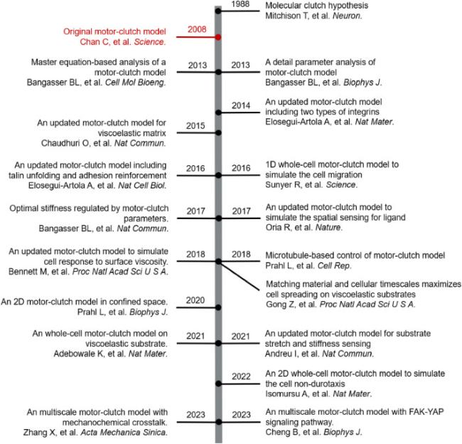
Fig. 1. Development timeline of motor-clutch model. |
Previous reviews on the motor clutch model have focused on elucidating its composition and describing how motor clutch models sense mechanical signals from the ECM, and transduce them into cells to influence cell traction on elastic substrates.6 However, the motor-clutch model and its extensions have also been widely used to study cell migration and cell spreading in different mechanical environments such as condensed spaces,7 viscoelastic substrates,8 and gradient stiffness substrates as well.9 These studies highlighted the importance of developing and understanding motor clutch models for cellular mechanobiology research, clinical detection, and treatments. This review thus provides a detailed description of not only how motor clutch modules sense mechanical signals from the ECM, but only how they influence cell behavior in a broader context, with the aim of providing profound insights into the theoretical and clinical applications of motor clutch models.
2. Physiology underlying the motor-clutch model
Integrins are critical elements of the molecular clutch system as they act as bridges between the extracellular and intracellular environments, and as vital receptors on the cell surface. Integrians control various cellular functions such as migration, differentiation, and survival by mediating relevant signaling pathways. The cytoplasmic domain of integrins interacts with adaptor proteins, whereas the extracellular domain binds to various ECM molecules (e.g., fibronectin, collagen, and laminin). Thus, integrins are essential for the transmission of mechanobiological signals between cells and the ECM.
Integrins activate critical intracellular signaling pathways through a cascade of downstream signaling proteins.10 Integrins initiate this process by binding to intracellular adaptors such as talin.11 Upon integrin-talin interaction, focal adhesion kinase (FAK) is phosphorylated and activated, and a cascade of signaling events is initiated. The activated FAK/Src complex phosphorylates paxillin and Cas, which then bind to C3G via Crk, thereby activating the Ras and MAPK cascades.12 This signaling pathway is essential for the control of anterior cell migration by actomyosin tension, which is regulated by Ras family proteins RhoA and Rac. RhoA activates the downstream Rho-kinase (ROCK), leading to myosin dephosphorylation, which allows myosin to interact with actin.13 Rac and RhoA have antagonistic roles, with Rac activating PAK, leading to the phosphorylation and inactivation of MLC kinase, reducing cell contractility, and preventing cell extension.14
FAK is another signaling adaptor protein that has been extensively studied in the context of malignant tumors. FAK phosphorylation results in the activation of key cellular proteins, such as PI3K and RAS, thereby activating the MAPK pathway.15 The activated MAPK pathway regulates cell proliferation, survival, and apoptosis, and thus influences cell fate.16 Integrin-talin interaction can also activate numerous intracellular signaling pathways and target proteins. When mechanical signals are overloaded and transmitted to talin via integrins, talin senses the increased load, unfolds, and exposes its binding sites for vinculin interaction. The activated integrin-talin-vinculin complex promotes integrin clustering, which facilitates cell adhesion and growth.17 Integrin clustering allows integrins to remain in adhesive structures, with β3 integrin clustering requiring the presence of talin.18 In addition, activated vinculin can also bind to actin and thus strengthen junction interactions.19
3. Original motor-clutch model
In 2008, Chan and Odde proposed a model to describe the effects of substrate stiffness on the traction dynamics of filopodia in neurons.5 Chan and Odde showed that integrins trigger two separate responses within integrin-mediated coupling upon sensing a stiffness signal. On high-rigidity substrates, rapid tension build-up leads to disengagement between the junction and actin filaments and a so-called “frictional slipping”. On the other hand, low-stiffness substrates prolong the interaction time due to slower tension development within the coupling, and thus increase the total tension generated and the so-called “load-and-fail”. Accordingly, maximum traction force occurs when the attachment time of the new coupling to the actin filaments is equal to the cycle time of the entire molecular clutch coupling, corresponding to optimal substrate stiffness.
However, modeling these phenomena using the traditional motor-clutch model requires extensive computational power, particularly for large-scale events. To address this issue, Bangasser (2013)20 proposed an ordinary differential equation (ODE) description of the motor-clutch model that reduces computational complexity, and expands the scope of simulations, allowing for larger spatial and temporal scales, such as whole-cell migration.
Some cell types, such as 3T3 fibroblasts and smooth muscle cells, showed a consistent increase in traction force with substrate stiffness,8,21 suggesting the existence of a detection limit in experimental settings. However, Bangasser22 also suggested that variations within the motor clutch components could alter a cell's perceived range of stiffness, potentially explaining the observed phenomenon. The authors performed a parameter sensitivity analysis, which showed that the optimal stiffness for cell migration can be manipulated by adjusting motor-clutch parameters. Increasing the clutch parameters shifted the optimal value toward a higher stiffness, whereas increasing the motor parameters resulted in the opposite.
4. Basic equations in the motor-clutch model
The motor clutch model incorporates various components, including myosin motor proteins, actin filaments, molecular clutches, and elastic substrates, as depicted in Fig. 2. In this model, myosin motor proteins exert forces on actin filaments, which lead to retrograde movement at a velocity of V0 even in the absence of external cellular forces. The model also accounts for the maximum pulling force produced by these motors, which is denoted as Fstall = nmFm, where Fm represents the maximum force generated by an individual motor. Since this force (Fs) is transmitted through all interconnected clutches, it results in a linear reduction in the retrograde flow velocity (Vr) of the actin filaments:
$V_{r}\left(F_{s}\right)=V_{0}\left(1-\frac{F_{s}}{F_{\text {stall }}}\right) \text {, }$
$F_{s}=K_{\text {link }} \sum_{i=1}^{N_{\text {link }}}\left(x_{i}-x_{s}\right),$
where V0 is the base flow rate of the actin filaments, and Fstall is the stall force of the myosin motors. Nlink is the number of engaged clutch bonds, xi is the elongation of spring i, xs is the displacement of the substrate.
$F_{i}=K_{\text {link }}\left(x_{i}-x_{s}\right),$
In this model, the clutch bonds are represented by parallel springs, where each spring is described with a spring constant denoted as Klink. As outlined by the Bell model, the force (Fi) applied to each bond accelerates the effective unbinding rate (koff) of the individual clutch bonds.
$k_{o f f}=k_{o f f 0} e^{F_{i} / F_{b}},$

Fig. 2. The key equations in original motor-clutch model. |
Here, Fb is the rupture force of the clutch bond, and koff0 indicates the initial unbinding rate of the clutches. It should be noted that the binding and unbinding of clutches are exceedingly dynamic processes taking place concurrently. The rate of clutch binding (kon) is also modulated by the integrin content.
$k_{\text {on }}=k_{o n}^{0} C_{\text {int }},$
Here, kon0 represents the initial binding rate of the clutches, and Cint is the integrin density on the cell membrane. Conversely, as the tension experienced by each clutch bond increases, unfolding of the talin protein is induced. Then, talin unfolding recruits integrins, thereby enhancing their adhesion. A threshold force, Fcr, was established to simulate this process. When the force (Fs) exerted by the clutches surpasses this threshold, integrin density Cint gradually increases with Dint. In contrast, when Fs is below Fcr, the integrin density Cint decreases with Dint.
5. Cellular mechanosensing of elastic ECM with uniform stiffness
Different cells respond differently to changes in the mechanical properties of the ECM (Fig. 3). The mechanical response to substrate stiffness varies significantly between the cells. Studies suggest the presence of higher levels of integrin and myosin expression in glioma cells than normal cells, increasing their sensitivity to substrate stiffness and resulting in a more pronounced response. In addition, differences in integrin types between different cells and their respective affinities for the substrate fibronectin contribute to variations in cellular responses to substrate stiffness. Determination of the clutch and motor parameters is critical to simulate the movement patterns of the different cells accurately. To this end, the mechanical response to the substrate stiffness in different cells and the component parameters that lead to these differences must be understood.

Fig. 3. The differences in mechanical response to the matrix stiffness in different types of cells. |
Recent studies have further highlighted the effects of integrin subtypes on the internal dynamics of motor clutch components, and consequently on the cell's perception of stiffness.23 Integrins have multiple subunits which perform different functions. Therefore, the type of integrin can affect the relationship between cell traction force and substrate stiffness. In 2014, Elosegui-Artola24 examined the role of αvβ1 integrin (constitutively expressed) or αvβ6 integrin (selectively expressed in cancer and development) in the perception of mechanical signals by cells. Their experimental results showed that in human breast epithelial cells containing only αvβ1 integrin, cell traction force peaked at a stiffness of 1 kPa, while αvβ6 integrin peaked at a malignant stiffness of 5 kPa. When both were present, no optimal stiffness was detected. Instead, a monotonic increase in the stiffness and cell traction force was observed. Furthermore, it was observed that αvβ6 integrin, which binds to fibronectin, had a fivefold higher density compared to αvβ1 integrin, and αvβ6 had a significantly higher affinity for FN than αvβ1. Thus, by regulating the expression of different types of integrins on the cell membrane, cells can regulate force generation and alter stiffness.
In addition to the integrin subtypes, the adaptor protein talin affects the optimal stiffness threshold of the cell's perception of substrate stiffness as well.25 Signal reception and transmission within the extracellular matrix is carried out by the mechanical link between integrin-talin and vinculin-fibronectin (FN). At low stiffness, the coupling experiences less loading, leading to dissociation of integrins from the FN and high actin flow. However, a high stiffness level leads to excessive loading, which leads the talin to sense the increased load and unfold. Unfolded talin then binds vinculin. The integrin-talin-vinculin complex can aggregate integrins through an active conformation, leading to cell adhesion and growth and lower actin flow. The authors also used talin2 shRNA to inhibit talin activity in mouse embryonic fibroblasts, and observed a significant decrease in cell traction force and a significant increase in retrograde actin flow on high-stiffness substrates (i.e., a biphasic relationship), in line with the results of the original motor clutch simulation.
The optimal stiffness for cell migration was then investigated by performing experiments and using a multiscale motor-clutch model (cell migration simulator, CMS) of U251 glioblastoma cells and embryonic chick forebrain neurons (ECFNs).26 The experimental results were found to agree with model predictions. Matrix mechanical responses of ECFNs and U251 glioma cells were also investigated. Accordingly, a ten-fold difference between the optimal stiffness levels of ECFNs and U251 cells was detected. Until the optimal stiffness was reached, the cell adhesion increased with increasing stiffness. Once the optimal stiffness is reached, excessive tension on the coupling can cause cells to detach from the substrate, leading to reduced adhesion.27 In addition, adhesion strengthening phenomena have been observed in certain cell types, in which adhesion is continuously strengthened with increasing stiffness. This phenomenon is attributed to the fact that, when the stiffness is too high, the average force on the linkage exceeds the threshold of the talin protein, causing it to unfold. Unfolded talin then recruits additional integrins, and thereby increases adhesion. In cases where myosin motors provide constant traction, adhesion enhancement compensates for the traction, resulting in a reduction in actin flow rate.
The CMS was used to validate these results. The researchers adjusted the number of motors and clutches to simulate the behavior of the two cell types, with the high-adhesion group having 100 times more motors and clutches than the low-adhesion group. The results validated the predicted mechanical responses of the two cell types by the model, suggesting that CMS can extend previous simulations into a third dimension, and thus can be used for computational diagnosis and treatment of disease.
6. Cellular mechanosensing of elastic ECM with gradient stiffness
Gradient stiffness of the ECM affects many biological mechanisms significantly, including wound healing, cancer progression, and morphogenesis. Gradient stiffness is also controlled by focal adhesions.28 The frictional forces for cell migration are provided by adhesion plaques formed between the cell and substrate, and are composed of integrin proteins, overlying adhesion proteins, and fibronectin. Adhesion plaques form continuously at the leading edge of the cell and disassemble at the trailing edge. Recently, we found that U-251MG glioblastoma cells can migrate to substrates with lower stiffness, which was attributed to lower levels of myosin contractility and talin.9 Experimental evidence has shown that cells treated with talin inhibitors exhibit reduced focal adhesion numbers and altered morphology, and thereby shift their durotactic response to softness. This confirms that cell contraction and adhesion mechanisms are the molecular determinants of cell durotaxis or non-durotaxis. To explain the sensitivity of cell migration to stiffness, Bangasser26 developed a cell migration simulator (CMS) based on a motor-clutch model that incorporates multiple randomly generated clutch systems to simulate cell protrusion. The model correctly predicted the mechanical response of the matrix at the whole-cell scale. The modules within the model are in force equilibrium, and the total amounts of actin, motors, and clutches remain constant to maintain mass conservation.
7. Cellular mechanosensing of viscoelastic ECM
Previous studies have focused on purely elastic substrates, where the substrate stress depends solely on deformation. Viscoelastic materials exhibit both viscous and elastic properties when deformed. Certain natural ECMs also exhibit viscoelastic behavior (e.g., a matrix composed of collagen I and fibronectin molecules), indicating that relationship between force and deformation is highly dynamic and time-dependent. In certain cases, viscoelastic solid materials exhibit stress relaxation, in which a stepwise constant strain leads to a decrease in stress. However, in other cases, the stress in the material cannot relax, as in the Kelvin-Voigt model. This model consists of linear springs and dampers arranged in parallel. This parallel arrangement causes the model to exhibit creep behavior, primarily because the springs and dampers cannot expand independently. Consequently, the model does not demonstrate steady-state creep or stress relaxation. After releasing the stress, the spring does not immediately return to its original position. Instead, the spring applies force to the damper, resulting in compressive creep under external stress. Eventually, after a finite time, all creep strains recover.
In our updated motor clutch model,29 we used a standard linear solid (SLS) model to represent a viscoelastic solid, which consists of a spring and a dashpot (as in the Kelvin-Voigt model). In this model, the stress relaxation time is $\tau_{s}=\eta / E_{a}$, where η is the matrix viscosity and Ea is the additional stiffness, i.e., the decrease in stiffness as the SLS element relaxes. Thus, due to this mechanical element, the matrix viscosity and stress relaxation time form a linear relationship. Hence, an increase in the viscosity causes an increase in the stress relaxation time.
Updated motor clutch models have been then proposed to predict cell mechanotransduction on viscoelastic substrates.30 When human osteosarcoma cells (U2SO) were seeded on elastic and viscoelastic substrates, a larger cell spreading area and stress fiber formation were observed on viscoelastic substrates than on pure elastic substrates at a low initial modulus, suggesting that the viscous behavior in a viscoelastic substrate can directly compensate for the decrease in the initial modulus.
Later, the effects of substrate surface viscosity (i.e., ligand mobility on supported lipid bilayers) on cell mechanosensing were investigated using a motor clutch model.31 For this purpose, C2C12 mouse myoblasts were placed on substrates with varying viscosities, ranging from low to high, such as DOPC and DPPC. The results revealed that as viscosity decreased, the cell morphology changed to a smaller and rounder shape, accompanied by a decrease in cytoskeletal tension. The actin flux also decreased with increasing viscosity, similar to that observed for rigid substrates. In the motor-clutch model, lower viscosity leads to a low tensile loading rate, which prevents the exposure of mechanosensitive sites in talin, ultimately leading to fewer connected clutch bonds and an increase in the actin flow rate. However, a higher viscosity causes a higher force loading rate, leading to an increase in the clutch bond on-rate and adhesion size and ultimately a decrease in actin flow.
The relationship between the adhesion size and ligand density on substrates with different viscosities was also investigated. Accordingly, at extremely low viscosities, the adhesion size is independent of ligand density. However, at extremely high viscosities, it increases with increasing ligand density. In the motor-clutch model, at extremely low viscosities (i.e., the ligands move rapidly on the substrate surface), the clutch bonds cannot establish effective tension due to the rapid dissipation of the tension generated by actomyosin. Therefore, talin unfolding does not occur regardless of the number of ligands. However, at relatively high viscosities, the associated clutch bonds generate effective tension. The force is distributed over fewer bonds in less dense substrates (low ligand number), leading to bond rupture and eventual collapse before bond reinforcement, resulting in a small adhesion size. However, as the number of ligands increases, the tension in the bonds increases the bond to strengthen, resulting in a larger adhesion size.
In addition to stress relaxation, the accumulation of matrix residual stress in cell clusters is another crucial factor that influences cell migration on viscoelastic substrates.32 Accumulation of residual stress is caused by both intrinsic and extrinsic cellular processes. Intrinsic processes include differences in cell signaling, the remodeling of subcellular structures, and their interactions.33 In active materials, such as tissues, the accumulation of residual stress can lead to irreversible structural changes at the subcellular level, such as adhesion and cytoskeleton, and thereby regulate cell migration.27,32,34 The extrinsic processes that contribute to the accumulation of residual stress primarily include the mechanical properties of the substrate and/or adjacent tissues. The cumulative effect of these mechanical and biochemical processes influences the configuration and rate of change of migrating cells, and subsequently, the behavior of multicellular systems.32,35
8. How do viscoelastic ECM affect mechanical properties of cells?
Eukaryotic cells are complex viscoelastic entities whose mechanical properties or phenotypes can undergo various changes during physiological and pathological processes. For example, pluripotent stem cells exhibit increased resistance to deformation as they differentiate.36,37 While the deformability of cancer cells is correlated with their invasive potential.38,39 Consequently, cellular mechanical properties are emerging as label-free biomarkers for delineating different cellular and pathological states. The concept of cell viscoelasticity encompasses several timescales, each of which plays a distinct role. At shorter timescales, rapid intracellular biomechanical events and consequent changes in the cell shape are prominent. Interactions within intracellular protein chains and cumulative effects of bilayer bending, together with changes in cell shape, contribute to the viscoelastic relaxation of cells within seconds. At intermediate timescales of minutes to hours, the focus is often on single-cell migration, whereas longer timescales of tens of minutes to hours are associated with collective cell migration. Finally, days or longer timescales are often influenced by biological processes such as cell growth, differentiation, and proliferation.40,41
Pajic-Lijakovic and Milivojevic42 developed a linear viscoelastic constitutive model to describe stress relaxation and formalized residual stress during collective cell migration. During this process, cell clusters exhibit viscoelastic behavior on both short and long timescales. Consequently, the model simulates cell-substrate interactions as an interplay of two viscoelastic systems. The model treats both systems as thermodynamically closed, and uses the minimum energy principle to maintain energy equilibrium within the systems. Within the system, the energy of the cell cluster, energy of the boundary layer of the cell cluster, and energy generated by the interfacial tension of the cell cluster are conserved. Thus, the interaction of cell clusters with the surrounding boundary layer during collective migration are simulated.
The effect of matrix viscoelasticity on the viscoelastic behavior of cells should be considered at the level of cellular adhesion. Changes in the viscoelastic properties of the extracellular matrix can lead to changes in cell adhesion density. Adhesion density is modulated by the products of various biochemical processes within the cell, such as cell signaling and gene expression. Changes in adhesion density subsequently induce variations in cellular stress, with a particular emphasis on the direction of adhesion contacts. In the context of the multicellular migration model presented by Pajic-Lijakovic,42 if the normal residual stress within migrating cells significantly exceeds the shear stress, adjustment of the shear stress within migrating clusters can be achieved by modifying the adhesion densities perpendicular to the migration direction and horizontal adhesion densities. However, the model had certain limitations as well. Although cells can endure normal stresses on the order of several kilopascals, they exhibit high sensitivity to shear stress. Shear stress as low as tens of pascals can trigger inflammation or even lead to cell death in epithelial cells.
9. Cellular mechanosensing of mechanical stretching
Perception of external mechanical signals is a key process in the regulation of cell migration and disease progression. Cells are considered not to respond directly to forces, but rather to the associated deformation of the extracellular matrix (ECM).43 The rate of loading is the product of the rate of ECM deformation and the effective stiffness of the substrate. Therefore, variations in stiffness, strain amplitude, and frequency affect the rate of force loading.
Andreu (2021)44 found that the loading rate affects cell mechanotransduction. In their study, the authors incorporated an experimentally derived force threshold of 142 pN acting on the substrate into the motor clutch model. Forces above this threshold resulted in the softening of the actin cytoskeleton and impaired force transmission, but only in the talin-stretched clutch. This study proposed that at moderate loading rates, talin unfolds on integrins, recruits more integrins, and enhances their adhesion and growth. However, at higher loading rates, the actin cytoskeleton is disrupted, thereby inhibiting reinforcement. Experimental results in human lung embryonic fibroblasts showed that at moderate strain rates, YAP nuclear translocation and adhesion size are maximal, but rapidly decrease with further increases in strain rate, indicating a reduction in cell contractility and decreased adhesion.
10. Cellular mechanosensing of extracellular fluid viscosity
Cells sense the properties of the extracellular matrix and respond to stimuli from the extracellular fluid (ECF). Here, the viscosity of ECF serves as a critical physical cue that changes under various physiological and pathological conditions. However, the mechanisms by which cells perceive and respond to this viscosity remain insufficiently understood.45,46 Integrins are considered to play a critical role in the response to ECF viscosity. During cell migration, F-actin pulls the cell edge and causes membrane ruffling, whereas viscous fluid can inhibit peripheral ruffling by increasing integrin-matrix contact.47 Recently, we also found that ECF viscosity and substrate stiffness synergistically affect cell migration. The motor-clutch model, incorporating the influence of the ECF, was updated to include the effect of viscosity on the rate of integrin engagement. The results showed that viscosity regulated the mechanical response of cells differently under different substrate stiffness values. Specifically, at high substrate stiffness, the ECF viscosity effectively enhances the mechanical response of cells, whereas at low substrate stiffness, only a higher range of viscosity can stimulate a higher degree of mechanical response.48
In the presence of flowing extracellular fluid (ECF), cells are subjected to shear stress generated by the motion of the fluid along their surface, which in turn alters their behavior. The theoretical model provides insights into this dynamic interaction. The flow velocity of the ECF directly affects the shear rate experienced by the cells, and the shear rate and viscosity influence the fluidic shear forces acting on the cells. Consequently, changes in the ECF flow rate or viscosity, while keeping the cell-ECF contact area constant, result in increased shear forces that affect cell behavior. Notably, cells are more sensitive to shear stress than to compressive stress.49 Shear stresses greater than a few Pascals significantly affect the state of epithelial cells.50 Elevated shear stresses, particularly over long periods, can be detrimental and cause severe damage to intermediate filament networks such as vimentin and keratin.51 A shear stress of 60 Pa was observed to induce inflammation in epithelial cells in as little as 5.5 h. ECF viscosity plays an important role in regulating a wide range of cell behaviors. This model contributes to a deeper understanding of the underlying mechanisms governing these phenomena.
11. Cellular mechanosensing of ligand spatial distribution
Oria (2017)21 found that adhesion strength is not only related to stiffness but also to the distance between integrin ligands. The adhesion formation has an optimal stiffness that decreases with increasing ligand spacing. As stiffness increases, adhesions with greater spacing are more likely to reach the force threshold, resulting in increased integrin recruitment. However, there is also a limit to integrin density. When the force load cannot be further compensated for by additional integrin recruitment, the adhesion collapses, significantly reducing the length of the adhesion.
12. Updated motor-clutch model for mechanochemical conversion and crosstalk
In the original motor-clutch model, the mechanisms behind the effect of clutch dynamics on the intracellular environment are not addressed. To this end, several updated motor-clutch models have been proposed to study the mechanochemical transformation and crosstalk at the integrin-ECM interfaces. In our updated motor clutch model,29 we resolved the relationship between the activation of FAK-YAP signaling and ECM mechanics. In a recent study by Zhang et al. (2023),52 the influence of factors on trail formation during neutrophil migration was analyzed using an improved motor-clutch model that included two mutually inhibitive signaling pathways: the PAK1/Rac/Arp2/3 and PAK/RhoA/myosin axes. The improved model includes the influence of chemical signals on cell migration. With the polarisation distribution of PAK1 and its downstream signalling molecules Rac and RhoA, integrin also accumulates at both ends of the cell migration direction, significantly increasing the migration speed and the number of trails formed. Subsequent experimental results also showed that the number of trials formed increased monotonically with increases in substrate hardness, integrin-ligand binding rate, motor-free speed, motor-free force, and number of junctions.
13. Motor-clutch model in mechanomedicine
Microtubules play a critical role in maintaining cell shape, division, signaling, and intracellular transport. Microtubule structure determines cell organization, organelle positioning, and functionality. Microtubules can regulate cell protrusion polarity and alter the cell traction force through the motor-clutch system. Prahl (2018)53 treated human glioma cells with microtubule-targeting agents (MTAs) to observe changes in cell spreading and updated the motor-clutch model to simulate cell migration. Experimental results showed a significant decrease in cell spreading area after MTA treatment, whereas simulation results indicated that altering the microtubule parameters does not affect the cell traction force. In contrast to the long protrusions in the control group, numerous short protrusions surrounding the cell periphery were observed in the MTA-treated cells. Subsequent analysis of whether microtubule proteins affect the unfolding of cell polarity revealed a decrease in the nucleation rate and loss of nucleation polarity in MTA-treated cells. The authors proposed that microtubules promote cell polarity by transporting inhibitory signals from the leading edge. MTA disrupts this link, leading to the accumulation of inhibitors, inhibition of protrusion, and formation of new protrusions in different regions of the cell. Ultimately, a change in the migration direction and a significant reduction in the cell migration distance are observed.
Cancer cells can modify the characteristics of the extracellular matrix (ECM) to enhance cancer cell migration and proliferation. Overexpression of fibronectin (FN) plays a key role in facilitating these processes within the remodeled tumor ECM. FN acts as a central organizer within the ECM. Upregulation of FN is linked to various aspects of cancer development, including angiogenesis, tumor progression, metastasis, and drug resistance. Consequently, numerous ligands targeting FN have been developed for use in cancer therapy. Weinberg (2017)54 developed a motor clutch model that incorporates the influence of the FN. This model was used to simulate the response of the FN to changes in ECM properties, and the feedback of this response to influence cellular behavior. The model suggests that the contractile forces of actin act on fibronectin (FN): when the force on FN exceeds a critical tension level, actin unfolds and exposes hidden FN-binding sites, promoting additional FN binding. As the substrate stiffness increases, the cell traction force also increases, leading to increased FN clustering and providing more ligands for integrins. Therefore, on rigid substrates, the cell traction force can continue to increase, which explains the phenomenon of monotonically increasing traction force with substrate stiffness.
Cells convert mechanical signals from the extracellular matrix (ECM) into chemical signals that influence the cell nucleus, and thereby determine cell fate (differentiation, migration, or apoptosis). This process is regulated by phosphorylation of focal adhesion kinase (FAK) at Y397. FAK phosphorylation affects a variety of downstream signaling proteins, leading to changes in cell behavior. Therefore, the analysis and prediction of these adhesion-related signaling proteins holds great promise for disease detection. Recent studies also indicated that an improved molecular clutch model can effectively simulate reciprocal interactions between various signaling proteins, including FAK, Rho, and YAP, and cell behaviors.55
Recent research has also revealed a fascinating mechanism of cell regulation in which the YAP/TAZ signaling pathway controls its own nuclear translocation. This process is influenced by the stiffness of the extracellular matrix (ECM), which promotes integrin binding, leading to aggregation and tension in the cytoskeletal contractile apparatus. The YAP signaling pathway plays a crucial role in determining cell fate. When dephosphorylated, YAP/TAZ enters the nucleus and triggers the transcription of genes associated with cell proliferation and migration. However, the dysregulation of this pathway has been observed in several solid tumors, contributing to cancer development, metastasis, drug resistance, and proliferation. Therefore, targeting YAP is a promising strategy for the treatment of gastric cancer (GC). Numerous YAP inhibitors and compounds have been developed and validated for potential therapeutic use.56
Cheng (2023)29 incorporated the effects of the YAP/TAZ signaling axis into a motor clutch model and established a computational model for YAP/TAZ nuclear translocation in matrix mechanosensing. Here, a computational model was used to simulate the responses of the cells to various matrix properties. The results showed a significant relationship between matrix stiffness and YAP/TAZ nuclear-cytoplasmic ratio, suggesting that these factors are positively correlated. This innovative model also provided valuable insights into the diverse mechanosensing behaviors of cells, and quantitative guidelines for mechanobiological transduction. The model also has the potential to identify promising targets for mechanomedicine, which may be crucial for the development of treatments that employ mechanical forces for therapeutic purposes. In addition, the predictive capabilities of the model allow the evaluation of existing inhibitor drugs, helping to estimate their efficacy in specific cellular contexts. Overall, this computational approach represents a promising avenue for advancing our understanding of cell behavior and for future biomedical applications.
14. Perspectives
The properties of the extracellular microenvironment, including the stiffness of the elastic matrix, viscosity, initial modulus of the viscoelastic matrix, and mechanical stretch, play significant roles in maintaining normal cell proliferation, differentiation, metabolism, and functional activities. The molecular clutch plays an essential role in communication between cells and the microenvironment as a key component of cell adhesion. The molecular clutch model is closely associated with disease and may serve as a therapeutic target for several conditions. Several potential targets related to molecular clutches and existing drugs are summarized in Fig. 4. Treatments targeting cell adhesion have been successfully used to treat cardiovascular diseases, viral infections, and multiple sclerosis.57

Fig. 4. Potential drug targets for modulating molecular clutch dynamics. |
Integrins are involved in multiple stages of cardiovascular disease, ranging from atherosclerosis to thrombus formation. For instance, integrin αvβ3 facilitates the homing and infiltration of inflammatory cells. Blocking this integrin specifically is effective in preventing NF-κB activation, which includes p21-activated kinase (PAK) signaling on fibronectin.58 Additionally, α4β1 and several β2 integrins have significant roles in the formation of atherosclerotic plaques.59 Activation of α9 has been shown to induce platelet adhesion, promoting platelet aggregation and thrombus growth. In a study of integrin α9 gene-deficient mice with high-fat aging, levels of various inflammatory factors, thrombus formation, and stroke severity indicated a significantly reduced likelihood of stroke and lower levels of inflammatory factors in α9-deficient mice.60 Multiple sclerosis is an autoimmune disorder characterized by inflammation of the brain and spinal cord, with a key mechanism involving the infiltration of autoreactive immune cells into the brain. Consequently, identifying adhesion molecules that contribute to leukocyte activation and mediate their migration is crucial for exploring potential therapeutic interventions against multiple sclerosis. Among these adhesion molecules, integrins, particularly α4 integrins, have emerged as significant players in the disease progression. These proteins facilitate the homing of leukocytes to the brain and expedite the inflammatory response, underscoring their importance as potential targets for treating multiple sclerosis.61
Tumor cells can alter their microenvironment to favor development by secreting inflammatory factors and immunosuppressive signals. In the early stages, tumors induce matrix fibrosis, which increases tumor stiffness and enhances integrin signaling and proliferation. During metastasis, cancer cells break integrin-mediated adhesions and detach from the extracellular matrix, resulting in a migratory phenotype. Metastatic tumor cells typically exhibit characteristics consistent with the epithelial-mesenchymal transition (EMT), allowing epithelial cells to migrate in a mesenchymal form. This enables cancer cells to invade a high-density ECM, facilitating their spread to distant sites.54 Therefore, understanding cell adhesion is fundamental for controlling tumor migration and its application in therapy. Currently, targeting the RGD motif binding integrin αv is a promising approach due to its role in cancer, viral infection, fibrosis, and other diseases. The stages of tumor development include growth, metastasis, and angiogenesis, which provide nutrients for tumor cell proliferation, with integrins playing an important role. Integrin αv has been identified as a biomarker for several cancers, including breast, liver, and gastric cancers.62 Therefore, targeting of cell adhesion holds great promise for cancer treatment.
The mechanism of wound healing involves re-epithelialization, in which epithelial cells close the wound gap. This is achieved by the proliferation and migration of epithelial cells, which form a barrier between the wound and surrounding environment. Epithelial cell closure in the wound gap is attributed to the purse-string contraction of the actomyosin filament and active cell migration.63 Recent studies have shown that altering cell density and wound shape can control the rate and polarity of wound cell migration.64,65 These phenomena arise from the reorganization of actin filaments and adhesion between cells and extracellular matrix. Therefore, understanding the principles of cell migration is crucial as it is a necessary step in the transition from basic research to clinical applications. This will not only help identify disease targets but also optimize existing techniques.
15. Conclusion
In this review, we provided an overview of the motor clutch model as a computational tool for studying the interaction between cells and the microenvironment, in addition to its development. Starting from the original motor-clutch model, updates to motor-clutch models under different external mechanical conditions have enabled broader applicability of this framework throughout mechanobiology as extensions from two-to three-dimensional frameworks. Current applications and prospects of the motor clutch model are driving further innovations in drug development and mechanomedicine, demonstrating its continued and growing value as a predictive framework in the field of mechanobiology.
Declaration of competing interest
The authors declare no competing interests.
