
1. Main
In recent years, the occurrence of various microbial infections caused by viruses and pathogenic bacteria has already posed a huge threat to human society. In general, mechanical adhesion of microorganisms to host cells is a critical first step for them to invade and infect the hosts. The strength of interfacial adhesion between microorganisms and the hosts largely determines the outcome of subsequent invasion and infection (Fig. 1).1 It is worth noting that the spatiotemporal dynamics of interfacial adhesion are not only modulated by mechanical microenvironments in which they live, but also involve a series of complex mechanobiological processes. There is increasing evidence indicating that the mechanical microenvironments play a non-negligible role in mediating the functions and behaviors of host cells, thereby subtly exerting an important influence on bacterial pathogen-host interactions.2,3,4 Simultaneously, it has been demonstrated that the interfacial adhesion can trigger a cascade of mechanotransduction pathways in the host cells, which may in turn regulate the interactions between bacterial pathogen and host cells.2 It is crucial for the prevention and treatment of infectious diseases to comprehensively investigate the mechanical interactions between microorganisms, host cells and their living microenvironments and thus reveal the underlying mechanobiological mechanisms.
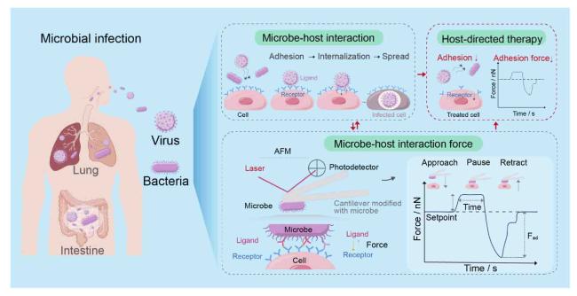
Fig. 1. Microbe-host interfacial interactions. During microbial infections, bacteria or viruses essentially undergo the processes of adhesion, internalization and spreading to interact with host cells. By test methods, such as AFM-based SCFS, individual microorganisms can be modified on the surface of the probe to quantify microbe-host interaction forces by approaching, staying and retracting from the host cell, where the adhesion force (Fad) directly reflects the degree of infection. Host-directed antimicrobial therapies by blocking the infection process, further combined with interaction forces as indicators provide new insights for the treatment of microbial infections. |
Single-molecule force spectroscopy (SMFS) based on atomic force microscopy (AFM) and single-cell force spectroscopy (SCFS) based on fluidic force microscopy (FluidFM) provide quantitative tools for detecting the interfacial adhesion between microorganisms and host cells, in which the regulatory role of the microenvironmental cues can also be explored. SMFS can accurately characterize the binding forces as small as piconewtons between a single receptor and the corresponding ligand on viruses, bacteria or host cells,1,5 whereas SCFS may quantitatively measure the interfacial adhesion forces between bacteria and host cells up to tens of nN or higher.6,7 With the aforementioned interfacial force measurement technology, for instance, a recent investigation has revealed that heparin-induced allosteric changes in the spike protein of severe acute respiratory syndrome-coronavirus-2 (SARS-CoV-2) can facilitate the binding of host cell receptor angiotensin-converting enzyme 2 (ACE2) and viral entry, which furthers the understanding of the SARS-CoV-2 infection mechanisms and uncovers the potential interventions targeting viral entry from a mechanobiological perspective.1 Likewise, a SMFS-based study has demonstrated that bacterial surface-located clumping factors can form strong catch bonds with annexin A2 expressed on surface of host cells, which not only identifies a force-dependent interfacial adhesion phenomenon during bacterial pathogen-host cell interactions, but also opens new ideas for designing new therapeutics targeting intracellular Staphylococcus aureus (S. aureus).5
Recently, we have employed the FluidFM-based SCFS to quantify interfacial adhesion forces between pathogenic bacteria, e.g., S. aureus and Escherichia coli (E. coli) and host cells, e.g., IEC-6 cells (rat small intestinal epithelial cell line-6) and HaCat cells (human keratinocyte cell line) (Fig. 1), and therefore developed an in vitro model to quantitatively study bacterial-host cell interactions mediated by mechanical microenvironments of extracellular matrices.7 Based upon a well-developed microcontact printing approach, we first established a series of host cell monolayers with different geometric shapes and sizes on collagen-coated polyacrylamide (PAAm) substrates whose rigidities were 10.14 kPa (soft), 32.29 kPa (medium) and 93.46 kPa (stiff), respectively. Subsequently, we explored the spatiotemporal dynamics of interfacial interactions between the geometrically confined host cell monolayers and the pathogenic bacteria expressing green fluorescent protein (GFP).7 Surprisingly, most bacteria always adhered near the edges of the geometrically micropatterned host cell monolayers, showing strong spatial heterogeneity during the bacterial-host interactions. Further, we revealed that bacterial (S. aureus) adhesion forces increased linearly along the radial direction of a circular IEC-6 cell monolayer (200 μm in diameter), approximately. These facts demonstrate that spatially geometric constraints on host cell monolayers intrinsically play a key role in modulating bacterial-host interactions. Also, our experimental results showed that the interfacial adhesion forces were directly related to the underlying substrate stiffness. Specifically, the peak adhesion force near the outer edges of the circular cell monolayers cultured on the soft substrates was ∼40 nN while it increased to ∼75 nN when the cell monolayers were cultured on the stiff substrates.7 These findings imply that the interfacial adhesion is susceptible to mechanical aspects of extracellular matrices as well.
To decipher the mechanobiological mechanisms behind the spatially heterogeneous bacterial-host interfacial adhesion, we performed single-cell RNA sequencing and Monte Carlo simulation analyses and thus identified that heterogeneous expression of type IV collagen in the host cell monolayers caused by the geometric constraints was responsible for the spatial location-dependent bacterial adhesion. Along this line, we screened collagen IV inhibitor, i.e., VU6015929, as antibiotic adjuvants, which could effectively reduce bacterial-host interfacial adhesion and hence significantly enhance the antibiotic efficacy, as confirmed in a rat wound infection model.7 These not only highlight the critical regulatory role of interfacial adhesion in bacterial infection, but provide a mechanobiology-inspired idea for the screening of new antibiotic adjuvants and the development of new host-directed anti-bacterial therapies.8 This point is of vital importance for substantially combating microbial infections induced by drug-resistant bacteria and highly pathogenic bacteria considering that there is currently a huge gap in antibiotic development and the corresponding clinical applications.
Once the pathogenic bacteria form a relatively stable interfacial adhesion on the host cell surface, they have the opportunity to invade and internalize into the host cells, during which the extracellular matrix (ECM) stiffness still plays a non-negligible regulatory role.9,10 As reported in our previous work,2 bacterial adhesion and invasion are closely associated with spatial distribution of ECM stiffness-regulated F-actin cytoskeletons in host cells, because the process of invasion and internalization of the pathogenic bacteria into the host cells involves rearrangements of the host cytoskeletons. More unexpectedly, as the ECM stiffness increases, there is an invasion mode transition from agminate invasion to dispersive one. At the same time, intracellular accumulation of antibiotics and therapeutic efficacy for elimination of internalized bacteria are ECM stiffness-dependent in nature. These findings offer a valuable reference for developing new antibiotic therapy against serious bacterial infections diseases in clinic. Overall, these studies not only demonstrate that the critical regulatory role of interfacial adhesion and mechanical microenvironments in microbe-host interactions, but also lay a preliminary foundation for in-depth investigations on microbial-host infection from a mechanobiological perspective.
Ethical approval
This study does not contain any studies with human or animal subjects performed by any of the authors.
Declaration of competing interest
The authors declare that they have no known competing financial interests or personal relationships that could have appeared to influence the work reported in this paper.
Acknowledgements
This work was supported by National Natural Science Foundation of China (Grant no. 12372175).
