
1. Introduction
Protein, a fundamental constituent of living organisms, plays a crucial role in physiological processes and pathogenesis of diseases.1 However, as depicted in Fig. 1, conventional biological detection techniques can only identify less than 30% of known human proteins,2 limiting the utilization of numerous potential biomarkers in both life science research as well as clinical diagnosis and treatment. Taking cytokines as an example, which hold significant value in immunological research, clinical diagnosis, disease progression monitoring, efficacy assessment, and treatment surveillance; studies have demonstrated that resting levels of cytokines in healthy individuals can reach concentrations as low as fM.3 In early stages of tumors and infectious diseases, the concentrations of protein biomarkers in blood may even down to aM,4,5 that is significantly below the detection limits of traditional enzyme linked immunosorbent assay (ELISA) methods (typically at pM levels,6 as shown in Fig. 1). Furthermore, these low-abundance proteins are prone to be obscured by other high-abundance proteins (such as albumin and globulin) in samples. This further exacerbates the challenges to detect these proteins and results in missed opportunities for early detection and diagnosis of major diseases, leading to delayed optimal treatment timing and unfavorable outcomes.

Fig. 1. The concentrations of biomarkers in human blood.7 |
At the same time, the regulatory network of proteins that participate in various physiological functions in the human body is extremely complex. Detecting one protein alone falls short of meeting the requirements for clinical diagnosis. Previous studies8, 9, 10 have already demonstrated that simultaneous detection of multiple analytes (ranging from dozens to nearly one hundred types) could significantly improve the diagnostic sensitivity and specificity of diseases, thereby assisting doctors in achieving more precise disease diagnosis and treatment, prognosis assessment, and relapse monitoring. Taking COVID-19 infection as an example, critically ill patients have shown significant increases in multiple cytokines known as a cytokine storm,11,12 such as interleukin (IL)-6, IL-10, IL-7, and tumor necrosis factor-alpha (TNF-α). It would be significant for rapidly observing the progression and deterioration of the disease, and further responding promptly if these inflammatory factors related to COVID-19 or immunotherapy-induced inflammation can be detected in time. However, traditional methods fail to simultaneously achieve multiplex detection in single-tube and high detection sensitivity. Hence, one critical challenge in the clinical application of protein biomarker detection is the ultra-sensitive multiplex detection for low-abundance proteins. Meanwhile, relying on central laboratory testing methods are often time-consuming, making it unable to meet the immediate testing needs for cytokine storms and other sudden illnesses. A portable, inexpensive, and high-performance point-of-care testing (POCT) solution capable of constantly monitoring biomarker changes awaits breakthroughs.
Microfluidic chips, owing to their compact size, high reaction efficiency, and minimal reagent consumption, exhibit great potential in POCT.13 Various immunoassays have been developed based on microfluidic platforms,14,15,16 which integrate sample pre-treatment, multi-step reactions, and analysis within small volume, enabling complex processing procedures while minimizing reagent waste and reducing the cost of detection. These advantages contribute to the cost-effective and rapid detection, as well as augment the potential application in POCT for immediate diagnostics.
Beads play a crucial role as carriers for the identification, capture, and manipulation of target molecules in protein immunoassays. Integrating beads with microfluidic platforms could combine high-efficiency capture and separation capabilities with advantages such as miniaturization and low reagent consumption, which is particularly suitable for addressing challenges in POCT for multiplex protein detection in clinical diagnostics. Beads, also known as microspheres and particles, are typically nano or micrometer-sized particles with a high surface area-to-volume ratio and multiple functional groups on the surface for efficient protein capture. The protein capture efficiency and resistance to nonspecific adsorption of impurities could be optimized by modifying the physicochemical properties of the bead surface. By incorporating magnetic materials such as Fe3O4 inside the microspheres to form magnetic beads, they can be easily manipulated and separated using a magnetic field. The enhanced relative motion between the beads and the sample fluids promotes contact and capture efficiency of antibodies, peptides, aptamers, or other ligands on the surface of beads with target proteins, that enables effective protein target capture and unwanted impurity removal through magnetic separation. In addition to as capture carriers, beads could also serve as signal labels like gold or silver particles or by incorporating fluorescent molecules within beads. Due to the larger size compared to single fluorescent molecule, more fluorescent molecules can be loaded inside these beads to enhance signal intensity while providing protection against the external environment through durable coatings on the outer surfaces.
In summary, with the development of clinical applications, there is an escalating demand for simultaneous detection of multiple low abundance proteins. This review presents a comprehensive overview of the progress in bead-based ultrasensitive multiplex immunoassays using microfluidic systems over the past decade, including encoding techniques, the role of microfluidic platforms, applications, and prospects. The key points of this review are shown in Scheme 1.
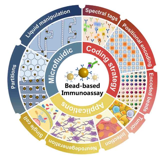
Scheme 1. Summary of the key points of bead-based immunoassay. |
2. Encoding techniques
The simultaneous detection of ultra-low abundance multiple proteins is an urgent requirement in order to address the changes in various biomarkers accompanying the occurrence and progression of diseases. This poses a challenge as dividing the sample into multiple tubes may introduce variations in target molecule concentrations between different tubes and further compromise detection sensitivity. In such cases, simultaneous detection of multiple protein targets in a single microfluidic chip offers advantages in terms of reducing sample consumption, improving detection sensitivity and throughput, as well as lowering labor and cost. Encoding techniques in multiplex detection within microfluidic chips can be categorized into three forms (Fig. 2): 1) spectral tags; 2) positional encoding; 3) encoded beads.

Fig. 2. Multiplex encoding methods: (a) spectral tags; (b) positional encoding; (c) encoded beads. |
2.1. Spectral tags
Different spectral tags are molecules with distinct fluorescent properties. The simultaneous detection of different target proteins is achieved by labeling different spectral tags on the corresponding detection antibodies and distinguishing their spectral information. For instance, Gilbert et al.17 demonstrated the simultaneous detection of three cytokines by three fluorescent tag labeled antibodies within microfluidic channel in the Singulex system; Stambaugh et al.18 labeled SARS-CoV-2 and influenza A antigens by two bright fluorescent probes, then released them from the magnetic beads, and introduced into a multimode interference waveguide microfluidic platform for detection. However, due to limitation of non-overlapping signal spectra, multiplexing capability of spectral tags is restricted, usually less than five targets in single microfluidic chip.
2.2. Positional encoding
Positional encoding is immobilizing distinct capture antibodies at specific locations in microfluidic channels. By leveraging known types and designated positions of each capture antibody, multiplex detections can be achieved on microfluidic chips through different physical locations.19,20 Higher encoding multiplicities could be easily attained by expanding the spatial dimensions, enabling analysis of hundreds of detection targets in a single assay. However, due to the reliance solely on molecular diffusion for analyte capture, fixed surfaces exhibit relatively low reaction kinetics, necessitating the utilization of additional signal amplification methods to enhance detection sensitivity. For instance, Dhanapala et al.21 used enzyme-labeled beads as signal amplification units and simultaneously detected samples on 56 microfluidic electrochemical sensors with sub-fg/mL (0.08-0.22 zmol) sensitivity. Moreover, positional encoding methods shows extensive application in POCT of nucleic acid, particularly in microfluidic platforms with magnetic bead pre-processing and isothermal amplification in multi-reaction wells.22,23,24 Li et al.,25 for example, developed a microfluidic system that integrated bacterial lysis, magnetic bead based DNA extraction, and multiplex loop-mediated amplification (LAMP), allowing simultaneous sample distribution into eight different partitions for 8-plex LAMP amplification and analysis.
2.3. Encoded beads
Encoded beads are also known as nano- or microscale suspended chips that carry coding signals, which require pre-labeling with specific capture antibodies for each type of encoded beads during the execution of the immunoassay process. Multiple types of encoded beads could simultaneously capture their corresponding target proteins.26,27,28 Using detection devices such as flow cytometers or microscopes, both the bead types and signal intensities can be identified simultaneously, enabling multiplex detection of target proteins in one reaction. This technology could easily increase the multiplexing capability from less than five targets in traditional dye-based method to hundreds of targets, which is currently primary multiplexing form in microfluidic digital ELISA methods29,30 and is considered as the technology most likely to be applied in actual clinical practice.26,31 For instance, Yi et al.32 employed encoded beads as capturing carriers for five target cytokines and achieved ultra-sensitive detection on a droplet-based microfluidic chip with sub-fM sensitivity.
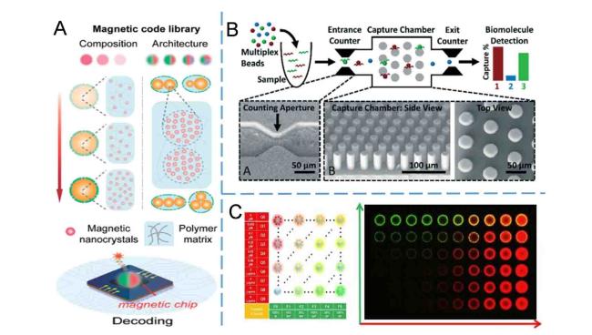
According to the encoding signals and decoding devices employed, encoded beads can be classified into three types: magnetic field,33 electric field,34 and optical spectra17 (as shown in Fig. 3). Magnetic field encoding is achieved by adjusting the magnet composition to control their magnetic responsiveness. Liu et al.33 fabricated encoded beads by microfluidic chips through optimization of the position and composition of magnets within the beads, which allows for 100-10,000-fold encoding capacity theoretically (Fig. 3A). In electric field encoding, Cowell et al.34 devised hydrogel beads exhibiting diverse electrical impedance characteristics for target capture and detection (Fig. 3B). The beads capturing the corresponding targets were confined within a columnar-shaped microfluidic chip, while other beads remained unconfined, enabling quantification of target concentrations by comparing the bead count before and after trapping in the microfluidic chip using a Coulter counter. Due to the superior stability and lower magnetic background noise in samples compared to fluorescence and electrical methods, magnetic field encoding method holds better prospects for applications. However, since the current mainstream reporting signal typically relies on optical detection, the decoding equipment used for magnetic field and electric field methods cannot be universally applied to interpret the reporting signal, thereby increasing the complexity of detection equipment. Consequently, these two encoding methods are not as extensively employed as optical spectra methods.
Optical spectra encoded beads could be further categorized into four groups according to the diverse optical signals used:17 Raman,36,37 time-resolved spectroscopy,38 reflectance spectroscopy,39,40 and fluorescence spectroscopy.41,42 Among these categories, Raman and time-resolved encoding techniques have the potential to achieve high coding capacity of up to tens of thousands by utilizing information such as Raman frequency or fluorescence lifetime, intensity, and wavelength. For instance, Hu et al.36 engineered ten distinct Raman frequencies on beads with a diameter of 3 μm through manipulation of conjugation length, bond-selective isotope doping, and end-capping substitution of polyynes. This approach could generate unique spectral barcode beads numbering up to 59,048, theoretically. Reflectance spectroscopy typically employs photonic crystals as the material for beads. These dielectric materials with periodic structures could result in specific frequencies of light reflection under illumination. Liu et al.,39 for example, employed size-controllable silica hybrid photonic crystal beads constructed via microfluidics technology. However, their sizes often exceed 100 μm in comparison to other encoding beads with diameters below 10 μm, potentially resulting in compromised reaction kinetics. Benefiting from the widely used flow cytometry platforms, fluorescence encoding becomes the most commonly used technique in encoded beads due to its ease-of-handling and accuracy.41,42
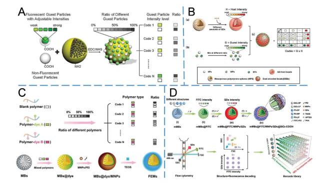
According to the specific fluorescent materials employed, fluorescence encoded beads can be further categorized into organic dyes, quantum dots, and aggregation-induced emission materials.43 To augment the coding capacity, apart from modulating the intensity of monochromatic fluorescence, multiple hues of the same material or diverse materials with distinctive colors can be co-doped within a single bead. This strategy has been adopted by commercialized suspension array platforms such as Luminex and BD. However, in the pursuit of increasing the number of codes through multi-color doping, potential interactions between dyes or quantum dots (such as fluorescence resonance energy transfer) may introduce complexities in the distribution of individual clusters or the final fluorescence spectra of beads, thereby complicating the encoding process and reducing the multiplex capability.44,45,46 To address this issue, some groups47,48 have employed aggregation-induced emission materials that leverage their characteristic aggregate luminescence to overcome signal saturation and quenching issues when heavily loaded. Our research group also integrated organic dyes and quantum dots into two independent beads (Fig. 4A and B), forming a host-guest structure42 to effectively mitigate optical interference.49 By employing beads of different sizes, a 3D barcode library with an encoding capacity of 100 were successfully prepared using this approach35 (Fig. 3C). Moreover, the guest beads were enhanced by implementing a spherical polyelectrolyte brushes structure with increased dye-carrying capacity,50 resulting in an encoding capability of 10151 (Fig. 4C). Furthermore, specific structural coding enabled a further increase to 300 encoding capability52 (Fig. 4D), currently the highest fluorescence decoding multiplicity achievable with a single laser.
The combination of these encoding beads and microfluidic platforms is expected to improve detection throughput, reduce costs and shorten detection times for ultra-sensitive protein detection.
3. The role of microfluidic platforms
In bead-based multiplex immunoassays, microfluidic platforms typically play two roles. One is as partition tools, often combines with encoded beads in digital ELISA detection, utilizes microfluidics to efficiently and conveniently encapsulate encoded bead-target protein-enzyme complexes into tens of thousands or even millions of small partition chambers for ultrasensitive detection. The other role is mainly as platforms for liquid manipulation, providing pre-treatment methods and platforms for subsequent electrochemical and fluorescence detection methods, so as to make the whole system more integrated and conducive for POCT applications.
3.1. As partition tools
Depending on the method used as partition tools, microfluidic platforms can be further classified as either microwell array-based microfluidic platforms or droplet-based microfluidic platforms.
3.1.1. Microwell array-based microfluidic platforms
Digital ELISA based on beads and microfluidic well array was first proposed by David R Walt's research group in 201053,54 for low abundance protein detection (as shown in Fig. 5A) and subsequently commercialized by Quanterix company55 known as Simoa™ with a limit of detection (LOD) ranging from aM to fM. This method utilized magnetic beads as carriers, which were coupled with antibodies to specifically capture target proteins. Subsequently, detection antibodies labeled with enzymes were further linked to form an immune sandwich complex after capturing the targets. The beads were mixed with substrate solution and introduced into an array of microwells with a volume of 50 fL, where each well was separated and contains no more than one magnetic bead. By quantifying the presence or absence of the enzymes on the beads, the concentration of target proteins in samples could be calculated using Poisson distribution.56 The multiplex digital ELISA was implemented by substituting beads with encoded beads. The Simoa platform currently enables the analysis of up to ten targets in a single reaction.57 To address the issue of cross-reactivity in multiplex assays, Duffy's group58 further developed a sample re-utilization method wherein three types of magnetic beads, each capturing specific targets, were sequentially introduced and removed from the same sample before being mixed together into the microfluidic well chip for detection. The results have demonstrated that this procedure effectively mitigates interference in multi-detection assays while enhancing the dynamic range.
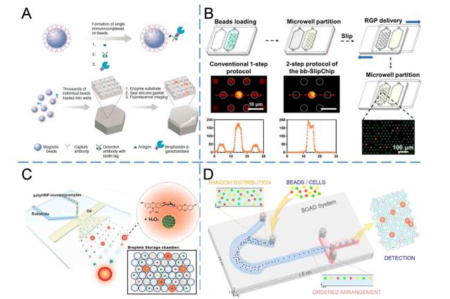
In addition to the aforementioned challenges, achieving a certain statistical quantity of beads is imperative to ensure the dynamic range and sensitivity, as well as reduce statistical errors caused by Poisson distribution. However, the proportion of microwells containing one bead is typically limited to approximately 10%, thereby impeding advancements in both detection sensitivity and multiplicity. To tackle this issue, Duffy's group61 integrated a magnet at the bottom of the microfluidic well chip to enhance the proportion of microwells containing one bead. However, despite these efforts, the pre-mixing of enzyme substrates and beads prior to loading into the wells still leads to elevated background signal. Decrop et al.62 employed a magnet and repeated loading of beads on an open microfluidic well array chip to enhance well utilization efficiency up to 96%, while concurrently mitigating background noise through a secondary injection of substrates. In our study,59 the background signal was effectively reduced through a two-step loading process, while achieving a high bead loading efficiency of over 90% combining slipchip and microwell array techniques (as illustrated in Fig. 5B). As a result, the detection sensitivity of IL-6 and IL-10 was improved by 2.0-2.6 times compared to the Simoa system.
3.1.2. Droplet-based microfluidic platforms
Compared to the aforementioned microwell platforms, droplet-based platforms for multiplex digital ELISA offer enhanced detection throughput and sensitivity by overcoming partition number limitations as well as enabling analysis of a larger number of beads (as illustrated in Fig. 5C). Shim et al.63 developed a bead-based digital ELISA platform capable of generating droplets with volumes ranging from 5 to 50 fL at a frequency exceeding 1 MHz, enabling the formation of millions of droplet partitions within a short period of time, surpassing the capacity of traditional microwell chips. Utilizing this chip design, a LOD of 46 fM was achieved for prostate-specific antigen (PSA) after 10 min in-chip incubation. In the context of multiplex detection, Yelleswarapu et al.64 employed dual-encoded beads for the simultaneous detection of granulocyte-macrophage colony-stimulating factor (GM-CSF) and IL-6. By leveraging the parallel operation of one hundred droplet generators and time domain-encoded mobile phone imaging, they achieved a remarkable enhancement in both droplet generation and detection speed by a hundredfold compared to conventional single-channel methods, while attaining a LOD as low as 300 aM. Our research group32 has also developed a fluorescence-encoded bead-based multiplex digital ELISA method with rapid amplification signals within 5 min using poly-horseradish peroxidase (poly-HRP) as enzyme labels. As a result, simultaneous detection of five cytokines was achieved with 100-10,1000 times improvement in LOD compared to conventional multiplex suspension array chip technology.
These droplet-based digital ELISA methods effectively address the bottleneck issue of pre-catalysis concerns by delivering the substrate solution and beads separately through two microfluidic channels, and then encapsulating the two phases within individual droplet simultaneously, thereby addressing the limitation inherent in microwell-based digital ELISA methods. Additionally, this approach leads to a significant increase in the number of detected beads through a substantial increase in partitions. Cohen et al.65 conducted a comparative study of the detection performance between microwell-based digital ELISA (Simoa) and droplet-based digital ELISA (ddELISA) with varying bead utilization ratios. The ddELISA achieved a bead utilization ratio of approximately 60% by mitigating the loss of beads during both the generation and detection processes, while Simoa exhibited a mere 5% bead utilization ratio. Results demonstrate that ddELISA can enhance detection sensitivity by 25-fold compared to Simoa when employing identical target proteins, revealing the importance of enhancing bead utilization ratio in digital ELISA. However, the encapsulation of individual beads in droplets is inherently stochastic. Constrained by the Poisson distribution,56 less than 10% effective partitions contain a single bead, while approximately 90% of the droplets remain unoccupied. The aforementioned literature enhances the bead utilization by generating a huge number of droplets but does not effectively mitigate the presence of empty droplets. These unoccupied empty droplets consequently lead to significant wastage of analytical resources during subsequent detection and analysis. For example, achieving digital detection with statistical significance and linear range in multiplex detection requires a minimum of 10,000 effective partitions per target. However, in the conventional system, single target detection necessitates 100,000 droplets at least due to Poisson distribution encapsulating restrictions. Apparently, a system with 100% effective partitions can simultaneously detect ten targets using the same number of droplets. In essence, increasing the ratio of effective partitions under limited droplet numbers undoubtedly enhances detection multiplicity while reducing both detection time and reagent consumption. To address this issue, our research group has proposed a bead ordered arrangement droplet (BOAD) system60 in order to overcome the limitations imposed by the Poisson distribution (as shown in Fig. 5D). By achieving a precise alignment of beads within the microfluidic channel, the efficiency of single-bead encapsulation was successfully enhanced up to 86%, representing a remarkable nine-fold increase compared to conventional detection systems. Simultaneously, by encapsulating over 50 kinds of fluorescent encoding microspheres into the generated droplets, the potential of multiplexed ultrasensitive detection is demonstrated.
According to the authors' perspective, for digital ELISA devices, if the current manual or large automated instruments used in the reaction process could be integrated into a compact microfluidic chip and the imaging module could be further miniaturized, it would enable enhanced utilization of microfluidics' advantages in terms of miniaturization and integration. On the other hand, irrespective of whether microwell-based or droplet-based partitioning methods were employed, it remains challenging to completely avoid the loss of beads during the partitioning and detection processes, even with an increased proportion of effective partitions. These losses not only escalate the consumption and cost of beads in tests but also diminish detection sensitivity.65 Moreover, the manufacturing cost and operational complexity of microfluidic chips are relatively high. By employing beads as partitions66,67 in digital ELISA instead of microwell or droplet partitions, the issue of bead loss during the partitioning process will be eliminated, thereby potentially enhancing the detection ratio of beads and the sensitivity, simplifying reaction processes, and reducing reagent consumption. Furthermore, the integration of cost-effective and portable imaging systems such as smartphones could potentially facilitate advancements in next-generation digital ELISA for POCT.
3.2. As liquid manipulation tools
In addition to serving as a partitioning platform for dELISA, offering the advantages of easy manipulation and compatibility with various detection methods, microfluidic platforms could also be utilized as tools for liquid manipulation during immunoreaction and washing steps within microchannel. The use of beads in microfluidic platforms as capture or labeling carriers further enhances detection sensitivity and throughput. Additionally, by carrying out all processes within the microchip, these platforms effectively harness the benefits of miniaturization and integration offered by microfluidics. In the following sections, relevant literature will be classified according to detection methods: optical detection and electrochemical detection.
3.2.1. Optical detection
Integrated with microfluidic chips, the effects of beads in optical detection can be broadly categorized into two types: capture carriers and label carriers.
As capture carriers, the beads' substantial specific surface area could expedite the target capture process. Moreover, their facile conversion into magnetic beads enables convenient removal of non-target impurities through manipulation of a magnetic field. For instance, Stambaugh et al.18 employed magnetic beads to selectively capture target antigens labeled with brightly fluorescent reporter probes and eliminated impurities outside the microchip. They then employed ultraviolet light to break apart the two types of probes from the beads and introduced them into a multimode interference waveguide microfluidic platform for ultra-sensitive detection at the single protein level. Furthermore, except taking advantage of their large surface area in solution with high reaction kinetics, these beads could also function as capture carriers for multiplex detection that are conveniently customized and immobilized at specific positions within microfluidic channels for positional encoding. Tian et al.68 used three 46 μm-diameter microbeads as individual capture carriers for the respective proteins, and isolate these beads in separate units by specially designed microfluidic channels for multiplex analysis (as depicted in Fig. 6A). The microfluidic chip enabled single bead manipulation, sample capture, FITC labeling, multi-step washing, and detection of three cancer biomarkers at concentrations as low as pg/mL. Similarly, Soares et al.69 employed a three-dimensional structure formed by assembling beads within channels to enhance the capture efficiency of target molecules. They packed four different types of 90 μm agarose gel beads as capture carriers in distinct channel positions and integrated an array of a-Si:H thin-film photodiodes to acquire fluorescence signals within the microfluidic chip. By employing competitive immunoassays with fluorescent labeling, three types of fungal toxins were quantified within 1 min after mixing the sample with fluorescent conjugates.

In addition to serving as capture carriers, beads can also be utilized as labeling carriers by utilizing their inherent characteristics (i.e., gold nanoparticles in surface-enhanced Raman scattering (SERS) detection) or the capacity to accommodate signal molecules. Reza et al.73 used gold nanoparticles as SERS markers and enhanced the reaction kinetics by alternating current electrohydrodynamic-induced surface shear forces. Simultaneous detection of four cancer-specific proteins and a control were achieved in five parallel microfluidic channels with LODs as low as 10 fg/mL. Utilizing a microfluidic device for sample pre-treatment involving filtration, solid-phase extraction, and enrichment, Guan et al.70 integrated 5-FAM loaded mesoporous silica nanoparticles for sample labeling and immuno-chromatographic assay strips to enable multiplex detection of 11 types of sildenafil and tadalafil adulterants with LOD of 0.027-0.066 ng/mL using a smartphone (as depicted in Fig. 6B). Gan et al.74 immobilized three endonucleases onto gold nanoparticles conjugated with specific antibodies, enabling the formation of antigen-sandwich immunocomplexes in the presence of target proteins. These complexes selectively cleaved corresponding DNA substrates, resulting in the generation of DNA fragments with varying lengths. The concentrations of target proteins were quantified by analyzing the specific DNA fragments using microfluidic chip capillary electrophoresis analysis, achieving LODs of 0.35 pg/mL for alpha fetoprotein (AFP), 0.3 pg/mL for carcinoembryonic antigen (CEA), and 0.36 U/mL for Carbohydrate antigen199 (CA199).
Due to the non-contact nature of most optical detection methods, interference from the sample medium is relatively minimal.69 Integration with microfluidic systems further enhances the advantages of miniaturization, making these methods more robust and suitable for POCT. However, current optical detections predominantly rely on bulky devices such as fluorescence microscopes. A prominent trend in microfluidic chip-based multiplex optical detection might be the miniaturization of optical detection equipment or utilization of existing smartphones as imaging modules, thereby maximizing integration benefits and facilitating their application in clinical POCT.
3.2.2. Electrochemical detection
Electrochemical detection has been extensively employed in microfluidic devices75 for highly sensitive immunoassay in POCT due to its straightforward detection apparatus, easy integration, and potential for multiplexed detection. Beads are commonly employed as carriers for enzyme labeling in electrochemical detection, while multiplex detection is typically accomplished by positional encoding within the microfluidic channels (Fig. 6C). Rusling's research group employed 1 μm superparamagnetic beads conjugated with antibodies and HRP labels for off-line target capture and reduction of nonspecific binding.76 Subsequently, the immune complex was introduced and immobilized by corresponding antibodies labeled on distinct electrodes within the detection channel. The quantification of PSA and IL-6 proteins with LOD of sub-pg/mL was achieved by monitoring the amperometric signals generated during the catalytic process of HRP within an 8-electrode screen-printed carbon array microfluidic device. By augmenting the quantity of antibodies and enzymes in the beads, the detection sensitivity of IL-6, IL-8, vascular endothelial growth factor (VEGF), and VEGF-C was further enhanced to 5-50 fg/mL.77 Additionally, the integration of magnetic bead-based immune reaction and washing step in the microfluidic chip was achieved by incorporating a magnetic capture module and a mixing module at the bottom of the microfluidic chip.78 As a result, simultaneous detection of IL-6 and IL-8 with LODs ranging from 5 to 7 fg/mL was accomplished within 30 min. Similarly, utilizing the aforementioned methodology and platform, simultaneous detection of four biomarkers (TNF-α, IL-6, IL-1β, and C-reactive protein (CRP)) in 5 μL serum samples was conducted with LODs ranging from 10 to 40 fg/mL.79 Otieno et al.80 utilized this platform for the multiplex detection of intact parathyroid hormone-related peptide (PTHrP) 1-173, along with circulating N-terminal and C-terminal peptide fragments, achieving a LOD of 150 aM. Moreover, they applied this method in diagnosing clinical cancer patients, thereby demonstrating its potential application in POCT. In terms of the detection throughput, this research group replaced the original 8 sensors with eight modules consisting of 32 sensors each in a miniaturized electrochemical module, enabling simultaneous analysis of 256 measurements within 1 h.81 To explore the limits of detection sensitivity in this system and further enhance reaction kinetics, Rusling's group21 subsequently replaced the magnetic bead-based enzyme signal amplification unit with a smaller polyHRP with higher enzyme load per unit. Consequently, improved LODs of sub-fg/mL were achieved for four proteins using gold nanoparticle coated sensors.
In POCT, cost optimization is also a major consideration for microfluidic based electrochemical detection. Conventional electrochemical methods typically employ microfluidic channels made of polymethyl methacrylate (PMMA) or polydimethylsiloxane (PDMS) with screen-printed electrodes, necessitating intricate assembly involving screws and other components, resulting in a module cost of approximately $10. Uliana et al.71 optimized the electrochemical sensor chip material to polyester and adhesive vinyl sheet, enabling the production of numerous microfluidic electrochemical devices within less than 2 h using a home cutting printer. The material cost for each device is below $0.20 with a LOD of 10.0 fg/mL for ERα. In addition, paper-based microfluidic chips have emerged as a significant development tendency due to their remarkably low cost. Since Henry's group82 first integrated electrochemical methods on paper-based chips in 2009, recently Wang et al.72 employed aptamers as capture units and gold nanoparticles as carriers to enhance the coupling capacity of aptamers and improve electron transfer ability (as shown in Fig. 6D). Positional encoding was utilized for determining two tumor markers with respective LODs of 2 pg/mL for CEA and 10 pg/mL for NSE.
In summary, electrochemical detection demonstrates extensive applications in bead-based microfluidic multiplex immunoassays due to its scalability, ease of miniaturization, and straightforward operation, thereby exhibiting high suitability for POCT. However, certain challenges persist including non-specific adsorption, electromagnetic interference, and chemical interferences that contribute to inadequate detection stability.83 This may be the reason why the microfluidic electrochemical immunoassay platform, despite its apparent suitability for POCT, remains predominantly utilized in academic and research settings rather than being adopted commercially. Further optimization efforts should prioritize the reduction of non-specific and other physicochemical interferences, in order to improve the detection stability.
4. Applications
The application of bead-based microfluidic multiplex immunoassays primarily focuses on areas requiring simultaneous measurement of multiple protein biomarkers, such as cytokines, tumor markers, infectious disease markers, neurodegenerative disease markers, and single-cell secreted proteins. The integrated and miniaturized features of microfluidic platforms make the first four applications mentioned above suitable in primary hospitals and home monitoring scenarios. The final application utilizes the micro-scale manipulation capability of cells within micro-channels and detect proteins secreted by individual cells, thereby investigating cellular heterogeneity. Each application will be introduced individually below.
4.1. Cytokines
The cytokines serve as valuable biomarkers for numerous diseases and play crucial roles in orchestrating and modulating immune and inflammatory responses through intricate networks. Given the interconnectedness of cytokine function within a network of activation and inhibition, the simultaneous measurement of multiple cytokines has the potential to offer valuable information on physiological and pathological mechanisms, with applications in inflammatory conditions,84 cancer diagnosis,85 and immunotherapy,86 which is a highly sought-after tool in both clinical biology and medicine.87 Due to their involvement in various diseases and inflammations, cytokines are presented as a distinct section here.
The majority of the relevant literature on cytokine application focuses on detection using standard samples to demonstrate the feasibility of the multiplex methods, typically involving a combination of several cytokines. For example, Chikkaveeraiah et al.76 simultaneously detected PSA and IL-6 in diluted serum mixtures; Otieno et al.78 analyzed IL-6 and IL-8 in cancer cell lines; Krause et al.79 quantified TNF-α, IL-6, IL-1β, and CRP concurrently in 5 μL serum samples. In our group, we have developed a droplet-based digital ELISA platform32 for the simultaneous detection of five cytokines: TNF-α, IL-6, interferon (IFN)-γ, IL-17A, and IL-10. Furthermore, by integrating traditional luminescent oxygen channeling immunoassay (LOCI) with suspension array technology, a multi-LOCI platform88 was constructed and demonstrated the simultaneous detection of IFN-γ, IL-17A, IL-10, as well as IL-6, while the whole immunoreaction and signal measure process no longer requires any washing steps, achieving a minimalist mode of mixing and measuring. Malhotra et al.77 quantified levels of IL-6, IL-8, VEGF, and VEGF-C in serum samples obtained from 78 oral cancer patients and 49 healthy participants with a clinical sensitivity of 89% and specificity of 98%, highlighting its remarkable diagnostic utility for oral cancer diagnosis. However, solely aforementioned study has applied their methodology to clinical cancer patient diagnosis, while other methods are still in the preliminary stage of sample quantification.
4.2. Tumor markers
Tumor markers are substances synthesized and secreted by tumor cells during their growth and proliferation, or abnormally produced by the body in response to tumors. They serve as indicators of the presence and progression of tumors and can be detected in various bodily fluids, cells, or tissues.89 The ultrasensitive detection of these markers is expected to be applied in early detection, prognostication, and treatment response.90 For instance, changes in postoperative PSA concentration could monitor the recurrence of prostate cancer,53 while the ultrasensitive detection of circulating LINE-1 ORF1p could facilitate early-stage screening of multiple cancers.91 In addition, due to the limited specificity of individual tumor markers, accurate diagnosis in clinical practice often requires the combined detection of multiple markers. The simultaneous detection of these biomarkers holds significant clinical value for auxiliary diagnosis, therapeutic efficacy observation, recurrence monitoring, and prognosis evaluation of tumors. Microfluidic platforms offer a potential POCT solution for the rapid detection of these biomarkers in scenarios such as primary hospitals where immediate testing is readily available upon arrival.
Similar to cytokines, the majority of studies on tumor marker detection primarily focus on validating the feasibility of detection methodologies using standardized samples. The selection of multiple markers often involves combinations of widely employed tumor markers, such as the simultaneous detection of CEA and neuron-specific enolase (NSE),72 along with PSA, IL-6, prostate-specific membrane antigen (PSMA), and platelet factor-4 (PF-4).81 Among them, Wang et al.52 achieved the highest multiplex capability utilizing encoded beads as multiple carriers for simultaneous detection of ten tumor markers including CEA, AFP, NSE, CA199, carbohydrate antigen 125 (CA125), squamous cell carcinoma antigen (SCCA), human choionic gonadotophin (hCG), thyroglobulin (TG), total prostate specific antigen (tPSA), and free prostate specific antigen (fPSA). Using positional encoding and magnetic beads as enzyme-labeled units, Otieno et al.80 successfully quantified PTHrP isoforms and fragments in 5 μL patient serum samples. Their findings exhibited a strong correlation with immunoradiometric assay (IRMA) (n = 57) and demonstrated a validated area under the curve (AUC) of 0.96, along with clinical sensitivity ranging from 80% to 83%, for the detection of solid tumors in cancer patients compared to healthy individuals. Bead-based multiplex immunoassays on microfluidic platforms exhibit promising potential for integration, rapidity, and high sensitivity in POCT and clinical diagnostic applications. However, its utility for cancer sample testing remains limited, akin to cytokines.
4.3. Infectious disease markers
In the context of infectious diseases, the presence of pathogenic proteins, nucleic acids, or phospholipids elicits a specific immune response leading to the production of targeted antibodies. By detecting pathogen-specific antibodies or antigens in patient blood samples or other specimens, the presence of infectious diseases can be ascertained and appropriate targeted interventions can be implemented. Importantly, due to the abrupt emergence, elevated transmission rates, and rapid progression of infectious diseases, there is a pressing need for expeditious and user-friendly multiplex testing methodologies. Moreover, it is undeniable that owing to the substantial caseloads, decentralized testing at the grassroots level becomes imperative, which coincides with the most suitable application for microfluidic chip based POCT. For instance, Stambaugh et al.18 employed a multimode interference waveguide microfluidic platform to detect severe acute respiratory syndrome coronavirus 2 (SARS-CoV-2) and influenza A antigen in clinical nasopharyngeal swab samples. Ou et al.92 developed an ultra-sensitive method utilizing Simoa technology based on the receptor-binding domain of the coronavirus and SARS-CoV-2 S2 protein derived 12-aa peptide, enabling detection of vaccine-induced immune antibodies for SARS-CoV-2 in advance by 14 days and 16 days respectively before conventional methods could identify; thus demonstrating the feasibility of employing ultra-sensitive microfluidic methodologies in epidemiology.
The detection of biomarkers for emergent infectious disease diagnosis necessitates a delicate balance between the sensitivity of detection, clinical throughput, and practicality, which is the most suitable application scenario of microfluidic chip based POCT. Future advancements could focus on enhancing detection sensitivity and throughput.
4.4. Neurodegenerative disease markers
The neurodegenerative disease is characterized by progressive loss of neurons in the central or peripheral nervous system, resulting in subsequent impairment of memory, cognitive, behavioral, sensory, and motor functions among patients, which significantly impacts millions of individuals worldwide. Taking Alzheimer's disease (AD) for example, revised diagnostic guidelines for AD were introduced at the Alzheimer's Association International Conference on October, 2023,93 recommending the utilization of blood biomarkers, including amyloid β-40 (Aβ40), amyloid β-42 (Aβ42), plasma phosphorylated tau at threonine 181 (p-tau181), and p-tau217, for AD diagnosis. Additionally, Leqembi drug has been approved by the U.S. food and drug administration as a treatment option for AD, demonstrating efficacy in alleviating symptoms among early-stage patients.94 Consequently, the treatment of AD will no longer pose a dilemma, and the imminent implementation of large-scale blood biomarker screening for AD is anticipated. However, due to the restricted permeability of the blood-brain barrier, concentrations of neurodegenerative biomarkers in blood are extremely low, even as low as aM level,4 necessitating the simultaneous detection of multiple markers. Therefore, there is an urgent demand for highly sensitive multiplex protein detection methodologies.
Previous studies have demonstrated the correlations between neuro-proteins in blood and cerebrospinal fluid using the aforementioned Simoa system.95,96,97 Furthermore, the diagnostic feasibility of neurodegenerative diseases was investigated, such as plasma p-tau181 which achieved an AUC of 90% in distinguishing AD patients from cognitively normal patients.98,99,100 Other blood biomarkers that have demonstrated potential for AD diagnosis by Simoa system include Aβ40, glial fibrillary acidic protein (GFAP), neurofilament light chain (NfL), and so on.101,102,103 In our study conducted on a Chinese population using the Simoa system, a diagnostic model that incorporates plasma p-tau181, Aβ42, and clinical features was developed, achieving a high diagnostic performance with an AUC of 93.3% in distinguishing AD patients.104 Furthermore, subsequent analysis105 of these markers revealed that Aβ40, Aβ42, Aβ42/Aβ40 ratio, t-tau, NfL, and p-tau181 have correlations with different cognitive domains. Notably, p-tau181 demonstrated associations with a broader range of cognitive functions.
All these results demonstrate that the ultra-low detection sensitivity achieved by this bead-based microfluidic multiplex immunoassay has the potential to overcome the effects of blood-brain barrier, thereby facilitating the identification of neurodegenerative markers in blood samples. However, despite the utilization of miniaturized microfluidic chips as building blocks, the corresponding control and detection modules remain bulky and complex, thereby posing challenges for overall instrument miniaturization and application in community settings and primary cares. For applications requiring early screening and daily monitoring such as neurodegenerative diseases, POCT devices accessible at grassroots levels are essential. Therefore, it is crucial to develop an integrated ultra-sensitive detection methodology suitable for grassroots applications. If further cost reduction and miniaturization could be achieved, the widespread implementation of this approach in community-based screening for degenerative diseases at an early stage appears promising.
4.5. Single cell secreted protein
Secretory proteins are soluble functional protein molecules actively released by cells, playing a pivotal role in facilitating intercellular communication. Analyzing secreted proteins at the single-cell level provides valuable insights into the functionality and status of individual cells as well as revealing distinctions and connections between cells that cannot be discerned by conventional multicellular methods. Therefore, this technique is a crucial tool in acquiring a comprehensive understanding of diverse biological processes, including immune signaling, stem cell renewal, and tumor microenvironment.106 Numerous approaches have been developed for the assessment of protein expression at the single-cell level, such as fluorescence-based flow cytometry107 and cytometry by time-of-flight,108 which enable single-cell phenotyping by detecting intracellular and cell-surface proteins. However, these methods do not allow for the detection of secreted proteins.109 Microfluidics offers an exceptional platform for the isolation and detection of individual cells and their secretions, providing a powerful tool for single-cell analysis. It enables efficient separation of cells into distinct compartments using microchambers or microdroplets, facilitating precise incubation and subsequent detection of secreted proteins at the single-cell level (as shown in Fig. 7). Microbeads can serve as versatile encoded carriers to capture target proteins, effectively concentrating the signal around the beads or immobilizing them in hydrogels. Alternatively, magnetic properties inherent in microbeads can be exploited to capture corresponding cells. Methods for analyzing single-cell secretion proteins without microbeads often employ a combination of microchamber arrays with fluorescent tags or positional encoding techniques.106,110,111 However, since this article focuses on approaches involving microbeads, introductions regarding multiple methods without microbeads will be omitted.
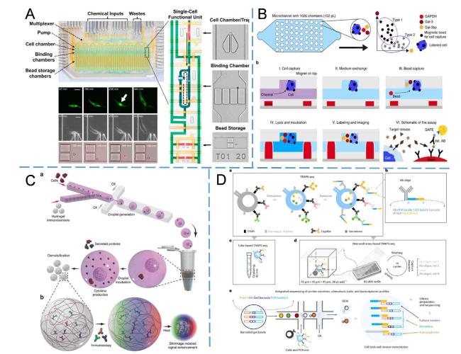
Among the techniques employing microchambers for individual cell separation, pump-valve structures are often utilized to drive cells and microbeads and form the microchambers, facilitating their co-localization within the microchip (as shown in Fig. 7A and B). An automated microfluidic system developed by Junkin et al.112 utilized a two-layer PDMS chip with pump-valve structures to precisely control the movement of individual cells. These cells were then trapped within the chambers for cultivation and stimulation. The products were transported to the binding chamber using pumps for single-cell secretion analysis. By employing encoded microbeads labeled with different antibodies, up to 40 single cells can be simultaneously analyzed for six types of secreted molecules. The use of pump-valve structures allows flexible material exchange required for long-term (days-weeks) cell cultivation within the microfluidic chip. Additionally, repetitive detection of cellular secretions at different time points can be achieved by replacing the detection beads. However, the implementation of simultaneous detection of hundreds of cells in this complex and challenging structure remains a formidable task. Optimizing the structure of microchamber can marginally enhance the capacity for single analysis, but typically limited to approximately one thousand chambers.116 In single-cell analysis using microchambers, beads not only serve as carriers for capturing target proteins during the detection process but also function as labels for the target cells, facilitating their manipulation into microchambers through a magnetic field. The microfluidic chip designed by Armbrecht et al. featured 1026 chambers113 (Fig. 7B), in which three mammalian cells were labeled with magnetic beads and a magnetic field was applied to enhance the trapping ratio of single cells. Approximately one-third of the trapping sites could be occupied by a single cell. Further incubation of three types of fluorescent encoded beads with single cells enabled the simultaneous quantification of triple secreted proteins in each chamber, achieving a minimum LOD of 1.8 × 10³ molecules/cell. However, for simultaneous detection of three indicators, it is necessary to introduce three different magnetic beads conjugated with corresponding antibodies into the same chamber containing single cells. Due to non-uniform distribution of magnetic beads within the chamber, the number of such chambers in experiments does not exceed 38, resulting in a significantly reduced analysis throughput. Despite the potential of providing material exchange in cell culture, the low probability of a single cell occupying a microchamber results in an analysis of only a few hundred cells, which fails to yield satisfactory statistical detection outcomes.
Droplet microfluidics, with an unlimited number of chambers, may serve as an optimal solution to address the flux problem. However, droplets also face challenges in terms of material exchange compared to microchambers. There are generally two approaches to tackle this problem: 1) employing a wash-free detection method, where cells and assay reagents are encapsulated together in droplets for incubation and on-site detection of secreted signals.117,118,119,120 Specifically, Wei et al.119 developed a wash-free multiplex detection method based on the phenomenon that the plasmon resonance peak shifts of gold nanorods in the presence of different targets. Eyer et al.,120 on the other hand, utilized microbeads as carriers for fluorescence signal aggregation to differentiate target cell droplets. 2) detecting the signal outside the droplet.114,121 Hsu et al.114 employed a droplet microfluidic chip to encapsulate single cells and hydrogels (as shown in Fig. 7C), forming solid hydrogel beads. Subsequently, off-chip immune reaction and fluorescent labeling were utilized to detect the substance enclosed within the hydrogel beads, enabling quantification of single-cell secretions. To address the challenge of low substance concentration for measurement, temperature-induced contraction of the hydrogel beads was employed to enrich the concentration of molecules in the beads. The quantification of three cell secretions in 6000 cells was achieved within 1 h, with a LOD ranging from 25.0 to 34.6 pM. However, due to its limited material exchange characteristics, the droplet system not only hinders prolonged cell incubation (often less than 12 h) but also poses challenges in achieving a protein detection multiplex capability exceeding three. On the contrary, due to its ability to rapidly and accurately detect tens of thousands of cells in a single experiment, the droplet system is particularly well-suited for antibody screening and other fields that necessitate large-scale cell secretion analysis.
To enhance the multiplexing capability of single-cell secretions, a potential strategy for increasing tag multiplicity is to use deoxyribonucleic acid (DNA) sequences.115 Zhao et al.122 employed DNA sequences to encode microbeads and affixed them onto the top of the microchamber. They utilized sandwich ELISA to quantify single-cell secreted proteins, followed by decoding the beads using complementary DNA sequences with distinct fluorescence signals. The assay successfully detected ten types of cytokines with LODs ranging from 12 to 103 pg/mL. However, due to limited labeling fluorescence, multiple detections necessitate a Quenching-Labeling process repeated n/3 + 1 times in order to decode the nucleic acid sequence on each bead, which is intricate and time-consuming, requiring precise imaging positioning. In addition to serving as an encoding marker for microbeads, DNA sequences can also be directly labeled on antibodies. Wu et al.115 utilized distinct antibodies with corresponding DNA sequence tags to label membrane and secreted proteins on cells (Fig. 7D). During library construction, the information of membrane protein tags, secreted protein tags, cell mRNA, and encoded gel beads is integrated for detection through second-generation sequencing. They demonstrated the application of this approach for simultaneous detection of three secreted cytokines, ten cell surface proteins, and sixteen genes, thereby elucidating the phenotypic and transcriptional determinants underlying pleiotropic T helper 1 cell cytokine secretion at a single-cell resolution.
However, the authors suggest that the constraints on enhancing throughput for single-cell protein detection may not solely arise from labeling molecules but also stem from reciprocal interactions between antibodies. The inclusion of additional antibody types in the system can lead to nonspecific enhancements and impact detection sensitivity. Presently, practical applications by antibodies in single-cell detection are limited to dozens of kinds,122 which falls short of characterizing the complete proteome within individual cells (comprising >20,000 proteins and >100,000 epitopes for humans123,124). While developing methods to measure secretions with higher levels of multiplexing beyond ten-fold or even hundreds-fold poses a challenge, their specific areas of application remain unclear. For applications such as antibody screening and specific secretion measurements, it might not be essential to quantify all proteins. Techniques such as single-cell mass spectrometry, which do not rely on immune reactions, may be better suited for applications like single-cell omics analysis that require simultaneous detection of hundreds or more markers with reduced consumption during antibody screening and minimized cross-interference effects.
4.6. Miscellaneous applications
In addition to the aforementioned categories, bead-based microfluidic multiplex immunoassays could also detect illicit additives in health food,70 anti-Müllerian hormone as a marker for premature ovarian failure,125 mycotoxins,69 and other proteins. The aforementioned targets also encompass other potential applications of bead-based microfluidic multiplex immunoassays, specifically in the prognostic monitoring of diverse diseases necessitating highly sensitive detection methodologies, expedited determination of food ingredients, quality control of healthcare products, etc., thereby demonstrating their extensive applicability in these domains that demand ultra-sensitive methodologies or POCT methods.
5. Future prospects
In light of the current clinical demand for the detection of multiple low-abundance proteins, this paper provides a comprehensive review of bead-based microfluidic multiplex detection methodologies for low-abundance proteins. Overall, bead-based microfluidic multiplex immunoassays currently encompass two major directions: ultrasensitive protein detection and POCT.
In the detection of ultrasensitive proteins, the Simoa instrument, which integrates automatic sample pre-treatment, remains the most widely used commercial microfluidic platform. This observation may also suggest that there are still some unidentified challenges in promoting bead-based microfluidic electrochemical ultrasensitive detection methods for practical applications. However, due to its requirement for a microfluidic platform to construct miniature compartments, Simoa still possesses certain limitations in chip fabrication and complex detection processes, necessitating high-precision control and imaging units. Utilizing beads as a partitioning carrier67,126 instead of microfluidic wells or droplets could enhance partitioning efficiency and detection sensitivity, streamline the reaction process, and reduce consumption and detection costs. Furthermore, coupled with portable and cost-effective imaging systems such as mobile phones may represent the next generation improvement direction for ultra-sensitive protein detection methodologies. Nevertheless, further exploration is required to find actual clinical applications where ultra-sensitive protein detection with aM or zM level sensitivities can be applied effectively. Currently, only certain neurobiomarkers and single-cell protein secretion fields have demonstrated the necessity of ultra-sensitive detection methodologies. However, no definitive conclusions have been drawn regarding the benefits in other domains such as cancer research or epidemiology applications. Correspondingly early-stage target screening is also under development and necessitates further observation.
In the field of POCT, although integrating magnetic beads offer ease of manipulation and the possibility of pre-processing within microfluidic chips, there is a limited number of methods in current literature that integrate bead capture and washing steps within the microchip. This may be attributed to the requirement for additional magnetic manipulation devices, which makes it less convenient compared to positional encoded detection methods that do not require supplementary magnetic fields for target capture and washing. Therefore, electrochemical detection appears to offer a more clinically viable solution owing to its straightforward chip structure, reduced consumable costs, and simplified implementation for multiplex detections. However, it is constrained by the slower reaction kinetics in solid-liquid systems compared to those in liquid-based systems, necessitating signal amplification and rendering it more susceptible to non-specific adsorption and electromagnetic interference.83 The observed variations in ETS-related gene protein levels among clinical samples21 may also suggest the presence of this phenomenon to a certain extent. Besides, most magnet bead-based detection approaches still necessitate external immunoreactions prior to on-chip testing, posing challenges in ensuring stability during multi-step reactions. Furthermore, the advantage of low sample consumption in microfluidic chips encounters difficulties due to volume disparities between pre-processing steps and chip steps. In response to the aforementioned requirements, one-step detection methods such as LOCI88 and other wash-free methods that eliminates the need for multiple washing steps may be an optimal choice. These approaches enable sample detection with just one incubation step, thereby mitigating the impact of external reactions. By integrating LOCI with encoded beads, high multiplex protein detection can be achieved. To address the issue of lower sensitivity compared to digital ELISA and similar approaches, further improvement in detection sensitivity and reduction in non-specificity can be accomplished through single-step washing, making it a promising direction for future development in POCT.
Ethical approval
This study does not contain any studies with human or animal subjects performed by any of the authors.
Declaration of competing interest
The authors declare that they have no known competing financial interests or personal relationships that could have appeared to influence the work reported in this paper.
Acknowledgements
This work was supported by the National Natural Science Foundation of China [grant numbers 32001067, 82272122, and 31927803], Interdisciplinary Program of Shanghai Jiao Tong University [grant number YG2022ZD028], and Major Projects of Special Development Funds for Shanghai Zhangjiang National Innovation Demonstration Zone [grant number ZJ2021-ZD-007].
