
1. Introduction
Cartilage defects as a result of aging and degenerative pathology, sports-related injuries, diseases, etc., have been noticed for more than 200 years [1]. The avascular structure with a relatively low density of chondrocytes inside causes the limited regeneration of cartilage. Natural wound healing in full-thickness cartilage defects leads to the formation of fibrocartilage, which is functionally and biomechanically inferior to the original hyaline cartilage, leading to further deterioration [2].
With the growth of the elderly population globally, it is predicted that more than 15% of people aged 60 years and older will develop cartilage-related problems [1]. Currently, many different cartilage repair-enhancing treatments, such as cartilage chondroplasty and microfractures, have been developed to treat cartilage defects but are unable to entirely repair the damaged cartilage. An urgent need for optimized functional restoration of defective cartilage has expanded the field of cartilage tissue engineering (CTE), in which scaffolds with materials that exhibit strong mechanical support, biocompatibility, biodegradability, and osteoinductive properties are constructed to develop new biomimetic cartilage that could properly replace the injured tissue [3].
The heterogeneity of the cartilage tissue, including areas with different cell morphologies and cell arrangements, as well as with different extracellular matrix (ECM) arrangements, constituents and distribution, poses challenges to reproduce functional cartilage [4]. Cartilage tissue is composed of ECM that consists of a unique and tissue-specific 3D environment of structural and functional molecules secreted by resident cells, which reciprocally interact with cells dynamically in response to the outer biochemical environment and mechanical cues [2,5]. The diversity of ECM components provides a number of advantageous physical properties and protein components that influence cell growth, differentiation, and migration [6], which endows the ECM with multiple roles. Therefore, CTE constructs based on natural ECM sources are likely better choices to produce a tissue with optimal functionality than those built from artificial compounds, bringing out better engineered cartilage for the treatment of cartilage defects.
The last few years have marked substantial progress in ECM-based CTE. This review considers the design and application of such CTE scaffolds based on ECM-derived biomaterials. We highlight the role of ECM-derived biomaterials, their applications in different forms of scaffolds and their processing approaches. The current clinical applications of these ECM-based CTE scaffolds are also reviewed in the last section. Therefore, the outstanding goal of the current review is to reinforce the importance of modification and improvement of multiple CTE constructions based on ECM-derived materials.
2. Material and methods
To obtain relevant articles on the field of ECM-based CTE, a literature search was conducted in different databases, such as PubMed and Web of Science, by using the following keywords: cartilage tissue engineering, extracellular matrix, decellularized extracellular matrix, cell-derived extracellular matrix, biomaterials, polymers, collagen, scaffold, fibrous scaffold, hydrogel, polymer films, application, and clinical application. We are confident that using combinations of such keywords has enabled us to identify relevant previous studies in the field. The articles were restricted to those written in English. Special mention of the role of ECM-derived biomaterials, their applications in CTE, and current clinical applications of ECM-based CTE are reported in this work. The goal is to provide the reader with a broad overview of this exciting research field that highlights the potential and benefits expected from the multiple CTE constructions based on ECM-derived materials.
3. Results and discussion
3.1. ECM-derived biomaterials for CTE
A large number of studies have shown the importance of selecting appropriate biomaterials as scaffolds for cell adhesion and supporting proliferation [7]. For the ECM plays an important role in living tissue development and regeneration, the ideal scaffold material should closely mimic the natural environment of the ECM [8]. The main sources of biomaterials for CTE are discussed as follows.
3.1.1. Nature-derived dECM scaffolds
Cartilage tissue has a specialized ECM structure and composition that modulates cell responses and benefits the survival of cells [9]. Due to its inherent properties that are difficult to replicate by synthetically designed materials, the use of nature-derived ECM for cartilage tissue regeneration has gained popularity. To reduce the risk of disease transmission and an immune response from the recipient, the cartilage tissue should be decellularized before application. Decellularization refers to the process of treating tissue with any combination of physical stress and chemical/enzymatic agents to remove cellular components, leaving behind only the noncellular ECM that can be used for therapeutic applications [10]. Decellularized extracellular matrix (dECM) retains components of the natural cell environment, including the complex biomolecular and physical cues for cell growth and viability in the ECM, while eliminating immune responses for decellularization techniques should ideally remove all cells and cellular antigens [2]. Therefore, incorporating dECM in CTE is believed to be a promising method to construct a suitable microenvironment for cell proliferation to promote the formation of new tissues [11]. The application of the functional decellularization of the extracellular matrix to tissue repair is schematically illustrated in Fig. 1.
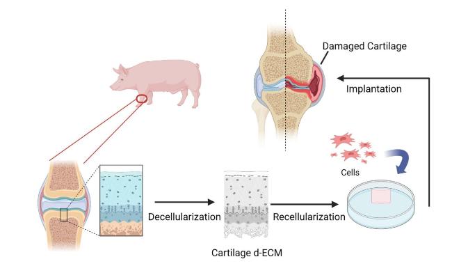
Fig. 1. Summary of extracellular matrix decellularization procedures (figure was created with Biorender.com) [12]. |
3.1.1.1. Tissue-derived dECM scaffolds
Cartilage tissue with rich ECM components is a direct source for decellularization to construct tissue-derived ECM scaffolds. Tissue-derived dECM scaffolds possess the natural 3D architecture from the whole tissue by eliminating immunogenic cellular components via a decellularization process and preserving non-immunogenic ECM. The resulting dECM scaffolds can be reseeded with specific cells to generate CTE grafts. Tissue-derived dECM scaffolds serve as reservoirs for site-specific bioactive molecules and cell-matrix interactions, offering native factors and cues that could drive tissue-specific differentiation [13,14]. In addition, their mechanical properties and microenvironment conditions are more similar to those of native ECM than cell-derived ECM, as discussed below [15].
Decellularization of tissues can be accomplished using various methods, which generally involve a combination of (a) physical, (b) enzymatic, and (c) chemical processes (Fig. 2) [10]. To achieve optimized decellularization, the three methods are often used in conjunction. Visscher et al. [16] freeze-thawed porcine auricular cartilage tissues and treated them with chemical agents such as Triton X-100 solution and enzymes such as nuclease. The sample was cryomilled into a powder form and subsequently processed into a hydrogel and mixed with chondrocytes to create a printable bioink for cell-based 3D bioprinting. The bioprinted construct exhibited a cartilage-specific microenvironment that could promote cellular activities and maturation of chondrocytes. Alternative physical treatments, including mincing, cryomilling, supercritical carbon dioxide (ScCO2), ultrasonic waves, osmotic pressure, hydrostatic pressure, chemicals including sodium laurylsulfonate (SDS), EDTA, acetic acid, PMSF and enzymes including nuclease and protease, have been applied in conjunction in many studies to properly decellularize cartilage tissues and construct an optimized microenvironment [11,17,18,19,20,21,22,23,24,25,26,27,28,29,30,31]. Several research groups have adopted a single decellularizing protocol among physical, enzymatic, and chemical processes or two of them [32,33,34,35,36,37,38,39,40]. Specifically, Xia et al. [32] minced meniscal tissue into cubes, stirred them in SDS solution and then washed them in EDTA solution before mixing with PCL to create a nanofibrous scaffold. Compared to the pure polymer construct, the dECM construct promotes cell attachment and spreading at early time points. Goldberg-Bockhorn et al. [39] used only chemical processes to decellularize porcine nasal septal cartilage. NaOH (1 N) solution, ethanol (70%), guanidine hydrochloride solution, sodium acetate and H2O2solution were performed successively. The author modified the scaffolds' surface by laser application to enlarge the surface of the dECM scaffolds. The examination revealed an increase of the scaffolds’ surface area with proliferation of cell numbers on the scaffolds. In particular, decellularization with only physical processes is named devitalization, for physical decellularization alone cannot completely remove cellular DNA or debris from the tissue but can effectively disrupt cellular membranes and nuclei.
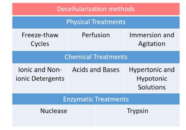
Fig. 2. Decellularization methods: physical treatments, chemical treatments, and enzymatic treatments. The typical processes for each treatment are cataloged [15]. |
Although various efficient protocols for treating cartilage tissue for CTE have been performed in studies, the approach is still relatively underexplored, and extensive research is required to optimize the decellularization techniques and ultimately the final repair tissue. Several persistent challenges and limitations are discussed as follows. First, different cartilage origins have various biomolecules, giving each type distinct properties. Therefore, different positions and ages of tissue origin are key factors that need to be considered. Exports have reported that immature cartilage dECM at different developmental stages would result in diversified effects in dECM-based CTE [41]. However, the optimized source of cartilage ECM for tissue engineering remains unclear and awaits further exploration. Second, rigorous decellularization induces loss of structural integrity of the ECM and of certain ECM compounds [2]. Nevertheless, it has been suggested that ineffectively decellularized ECM still induced similar host remodeling to that induced by effectively decellularized material [42]. Therefore, whether absolute decellularization is necessary, and whether milder decellularization protocols could realize the optimal effect of tissue-derived ECM in CTE should be further explored. Finally, due to the density of cartilage dECM, which is detrimental to cell growth and material exchange, its application forms are relatively limited, mainly in the form of particles or solutions. Lu et al. [43] suggested that dECM in solution form could provide a more favorable chondrogenic microenvironment for endogenous BMSCs than dECM particles. However, the differences in various aspects and effects of these two forms on cartilage repair have yet to be clearly compared. To summarize, the development of techniques of decellularization will be the key to facilitating the utilization of tissue-derived ECM in CTE applications and furthermore provide viable therapeutic applications for cartilage tissue regeneration.
3.1.1.2. Cell-derived dECM scaffolds
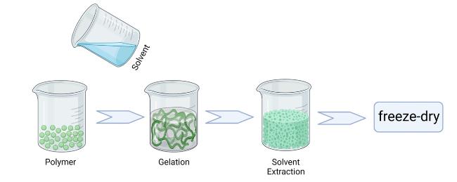
Although tissue-derived ECM has great prospects in CTE, intrinsic limitations, including its compactness, may not permit sufficient penetration of solutions applied to decellularization procedures [44], and the deficiency of native cartilage sources may limit its application. In contrast, ECM derived from cells (cell-derived ECM) can be easily obtained from autologous cells cultured in vitro with rich cell-generated bioactive signaling molecules, and has better plasticity with desirable porosity for penetration of decellular solutions and seeded cells [45], thereby avoiding the shortcomings of decellularized ECM derived from tissue. Therefore, cell-derived ECM has attracted increasing attention as a biomaterial to fabricate scaffolds for cartilage repair [46]. The basic procedure for preparing cell-derived dECM is illustrated in Fig. 3.
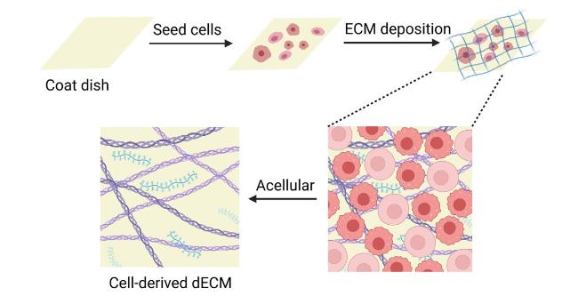
Fig. 3. Basic procedure for preparing cell-derived dECM (figure was created with Biorender.com). |
Many different cell types, including chondrocytes and mesenchymal stem cells (MSCs), can be used to secrete cartilage-like ECM for CTE. Previous studies have suggested that the chondrocyte-derived ECM promotes the proliferation and chondrogenic differentiation of stem cells, which may contribute to the formation of functional hyaline-like cartilage tissues and cartilage repair [45,47,48]. Jin et al. [47] evaluated the feasibility of a chondrocyte-derived ECM scaffold after inoculation of chondrocytes by implanting it in vivo in nude mice. Cartilaginous tissue was obtained successfully over a short-term period, and evaluations of biochemical and mechanical properties demonstrated that chondrocyte-derived ECM scaffolds could provide an ideal 3D environment for forming hyaline cartilage. The authors’ subsequent in vitro study showed that the ECM scaffold also supports chondrocyte attachment, proliferation, and cartilage matrix synthesis in cartilage formation, indicating that the chondrocyte-derived ECM scaffold functions as a promising biomaterial for CTE [48]. In addition to chondrocytes, MSCs are also promising cell sources for tissue engineering [49]. MSC-derived ECM, as a biomaterial with good bioactivity and biocompatibility, has been widely used as an expansion ground substance in CTE [50,51,52,53,54,55]. Specifically, Yang et al. [55] tested the applicability of using decellularized human bone marrow-derived MSC-ECM (hBMSC-ECM) as a scaffold for chondrocyte-based cartilage repair. The results showed a significant increase in the proliferation rate, better chondrocytic phenotype and chondrogenic differentiation profile, compared with chondrocytes grown on tissue culture plastic. The test of cartilage formation ability of composites of hBMSC-ECM impregnated with chondrocytes in vitro and in vivo both showed prominent cartilage formation with higher sGAG deposition. ECM deposited by nucleus pulposus cells (NPCs) was also investigated by scientists, exhibiting its function of presenting an NP-like microenvironment, which therefore provides the signals required to affect gene expression in human dermal fibroblasts and partially alters their phenotype toward that of an NP-like lineage [56]. Moreover, recent studies have also demonstrated more beneficial properties of cell-derived dECM, including its ability to maintain a stem cell niche for MSCs and retain their stemness [57,58].
Although the observations outlined above show promising results of cell-derived ECM in CTE, problems remain to be addressed, and further research is needed. A challenge is that the different effects of ECMs derived from different cell types are still unclear. Lu et al. [45] have reported that ECM scaffolds derived from different cell types have different components. Their study using MSC- and chondrocyte-derived ECM scaffolds to culture MSCs and stimulate chondrogenesis showed that the MSC-derived ECM scaffolds exhibited a stronger stimulatory effect than the chondrocyte-derived ECM scaffolds. Nevertheless, Hoshiba et al. [59] reported that the number of chondrocytes adhering to chondrocyte-derived ECM was markedly higher than that adhering to MSC-derived ECM. In addition to cell type, it has been suggested that ECMs derived from different cell aging periods may also have different effects on cell growth [6,60,61]. Ng et al. [61] reported that fetal MSC-derived ECM was superior to ECM derived from adult MSCs or human neonatal dermal fibroblasts for promoting adult MSC proliferation and multipotency. Specific mechanisms and the substances that play major roles in cell-derived ECM should be identified to optimize the application of cell-derived ECM in CTE. Moreover, shrinkage of cell-derived ECM scaffolds is often reported, suggesting inferior biomechanical properties of tissue-engineered cartilage [47,50]. Improvements in the mechanical strength of the cell-derived ECM construction should be further investigated. With more research regarding the mechanisms and breakthroughs in fabrication, cell-derived ECM is promising for clinical application as an ideal biomaterial with significant biophysical and biochemical properties.
3.1.2. Artificial fabricated ECM scaffolds
In addition to the application of natural ECM, CTE could also be implemented by fabricating structures that mimic the cartilage ECM with natural components of ECM. Natural-based polymers can provide a conducive structure similar to natural ECM, beneficial to cell attachment, proliferation and differentiation. Their degradation products in vivo are non-toxic and physiological [62]. Therefore, the addition of natural polymers, especially those ECM-derived natural polymers, such as collagen, hyaluronic acid, chondroitin sulphate and combinations of those compounds, is undoubtedly an attractive approach to mimic the native environment of cartilage ECM and provide substrates for CTE.
Collagen is a major component of ECM proteins that connects tissues and enhances strength and stability, which has been used extensively for cartilage regeneration and was reported as the most frequently used material among cartilage ECM derivatives [5,63]. Therefore, we take collagen as an example to elucidate the artificially fabricated ECM with natural polymers for CTE. Collagen has generally been utilized as a scaffold or carrier for chondrocytes and MSCs experimentally [64,65], for it has been proven to offer the advantage of a homogenous cell seeding density and is capable of promoting chondrocyte and MSC proliferation [66,67,68,69]. To enhance the multiple properties of collagen scaffolds, different fabrication methods have been applied. Mueller-Rath et al. [66] created a stabilized form of condensed collagen gel seeded with human articular chondrocytes by mechanical compression and filtration to improve its biomechanical capacity. A plastic compression technique was also used to produce dense fibrillar collagen scaffolds with controllable biomimetic structure and mechanical properties while allowing for direct cell seeding with high viability and growth potential [70,71]. Photo-crosslinking has also been applied to overcome the key problems of type II collagen, including the slow fibrillation process and inferior mechanical properties that hinder its utilization [72]. Moreover, the defective properties of collagen could be further rectified by crosslinking collagens with multiple natural and synthetic polymers to afford composite materials with superior properties [5]. Specifically, Choi et al. [73] incorporated Col II and CS into injectable chitosan hydrogels designed to gel and demonstrated that Col II hydrogels could be enhanced by chitosan with better cellular condensation, chondroid cluster formation, and chondrogenesis, which can lead to enhanced cell-matrix interaction and has a high potential for an injectable scaffolding system for cartilage repair. Chen et al. [74] fabricated a series of poly(ethylene glycol)/collagen (PEG/Col) double network hydrogel from PEG and collagen, which was examined to obtain enhanced strength and toughness while preserving the water-storage capability and favorable biocompatibility of collagen, thus providing the appropriate environment for the adhesion, growth, and proliferation of MC3T3-E1 preosteoblast cells. Similar to collagen, other natural polymers, such as hyaluronic acid [75,76,77], chondroitin sulphate [76,77], gelatin [78], silk fibroin [78,79], and alginate [79], could be fabricated separately or in conjunction with synthetic polymers via the methods outlined above.
Although artificially fabricated ECMs with natural polymers have been applied experimentally for decades and multiple properties could be improved through innovative fabrication methods, there are still problems to be solved. A current idea to enhance the properties of natural polymers is the addition of synthetic polymers, attempting to combine the advantages of both natural and synthetic polymers, as mentioned above. However, hybrid fabrications are also faced with the problem of comprising bilateral drawbacks. To mutually promote the advantages of natural and synthetic polymers while avoiding their disadvantages, studies of optimized assembly of proper kinds of natural and synthetic polymers and their proportion, as well as the crosslinking protocol, are critical. Moreover, clinical applications of new biomaterials can be limited by the cost and difficulty of passing safety and regulatory processes of those synthetic polymers, for materials that have not already been approved for use in humans have extensive requirements in quality control and safety [1]. Therefore, major efforts should be focused on the transformation of the obtained results into clinical applications. With new fabrication schemes being continually developed and their clinical utilization expanding, artificially fabricated ECM would be considered a robust vehicle for CTE applications.
3.2. Scaffold processing approaches for CTE based on ECM-derived biomaterials
To achieve desirable properties by applying the ECM-derived biomaterials outlined above properly, a multitude of porous ECM-derived scaffolds and scaffold processing approaches have been utilized in CTE [5]. To better integrate with ECM-derived biomaterials and mimic the natural environment of the ECM, an ideal scaffold should be designed with appropriate porosity, biodegradability and specific shapes and sizes with functions related to tissue regeneration [80]. The most frequently used types of scaffolds and their fabrication strategies are discussed below and some main researches are listed in Table 1.
Table 1. Some Main Researches on Scaffolds for Cartilage Tissue Engineering. |
| Author | Scaffold Composition | Fabrication Strategies | Advantages | Ref. |
|---|---|---|---|---|
| Liu et al. | poly(L-lacticacid)-copoly(ε-caprolactone) (P(LLA-CL))/collagen type I(Col-I)/hyaluronate hybrid/beta-tricalcium phosphate (TCP) | electrospinning | better cell infiltration, greatly improved repairing scores and compressive modulus | [90] |
| Munir et al. | PCL/collagen type I | thermally induced phase separation | equal distribution of collagen and collagen penetration throughout the scaffold, easily tuneable porosity and compressive properties | [95] |
| Tronci et al. | collagen-derived polypeptides | wet-spinning | adjustable mechanical properties and wet-stable morphology | [97] |
| Gupta et al. | collagen, alginate and oxidized alginate | Hydrogels based on interpenetrating networks (IPNs) | load bearing ability, biocapability for cellular growth and differentiation, unique self-healing character | [104] |
| Yu et al. | gelatin, hyaluronic acid and chondroitin sulfate | Diels-Alder “click” chemistry for tri-component IPNs | improved mechanical properties and great potential applications in CTE | [105] |
| Moreira et al. | chitosan, collagen and bioactive glass nanoparticles (BG) | an environmentally friendly processing route for producing hydrogels | bioactive thermogelling composite with promising potential biomedical applications in tissue repair and regeneration | [112] |
| Brito et al. | Col, HA, and polyelectrolyte | layer-by-layer (LBL) assembly | the physicochemical characteristics including surface zeta potential, thickness, layer growth, and wettability | [118] |
| Costa et al. | chitosan and a recombinant elastin-like recombinamer containing the cell attachment sequence RGD | layer-by-layer (LBL) assembly | acute and independent cyclic responses toward temperature, pH, and ionic strength | [122] |
3.2.1. Fibrous scaffolds
It has been documented that fibrous scaffolds could incorporate ECM-derived biomaterials to partially mimic the structure and function of natural ECM, as they present an increased surface area for cell attachment, improved pore architecture, and good mechanical stability [81,82]. Therefore, fibrous scaffolds have emerged as a preponderant option for CTE [83,84].
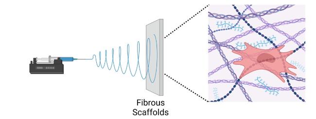
Recently, nanofibrous scaffolds seem to hold the leading position among various fibrous scaffolds as ideal candidates for CTE [86]. Nanofibrous scaffolds are generally fabricated by electrospinning (Fig. 4) [85], a highly versatile and promising method for developing 3D nanofibrous scaffolds with a range of biomaterials through the use of electrostatic force [3]. Electrospun CTE scaffolds have been widely fabricated from a multitude of ECM-derived polymers, and electrospinning techniques continue to progress [82,84,87,88,89,90,91]. Liu et al. [90] fabricated an oriented poly(L-lacticacid)-copoly(e-caprolactone) P(LLA-CL)/collagen type I(Col-I) nanofiber yarn mesh by dynamic liquid electrospinning to enhance the biomechanical strength of the scaffold. In vitro results show that the Yarn Col-I/HA hybrid scaffold (Yarn-CH) can allow the cell infiltration like sponge scaffolds, which was successfully used to repair the osteochondral defects in a rabit model with greatly improved repairing scores and compressive modulus, demonstrating that electrospinning is an effective technology for the preparation of fibrous scaffolds. Other methods are also applied for fabricating nanofibrous scaffolds, such as phase separation (Fig. 5) [83,86,92,93,94,95] and molecular self-assembly [96]. Specifically, Munir et al. [95] employed thermally induced phase separation to fabricate porous hybrid PCL/collagen type I scaffolds, which realized equal distribution of collagen and collagen penetration throughout the scaffold, exhibiting easily tuneable porosity and compressive properties. Moreover, fibrous scaffolds could also be constructed by wet-spinning [7,81,97,98,99,100]. Tronci et al. [97] investigated the formation of wet-spun fibres based on collagen-derived polypeptides with comparable chemical composition and varied molecular weight, revealing a reliable synthetic method that could be applied to the resulting fibrous system to ensure its adjustable mechanical properties and wet-stable morphology.
Although fibrous scaffolds have shown promising results for CTE application, their topography and morphology still present critical barriers to mimicking the natural ECM [3]. To provide a better environment for cell function, fibrous scaffolds are generally processed at the nanoscale nowadays [83], which causes a lack of macroscopic pores, making the diffusion of nutrients difficult. Therefore, further studies that focus on the development of interconnection between nanofibers and macropores would be crucial [3]. With the development of materials science and manufacturing technologies, more advanced fabrication strategies would certainly be used in fibrous scaffold construction, facilitating the solution of the clinical problem of cartilage defects in the future.
3.2.2. Hydrogel
The majority of polymeric scaffold types need invasive support for implantation. In contrast, hydrogels are 3D crosslinked hydrophilic networks with low viscosity that can be delivered via injection to the defect site, which meets the clinical demand for scaffolds that can be implanted with minimally invasive procedures [3]. Due to their interesting properties, including tunable elasticity and stiffness, high water content, excellent biocompatibility and biodegradation, and the ability to incorporate biological molecules, hydrogel scaffolds fabricated with ECM-derived materials have been widely used in CTE applications [1,3,101].
Hydrogels based on single-polymer networks usually exhibit very poor mechanical properties compared with native cartilage [1]. With the aim of increasing the mechanical properties of hydrogels to approach those of hyaline cartilage, the focus is shifting from conventional hydrogels to multi-material hydrogel systems, often including two or more independent networks [102]. Hydrogels based on interpenetrating networks (IPNs) are one of these multi-material hydrogels, comprising two or more separate crosslinked networks that partially intertwine [103]. The schematic patterns of IPNs utilized in hydrogels are depicted in Fig. 6 [102]. Gupta et al. [104] fabricated a 3D-printed scaffold with a self-healing IPN hydrogel-based construct with dual crosslinking capability using collagen, alginate and oxidized alginate, which caters to the load bearing requirements for total meniscus replacement as well as fulfils the micro environmental needs to provide a suitable niche for cellular growth and differentiation along with unique self-healing character that can heal after fractures. In addition to IPNs of two networks, tri-component IPNs were also investigated by Yu et al. [105], who synthesized a novel biological degradable interpenetrating network hydrogel from gelatin, hyaluronic acid and chondroitin sulfate by Diels-Alder “click” chemistry, showing improved mechanical properties and great potential applications in CTE. Other types of multi-material hydrogels, including Semi-IPNs [106], double networks [107], dual networks [108] and supramolecular hydrogels [109], have also been applied in CTE, attaining stronger mechanical properties than networks of single polymers and exhibiting superior integration with surrounding tissue in vivo [102]. Moreover, smart scaffolds that can respond to various stimuli have also been developed, enabling on-demand manipulation of cell microenvironments [1]. Stimuli-responsive hydrogels, such as temperature-responsive hydrogels, pH-responsive hydrogels and light-sensitive hydrogels, have gained great attention in CTE due to their capability to undergo physical or chemical changes in response to external alterations [110,111]. Specifically, Moreira et al. [112] synthesized a novel thermosensitive chitosan-based composite, chemically modified with collagen and reinforced by bioactive glass nanoparticles (BG), which resulted in a bioactive thermogelling composite with promising potential biomedical applications in tissue repair and regeneration.
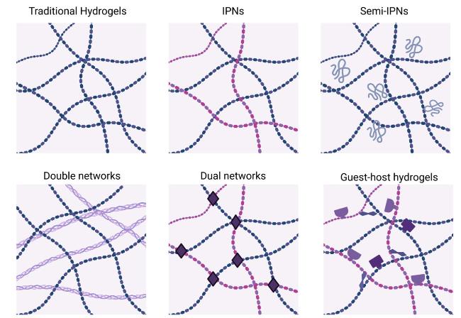
Fig. 6. Schematic depicting different designs utilized in hydrogels, from traditional single polymer networks to those that include multiple networks and mixtures of polymers (figure was created with Biorender.com) [102]. |
Although different types of hydrogel scaffolds that contain multiple ECM-derived polymers have been successfully fabricated with excellent biocompatibilities, some problems still need to be solved. For example, utilization of toxic cross-linking agents is required in multitudes of preparation processes, which would restrain clinical applications of these hydrogels [62]. In addition, although various strategies are currently available for the development of smart hydrogels with new properties, they do not meet all the specifications required for CTE [1]. Therefore, combining these high-performance hydrogels with valuable properties to achieve superior properties may be a promising approach that requires further investigation. With extensive research through collaboration of scientists from all fields, the development of biomimetic hydrogel scaffolds and their translation into the market will certainly be facilitated, supporting the treatment of human cartilage defects.
3.2.3. Polymer films
Polymer films, a kind of 2D scaffold, are widely used for CTE as a result of their good mechanical properties and readily available surface engineering methodologies. It is based on cycles of alternating adsorption of polyanions and polycations from aqueous solutions onto charged surfaces, and is composed of important coating ECM-derived biomaterials tethered with various functional groups that could tune the cell-scaffold interactions [113].
Bioactive polymer films could be produced by multiple methods, such as layer-by-layer (LBL) assembly (Fig. 7) [114], in situ polymerization on the surface [115], and chemical-vapor-deposited (CVD) coating [116]. LBL assembly has emerged as a very promising tool to prepare dynamic and responsive coatings owing to its versatility to easily change in the mode and in the strength of interactions between the interacting partners [117,118,119]. For example, Brito et al. [118] built an LBL surface coating that mimics the natural ECM of connective tissue by combination of Col and HA in polyelectrolyte multilayer (PEM) system. The multilayer film presents the physicochemical characteristics, such as surface zeta potential, thickness, layer growth, and wettability. Besides, LBL assembly is also widely used to produce multi stimulus-responsive films that can respond to a number of stimuli, including pH [114], temperature [120], or electrochemical potential [121], which are promising for diverse biomedical applications. Specifically, Costa et al. [122] fabricated biomimetic smart thin coatings using chitosan and a recombinant elastin-like recombinamer containing the cell attachment sequence arginineglycine-(aspartic acid) (RGD) through an LBL approach. The stimuli-responsive properties of the films were extensively studied, showing acute and independent cyclic responses toward temperature, pH, and ionic strength.
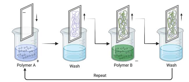
Fig. 7. Layer-by-layer (LBL) film assembly. A schematic of a (polymer 1/polymer 2)n bilayer film assembled using dip LBL assembly is shown. Repeating these steps n times allows the assembly of an LBL film containing n bilayers (figure was created with Biorender.com). |
Although multiple advantages, including biocompatibility, low immunogenicity, limited toxicity, and plasticizing properties, are presented in the films [123], serious drawbacks remain. Firstly, to better mimic the 3D environment, the construction of multilayers of ECM-derived polymer films is necessary, which leads to laborious and time-consuming fabrication processes that may hinder their practical utilization. In addition, multitudes of surface modification processes lead to residual chemicals, limited penetration depth, as well as imparting undesired chemical functional groups onto the surface [124]. There is thus a need to further the studies of better surface modification protocols with respect to optimizing the surface properties while maintaining the mechanical strength [124]. With further investigation and upgraded manufacturing technology in the future, the fabrication processes will be simplified, and performance will be optimized, which will be conducive to expanding the application of polymer films in CTE.
3.3. Clinical applications of CTE
It has been demonstrated that scaffolds containing ECM-derived biomaterials can enhance cell adhesion, proliferation, and differentiation because of their appropriate porosity, biodegradability and specific shapes and sizes. These promising features allow functional scaffolds to be applied in different kinds of CTE applications, and their clinical studies are discussed in this section.
Chondrocytes, considered the first candidate to be used for CTE with the ability to proliferate and form cartilage tissue, have been widely put into clinical use. Autologous chondrocyte implantation (ACI) has been conducted as an innovative therapeutic option for the treatment of chondral lesions and reported encouraging clinical results for a long time [125]. A nasal chondrocyte-based engineered cartilage tissue for the repair of articular cartilage defects was examined in a first-in-human trial by Mumme et al. [126] Chondrocytes isolated from the nasal septum were cultured onto collagen membranes and implanted into the femoral defects of 10 patients via miniarthrotomy, and safety was assessed up to 24 months after surgery. The results showed that cartilage tissue engineered from autologous nasal chondrocytes is feasible and safe for the clinical repair of traumatic knee cartilage defects. In addition, matrix-associated ACI (MACI) techniques have also been developed, based on the use of biodegradable polymers as temporary 3D scaffolds for the in vitro growth of living cells and their subsequent transplantation into the defect site [125]. Hyalograft C is one of the products based on MACI and has been widely reported for clinical applications [127,128]. In a prospective clinical case series with 2-7 years of follow-up, the treatment of full-thickness chondral defects in the knees of 53 patients with a hyaluronan-based scaffold seeded with autologous chondrocytes provided stable improvement in clinical outcome [128]. CaReS, composed of autologous chondrocytes seeded on 3D type I collagen gel, is another product based on MACI. A series with 116 patients implanted CaReS [129]. The IKDC score and average preoperative score were improved significantly, demonstrating that it is a safe and clinically effective treatment that yields significant functional improvement and improvement in pain level.
MSC is another source of chondrogenic cells in CTE that has been largely investigated for cartilage repair [130], and it has been clinically studied to utilize MSC-derived ECM as a naturally derived scaffold in CTE. Kazunori et al. [131] developed a 3D tissue-engineered construct containing autologous synovium-derived MSCs surrounded by only the ECMs synthesized by the cells. Five patients with knee chondral lesions were implanted with the construct and assessed up to 24 months postoperatively. Outcomes performed by self-assessed clinical scores, arthroscopy, tissue biopsy, and magnetic resonance imaging show secure defect filling and repair tissue with composition and structure that approach hyaline cartilage. Gobbi et al. [132] evaluated the medium-term effectiveness and regenerative capability of autologous adult BMSCs, along with a hyaluronan-based scaffold, in the treatment of ICRS grade 4 chondral lesions of the knee joint in patients older than 45 years. All scores significantly improved at the final follow-up, which suggested that the treatment was a viable and effective option, particularly a treatment that would not be affected by age, for it was capable to address the >45-year-old population with functional outcomes that were comparable to those of younger patients at the final follow-up.
Although the clinical applications of the CTE approach have been documented for different types of scaffolds, the limitations are transparent. Firstly, the clinical trials are mostly at short-term follow-up [125], and the results remain obscure without medium- or long-term clinical findings. Secondly, a large portion of CTE scaffolds that have already been constructed is far from clinical investigation, which will put off the creation of improved new scaffolds because the potential defects of older CTE scaffolds in clinical practice are still unknown. Therefore, larger clinical trials are needed to confirm these results, and more CTE scaffolds should be investigated clinically as early as possible. However, these early data are encouraging, showing a promising future for cartilage regeneration with CTE.
4. Conclusion
Osteoarthritis is one of the major chronic diseases that threaten human health, and the reconstruction of cartilage structure and function is a major topic in the field of orthopedics and regenerative medicine. As it has been reviewed, CTE provides a promising strategy to address this challenge, and CTE constructs based on natural ECM-derived biomaterials have achieved inspiring effect, including good biocompatibility, regenerative ability, low immunogenicity, biodegradability, stimuli-responsive property and so on. However, functional reconstruction of cartilage, which aims to restore the load bearing, load absorption and joint lubrication of the natural cartilage, should be given more attention in the future. In addition, the efficient and practical transformation from basic research to clinical application remains a challenge that needs further exploration.
Conflict of interest
The authors declare that the research was conducted in the absence of any commercial or financial relationships that could be construed as potential conflicts of interest.
Author contributions
Yuwei Wang and Mingze Du contributed equally to this work.
1. Conception and design of the study: D.J. Acquisition of literature: Y.W., M.D., T.W., T.S., and L.A.
2. Drafting of the manuscript: Y.W. and M.D. Revising it critically for important intellectual content: D.J.
3. Final approval of the version to be published: Y.W., M.D., T.W., T.S., L.A., and D.J.
Ethical compliance
Not applicable.
Data availability statement
All data that support the findings of this study are included within the article.
Acknowledgements
D J would like to acknowledge financial support from the National Key Research and Development Program of China (Grant No. 2019YFB1706900), National Natural Science Foundation of China (82072428), and Natural Science Foundation of Beijing Municipality (7212132).
