
1. Introduction
Biomechanics is a subject of the mechanical principles that govern the movement and structure of biological systems, which involves applying the principles of mechanics, medicine, and biology to explore the physiological and pathological processes of life responding to external forces [1]. Mechanobiology is a field of studies that focus on how mechanical forces and physical stimuli affect biological systems at the molecular, cellular and tissue level [2], which focuses on the study of the role and mechanism of mechanobiology microenvironments and stimuli in regulating biological processes such as migration [3], differentiation [4], proliferation [5] and apoptosis [1]. Apart from that, it reveals the mechanical response principles of cells inside tissues on the molecular level, explores the mechanism of how mechanical forces regulate shape and structures of tissue, and study the specific mechanism by which tissue maintain its own shape and structure to adapt to environmental changes through feedback of mechanical stimulation.
2. Mechanobiological structural basis for cell-matrix interaction
Changes in the mechanical microenvironment such as cell remodeling, extracellular matrix (ECM) architecture, shear stress, ion channel activation, hydrostatic pressure, stretching, substrate stiffness et, al. would produce tension and compression stimuli on cells [6] (Fig. 1). The cell could act as a bridge of adhesion molecules [7] between the matrix and the intracellular cytoskeleton. When the mechanical environment changes, it will affect the aggregation of integrin molecules [8] and the assembly of actin and myosin to form stress fibers [9], further affecting the ECM architecture by anchoring with the plaque adhesion complex [10]. The dynamic mechanical structure of the adhesion plaque-stress fiber microstructure enables the continuous transmission of mechanical stimuli [11], which then mediates the transport of intracellular signal molecules and the mutual movement of molecules, and then enters the nucleus to regulate the expression of related genes and affect the biological behavior of cells.
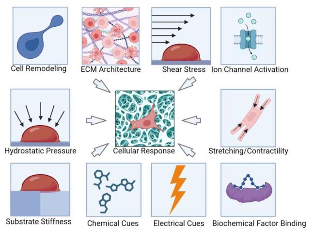
Fig. 1. The scheme for mechanobiology microenvironment influences the cellular response under loading conditions [12]. Microenvironment factors include cell remodeling, ECM architecture, shear stress, ion channel activation, hydrostatic pressure, stretching contractility, substrate stiffness, chemical cues, electrical cues and biochemical factors binding. All these Multiphysics would have an impact at cell level with biochemical cell response [12]. |
2.1. Extracellular matrix component and architecture
As a complex network of proteins and polysaccharides that surrounds cells in the tissue, the ECM composition such as fibrous proteins like collagen and elastin provide mechanical strength on cellular behavior and function, while glycosaminoglycans (GAGs) and proteoglycans contribute to the viscoelastic properties of the matrix. The fibrillar architecture for the ECM, where fibers of collagen or other proteins are organized into networks or bundles, could create different mechanical properties and guiding cellular behavior, further influencing cell migration, orientation, and tissue development.
2.2. Matrix stiffness and porosity
The mechanical stiffness or rigidity of the matrix is a critical factor in mechanobiology. Matrix stiffness is typically quantified by its elastic modulus, and it can impact cell adhesion, spreading, migration, proliferation, and differentiation. The porosity and pore size of the 3D matrix microenvironment influence cell migration and tissue infiltration, nutrient and oxygen diffusion, and the transport of signaling molecules.
2.3. Primary cilium
Primary cilium is a single, fixed, antenna-like structure with a cell membrane embedded in the extracellular matrix [13]. It primary cilium is composed of a microtubule-based axoneme, which extends from the basal body, a specialized structure derived from the centrosome. Localization of different proteins in the microdomains of the primary cilium enables the sensing and responding to a variety of environment cues, including mechanical, chemical and electrical stimuli [14].
2.4. Membrane-bound proteins
Membrane-bound proteins are key players in mechanobiology as they directly interact with the extracellular environment and transmit mechanical signals into the cell. These proteins can sense and respond to mechanical forces, mediate cell-substrate adhesion, and initiate signaling pathways that regulate cellular functions. Integrins are a family of cell surface receptors that play a crucial role in cell adhesion [15], migration and signaling. Integrins are composed of an alpha and a beta subunit, and there are over 20 different types of alpha subunits and 8 types of beta subunits, which can combine to form different integrin heterodimers with district binding specificities. They are transmembrane proteins that bind to extracellular matrix proteins, such as collagen, fibronectin and laminin, as well as to other cells [16]. The deformation of the matrix can be transmitted to the interior of the cell to cause a downstream signal response by activating integrins. Other proteins such as cadherins are calcium-dependent adhesion proteins that mediate cell-cell adhesion. They transmit mechanical forces across cell-cell junctions, contributing to tissue integrity and morphogenesis. The focal adhesions also known as adhesion plaque complexes, are specialized structures that form at the interface between cells and the extracellular matrix. They are composed of a variety of more than 100 species of talin, twistrin, paxillin, etc., one end of talin binds to the intracellular segment of the b subunit of integrin, and the other end can connect to the actin skeleton. Cell-matrix interactions typically occur in the immediate vicinity of focal adhesions and mediate interactions with signals through focal adhesion kinases [17]. Adhesion plaque complexes has been proved to play a crucial role in cell adhesion and migration, as well as in the transmission of mechanical forces between cells and the extracellular matrix [18].
2.5. Membrane ion channel
Membrane ion channel are porous membrane protein structure that span the lipid bilayer of cell membranes and regulate the flow of ions or charged particles across the membrane. Ion channels are important for a variety of cellular processes, including electrical signaling in muscle cells, regulation of fluid balance and cell-cell communications [19].
2.6. Motor protein
The motor protein, such as myosins and kinesins, are a class of proteins that play a central role in mechanobiology by converting chemical energy (hydrolysis of ATP molecules) into mechanical work (cytoskeletal filaments movements). Myosins are mainly involved in muscle contraction, cell migration, and intracellular transport. The Kinesins are involved intracellular transport, particularly in the movement of organelles, vesicles, and other cargo within the cell.
2.7. LINC complex
The LINC (Linker of Nucleoskeleton and Cytoskeleton) complex, also known as the LINC complex, plays a crucial role in mechanobiology by connecting the nuclear envelope to the cytoskeleton. It consists of two major components: the inner nuclear membrane protein SUN (Sad1p, UNC-84) and the outer nuclear membrane protein KASH (Klarsicht/ANC-1/Syne Homology) domain proteins. The LINC complex provides a direct physical link between the nucleus and the cytoskeleton, allowing for the transmission of mechanical forces and signals between these two compartments.
2.8. Clathrins and vesicles
Clathrin is a protein that plays a major role in the formation of coated vesicles. Clathrins are known to drive membrane remodeling events, particularly in clathrin-mediated endocytosis by assembling into a cage-like lattice structure that shapes and invaginates the plasma membrane to form clathrin-coated pits. Vesicles are small membrane-bound compartments within cells, involving in the transport of molecules, organelles, and signaling components, and their interactions with clathrins and cues can impact mechanical forces transduction and further cellular functions.
3. The construction of matrix mechanobiological structure by TPL
TPL is a technique that uses a high-intensity laser to selectively polymerize a photosensitive material in a layer-by-layer fashion. its versatility and ability facilities the production for complex geometries of ECM with high precision. The mechanism involves using a pulsed laser to activate a photosensitive material, which causes it to solidify and form a solid structure [20] (Fig. 2). Unlike traditional 3D printing techniques, which use a single photon to activate the photosensitive materials, TPL is a microfabrication technique exploiting the Two-Photon Absorption (TPA) process to induce the polymerization of a photosensitive polymeric material to achieve a higher resolution and more precise control over the shape and size of the printed structure [21,22]. The polymerization processes only occur in the focus of a femtosecond-pulsed laser voxel (volume pixel) because only in this ellipsoid 3D spot, the spatial and temporal density of photons is adequate to induce polymerization simulated by two photons absorption [23,24]. The technique allows for the creation of complex structures with features as small as a few nanometers in size.
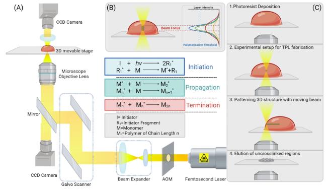
Fig. 2. (A). Schematic picture of an example TPL consisting of a femtosecond laser, acousto-optic modulator (AOM), beam expander, galvo scanner, CCD camera, 3D movable stage and controlling system (B). In TPL, the polymerization processes only occur in the focus of a femtosecond-pulsed laser voxel (volume pixel). But in one-photon direct laser writing, instead, the laser beam is absorbed all along the focalization cone, making it hard to realize complex 3D structures [27] (C). The generation mechanism of the procedure of TPL sample fabrication. controllable relative movement between the laser focus would cause the polymerization in a designed shape to form customized graphics [27]. |
Based on the above characteristics, higher machining accuracy and the hardness, surface roughness, porosity of the material matrix under physiological and pathological conditions can be accurately and stably simulated [25]. And the effects of different mechanical factors on cell behaviors such as cell viability, cell proliferation, and cell migration behavior and the behavior of biological tissues under different conditions, such as mechanical loading or exposure to different chemicals could also be studied [26].
4. The polymerization mechanism for the TPL
At present, the commonly used 3D printing polymerization system is photopolymerization crosslinking system. Among them, the photopolymerization system uses photo initiators to crosslink materials through light (UV, Vis and IR light) to initiate a crosslinking reaction, by which cleavage the chemical bonds (e.g., C-C, C=O, C-S, C-Cl) under light irradiation to form active species for photopolymerization [28,29,30]. The commonly used photo-initiators can be classified as Norrish type I and II [31] (Table 1). Up to now, A wide range of readily available and low-cost photosensitive resins have been used for TPL, including inorganic-organic hybrid materials (Ormocers) [32], urethane acrylate monomers [33], acrylic-based prepolymers [34], single-walled carbon nanotube-dispersed resins [35], gelatin hydrogels [36], zirconium sol-gels [37], and water-soluble materials [38].
Table 1. Photoinitiators with corresponding cross-linked hydrogels and the applications. |
| Name | Structure | Hydrogel Type | Application | Ref. |
|---|---|---|---|---|
| Benzophenone dimer | 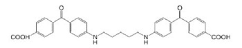 | BSA, Collagen | Drug Delivery, Scaffold | [39,40] |
| Flavin adenine dinucleotide | 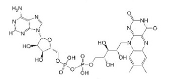 | BSA, Avidin, PMMA | Cell Delivery | [39,40,41,42] |
| G2CK(a) | 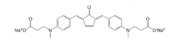 | GelMOD | Drug Delivery, Scaffold | [43,44] |
| Irgacure 369 | 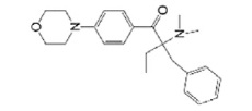 | PEGDA | Scaffold | [45] |
| Irgacure 651 | 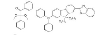 | PEGDA/HEMA | Scaffold | [46] |
| Irgacure 2959 | 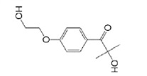 | GelMOD, HA | Scaffold | [47,48,49] |
| P2CK(a) |  | GelMOD | Drug Delivery, Scaffold | [43,44] |
| Rose Bengal |  | BSA, Fibrinogen, Collagen, Fibronectin, Concanavalin A | Drug Delivery, Scaffold | [39,40,50,51] |
| WSPI(a) |  | PEGDA, Gelatin Derivative | Scaffold | [43,52] |
BSA: Bovine serum albumin; PMMA: Poly(methyl methacrylate); GelMOD: Methacrylamide-modified gelatin; PEGDA: Polyethylene Glycol Diacrylate; HEMA (Hydroxyethyl) methacrylate; HA: Hyaluronic acid. |
When exposed to light (UV, Vis and IR light), type I photoinitiators can be photo-fragmented into radicals, but type II photoinitiators need the presence of a coinitiator, such as an amine or an alcohol [31]. After that, ketyl radical and a donor radical has been formed after hydrogen abstracted from a proton donor molecule. Then ketyl radical start radical couple to the growing macromolecular chain to expand the molecular weight, followed by the donor radical initiates the polymerization [28,29,30].
After verifying the optimal conditions for the polymerization, customized graphics can be designed through controllable relative movement between the laser focus, which finally allows the creation of 3D structures. The unexposed non polymerized material is then removed by solvent. In Table 1, the common photo-initiators, the hydrogel type, the cell type and the application has been listed as follow [27].
5. Recent mechanobiology studies made by TPL
The design of 3D polymer structures through TPL has been widely used in the field of mechanobiology (Table 2). By simulating the 3D microenvironment of cells, the force exerted on cells from the environment and the force exerted by the cells to the environment could be well-studied [27]. The common method to simulate the microenvironment is to create a mechanical structure that can be affected by cells to produce deformation. By studying the changes of geometric deformation under mechanical effects, the force of cells in this situation could be studied [53]. Klein et al. develop a new type of microstructured scaffold made of elastic materials for measuring cell forces. They first construct the spiderweb-like structures (Ormocomp scaffold) with TPL to support the attachment of cardiomyocytes seeding above. The contractile force exerted by cardiomyocytes was studied by changes in the spider web structure suspended above the substrate [54]. Then the contraction force of cardiomyocytes was calculated to be about 50 nN. They further fabricated two different photoresists composite-polymer scaffolds with distinct mechanical and protein-binding properties. And cell adhesion and shape could be fully controlled by these ECM functionalized parts [55]. Hohmann et al. discusses the use of direct laser writing to create 3D topographies for promoting the proliferation and differentiation of osteoblast-like cells, which demonstrated the 3D topographies can be used to control the orientation and alignment of cells, and that they can improve the adhesion, proliferation, and differentiation of osteoblast-like cells compared to cells cultured on non-patterned surfaces [56].
Table 2. Materials list for TPL techniques Realized for Mechanobiological Studies. |
| Materials | Photoinitiators | Structures | Cell Type | Resolution | Ref. |
|---|---|---|---|---|---|
| HDDA | Irgacure 819 | Star Shape | Oligodendrocyte Progenitor Cell (OPC) | 5 μm | [68]R |
| Gelma; PEGDA; PEGOA | N/A | Vessel | Human Breast Cancer Cells (MCF-7); HUVECs | 500 μm | [67]R |
| Acrylamide; Bis-Acrylamide | LAP | Strips | Rat Kidney Epithelial Cells (Nrk-52e) | 100 μm | [69]R |
| PEGDA | Irgacure 369 | Scaffold | Cardiomyocyte | 0.6 μm | [54]R |
| PEGDA | Irgacure 369 | Scaffold | Chicken Fibroblasts | 1 μm | [55]R |
| PEGDA | Irgacure 369 | Ridges | Rat Adrenal Pheochromocytoma Cell Line (Pc12); Human Neuroblastoma Derived Cell Line (Sh-Sy5y) | 0.3 μm | [20]R |
| IP-L 780 | N/A | Woodpile Structure | Mature Human Dendritic Cells (Dcs) | 1.3 μm | [58]R |
| IP-L 780 | N/A | Hexagonal Grid | Osteosarcoma Cells (Saos-2, MG-63, U-2 OS) | 0.45 μm | [56]R |
| PETTA | Irgacure 379 | Scaffold | Human Lung Carcinoma Cells (A549); Wildtype Mouse Embryonic Fibroblasts (MEF) | 0.75 μm | [64]R |
| TPETA | Irgacure 369 | Scaffold | Epithelial (A549); Fibroblast (3T3) | 1 μm | [57]R |
| IP-Dip | N/A | Tetrakaidecahedral Scaffold | Saos-2 Cell | 1 μm | [63]R |
| SZ2080 | Irgacure 369 | Nichoid Structure | Rat BMSCs | 2 μm | [70]R |
HDDA: Hexanediol Diacrylate, GelMA: Gelatin-Methacryloyl; PEGDA: Poly(ethylene glycol) diacrylate; PEGOA: 8-arm polyethylene glycol-octaacrylate; IP-L 780: Acrylatebased Resin; PETTA: Trimethylolpropane Ethoxylate Triacrylate; TPETA: Trimethylolpropane Ethoxylate Triacrylate. |
Moreover, the affinity of cells to different adhesion proteins could be detected by coating different biomaterials on micro-scaffolds. Richter et al. develop a microfabrication technique to selectively functionalize 3D micro scaffolds with two distinct adhesion proteins (fibronectin and biotin-vitronectin) for guiding cell attachment. The authors describe a novel method for creating 3D micro scaffolds with precise control over the functionalization of different regions of the scaffold with distinct adhesion proteins. They demonstrate that this new method can be used to guide cell attachment and migration, and that it can be used to create microenvironments that mimic the architecture of natural tissues [57]. Olsen et al. developed method for fabricating free-form 3D constructs for studying directed cell migration. The authors describe the TPL technique for creating 3D with woodpile topologies constructs prefilled with fibrillar collagen, which allows for the study of dendritic cell migration in complex environments. They demonstrate that this new method can be used to study the effects of substrate topography and stiffness on cell migration and turns under the chemoattractant concentration gradient [58]. Yang et al. studied the effects of substrate rigidity on stem cell behavior by describing a microfabrication technique for creating uniform array of closely spaced, vertical, elastomeric microposts, which allows for the precise control of substrate stiffness at the cellular level. They demonstrate this new method can be used to study the effects of substrate stiffness on stem cell morphology, traction force generation, focal adhesion organization and differentiation [59]. Marino et al. discussed the use of two-photon polymerization to create sub-micrometer patterned surfaces based on aligned ridges for studying cell-substrate interactions and improving the differentiation of neuron-like cells. The authors demonstrate that the sub-micrometer patterns can be used to increased molecular differentiation toward neurons of the SH-SY5Y cells on the submicrometric patterned substrates [60].
In addition, TPL can also accurately construct scaffold materials with controllable porosity, simulate the physical microenvironment of the matrix, and study the cell's attachment and migration inside [61,62]. Maggi et al. discusses the development of three-dimensional nano-architected scaffolds with tetrakaidecahedral periodic geometry for efficient bone tissue formation during early osteointegration. This new approach could create nano-architected scaffolds with precise control over stiffness, pore size, and geometry through TPL, which demonstrate that this new method can be used to create scaffolds with mechanical properties similar to natural bone tissue, and that these scaffolds can support the growth of bone cells in vitro [63]. Greiner et al. describe a microfabrication technique by TPL creating polymer scaffolds with precise control over pore size and shape, and the ability to incorporate chemoattractant gradients within the scaffold. Then this new method has been demonstrated to be used in studying the effects of chemoattractant gradients and pore size on cell invasion. The study also suggest that this new method could have important applications in the field of cancer research and tissue engineering, particularly for the development of new biomaterials that can promote cell invasion and tissue regeneration [64].
And the TPL technique could also integrate with organ-on-a-chip technology, which could control and simulate physiological phenomena and systems [65,66]. Cao et al. describe a new approach to creating a tumor-on-a-chip microfluidic system that can simulate the growth and metastasis of cancer cells. The device includes a bioprinted blood vessel and lymphatic vessel pair, which allows for the exchange of nutrients and waste between the tumor and the surrounding tissue. The authors demonstrate that this new system can be used to simulate the complex transport mechanisms of certain pharmaceutical compounds inside the tumor microenvironment [67].
6. Conclusions and future perspective
TPL is a powerful tool for fabricating 3D micro- and nanostructures with high precision and resolution. As a versatile technique, it has found widespread applications in various fields, including optics, photonics, materials science, and biology. In particular, TPL has gained significant attention in the field of mechanobiology, where it has been used to fabricate complex 3D microstructures for studying the mechanical behavior of cells and tissues, which allows researchers to create microenvironments that mimic the mechanical properties of native tissues and study the effect of mechanical cues on cell behavior. In addition to fabricating scaffolds with various geometries and stiffness values, TPL could be used to investigate how these parameters affect cell adhesion, proliferation, and differentiation. TPL also offers the advantage of creating structures with high aspect ratios and overhangs that are not possible with other fabrication techniques. Using this technology, researchers are able to design complex structures for drug delivery and tissue engineering, such as microfluidic channels and microneedles. This review discusses the various aspects of TPL and its applications in mechanobiology, as well as its potential to advance our understanding of cell mechanics and tissue engineering. For mechanobiology research, TPL has been used as a powerful tool for creating complex 3D structures (scaffolds, channels, and patterns). with high precision and resolution. Despite its many advantages, TPL also has some limitations.
However, in comparison to other techniques such as micro-milling and 3D printing, TPL's fabrication speed is relatively slow. This limits the throughput of TPL and makes it difficult to fabricate large structures. In addition, TPL has difficulty fabricating structures with high aspect ratios and overhangs without using sacrificial layers in this case, contamination of the final structure can occur and sacrificial layers may be difficult to remove.
Despite these limitations, TPL still has a bright future in mechanobiology research. One area of research where TPL is expected to have a significant impact is in the study of mechanotransduction for tumor biology, the process by which cells sense and respond to mechanical cues. TPL can be used to create tumor microenvironments that mimic the mechanical properties of the tumor tissues and study the effect of mechanical cues on tumor cell behavior, such as intravasation and extravasation. This can provide insights into the molecular mechanisms of mechanotransduction in cancer progression or regression and help develop new therapies for various cancers that are influenced by mechanical cues. The woodpile-based structure with different mesh size has been adapted to study the invasion of human lung carcinoma cells in the absence and in the presence of serum chemoattractant [64]. By changing the unchanged the pulse length and the repetition rate for the laser power, the materials Young's modulus and hardness could be controled. And the invasive behaviours of tumor cells under different stiffness of extracellular environment could also be studied [71]. This application would facilities the studies for the 3D mechanical interaction between the tumor cells and the surrounded extracellular microenvironment. The development of these next-generation 3D assay made by TPL would replace the 2D culture model to study the tumor cell's migration properties responded to the surrounding environment [72].
In addition to tumor biology, another area where TPL is expected to have a significant impact is in stem cell biology and tissue engineering. TPL can be used to fabricate scaffolds with complex geometries and stiffness values that can be used to guide the growth and differentiation of stem cells. This can be used to develop new tissue engineering strategies for the reconstruction of stem cell niche for tissue regeneration. Stem cell niche is the microenvironment in which stem cells reside and receive signals that control their behavior, such as self-renewal, differentiation, and proliferation. The niche includes a variety of physical and chemical cues, such as ECM components, growth factors, cytokines, and cell-cell interactions. In the context of stem cell research, TPL can be used to fabricate biomimetic scaffolds that mimic the structure and composition of the stem cell niche [73,74,75]. Recent studies try to provide a controlled microenvironment for stem cells to grow and differentiate. They further study the interactions between stem cells and their microenvironment made by TPL. The results show that the optimized the niche structures could maintain the multipotency of MSCs inside the specialized 3D microenvironment [20,76,77].
In conclusion, TPL is a powerful tool for the fabrication of complex 3D microstructures with high resolution and precision. It has found widespread applications in various fields, including mechanobiology. Despite its limitations, TPL has a bright future in mechanobiology research and tissue engineering. As the technique continues to evolve and improve, it is expected to play an increasingly important role in advancing our understanding of cell mechanics and tissue engineering.
Declaration of competing interest
The authors declare that they have no known competing financial interests or personal relationships that could have appeared to influence the work reported in this paper.
Acknowledgements
The study is supported by the Shanghai Pujiang Program (21PJ1409200) and the China Postdoctoral Science Foundation (2022M722122), Shanghai Frontiers Science Center of Degeneration and Regeneration in Skeletal System.
