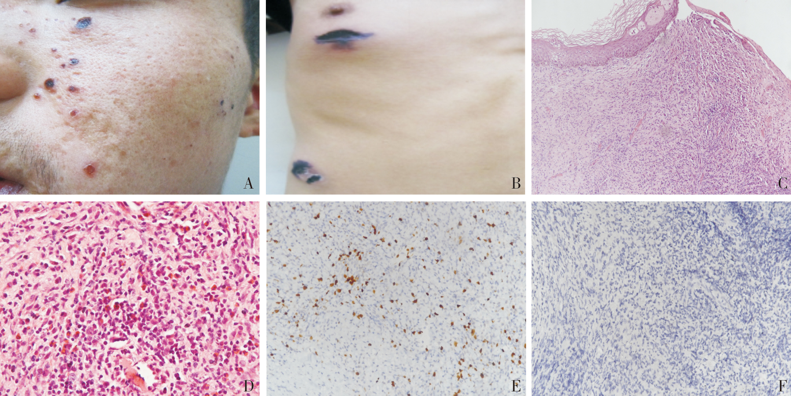
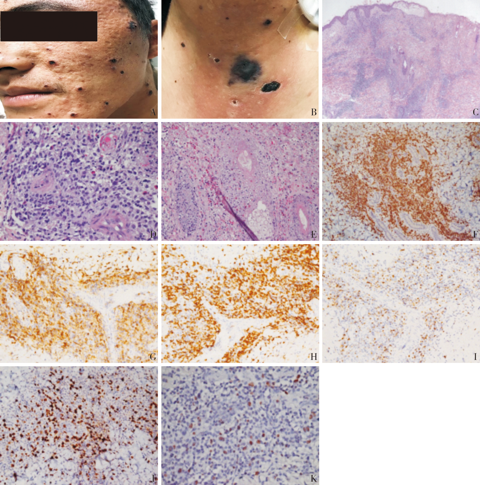
| [1] |
GUPTA G, MAN I, KEMMETT D. Hydroa vacciniforme: A clinical and follow-up study of 17 cases[J]. J Am Acad Dermatol, 2000,42:208-213.
|
| [2] |
QUINTANILLA-MARTINEZ L K Y, KO Y H, KIMURA H, et al. Hydroa vacciniforme-like lymphoproliferative disorder[M]// SWERDLOWS H, CAMPOE, HARRISN L, et al. WHO Classification of Tumours of Haematopoie-tic and Lymphoid Tissue. 4th ed. Lyon,France: IARC, 2017,360-362.
|
| [3] |
IWATSUKI K, SATOH M, YAMAMOTO T, et al. Pathogenic link between hydroa vacciniforme and Epstein-Barr virus-associated hematologic disorders[J]. Arch Dermatol, 2006, 142(5):587-595.
pmid: 16702496
|
| [4] |
QUINTANILLA-MARTINEZ L, RIDAURA C, NAGL F, et al. Hydroa vacciniforme-like lymphoma: a chronic EBV+ lymphoproliferative disorder with risk to develop a systemic lymphoma[J]. Blood, 2013, 122(18):3101-3110.
|
| [5] |
XIE Y, WANG T, WANG L. Hydroa vacciniforme-like lymphoproliferative disorder: A study of clinicopathology and whole-exome sequencing in Chinese patients[J]. J Dermatol Sci, 2020, 99(2):128-134.
doi: S0923-1811(20)30212-7
pmid: 32682634
|
| [6] |
MIYAKE T, YAMAMOTO T, HIRAI Y, et al. Survival rates and prognostic factors of Epstein-Barr virus-asso-ciated hydroa vacciniforme and hypersensitivity to mosquito bites[J]. Br J Dermatol, 2015, 172(1):56-63.
doi: 10.1111/bjd.2015.172.issue-1
URL
|
| [7] |
GRU A A, JAFFE E S. Cutaneous EBV-related lymphoproliferative disorders[J]. Semin Diagn Pathol, 2017, 34(1):60-75.
doi: S0740-2570(16)30093-4
pmid: 27988064
|
| [8] |
白玄业, 李文才, 王冠男, 等. 种痘水疱病样淋巴组织增生性疾病11例临床病理分析[J]. 临床与实验病理学杂志, 2018, 34(07):764-768.
|
|
BAI X Y, LI W C, WANG G N, et al. Clinicopathological analysis of 11 cases of hydroa vacciniforme-like lymphoproliferative disease[J]. Chin J Clin Exp Pathol, 2018, 34(07):764-768.
|
| [9] |
LIU Y, MA C, WANG G, et al. Hydroa vacciniforme-like lymphoproliferative disorder: Clinicopathologic study of 41 cases[J]. J Am Acad Dermatol, 2019, 81(2):534-540.
doi: S0190-9622(19)30081-7
pmid: 30654082
|
| [10] |
HAN B, HUR K, OHN J, et al. Hydroa vacciniforme-like lymphoproliferative disorder in Korea[J]. Sci Rep, 2020, 10(1):19294.
doi: 10.1038/s41598-020-76345-2
pmid: 33168864
|
| [11] |
王新华, 梁远征, 周志远, 等. 种痘水疱病样淋巴组织增殖性疾病18例临床分析[J]. 郑州大学学报(医学版), 2020, 55(02):166-172.
|
|
WANG X H, LIANG Y Z, ZHOU Z Y, et al. Clinical analysis of 18 patients with hydroa vacciniforme-like lymphoproliferative disorder[J]. J ZHENGZHOU UNIVERSITY (MEDICAL SCIENCES), 2020, 55(02):166-172.
|
| [12] |
周小鸽, 张燕林, 谢建兰, 等. 种痘样水疱病的临床病理特点及性质分析[J]. 临床与实验病理学杂志, 2017, 33(5):544-546.
|
|
ZHOU X G, ZHANG Y L, XIE J L, et al. Analysis of clinical pathological characteristics and properties of hydroa vacciniforme[J]. Chin J Clin Exp Pathol, 2017, 33(5):544-546.
|
| [13] |
CHEN C C, CHANG K C, MEDEIROS L J, et al. Hydroa vacciniforme and hydroa vacciniforme-like lymphoprolife-rative disorder: a spectrum of disease phenotypes associa-ted with ultraviolet irradiation and chronic Epstein-Barr virus infection[J]. Int J Mol Sci, 2020, 21(23):9314.
doi: 10.3390/ijms21239314
URL
|
| [14] |
XUE R, ELBENDARY A, LIU H, et al. Hydroa vaccini-forme-like lymphoma: clinicopathological description, treatment and outcome[J]. J Am Acad Dermatol, 2021, 85(3):752-755.
doi: 10.1016/j.jaad.2019.11.031
URL
|
 ), 肖立1a(