诊断学理论与实践 ›› 2025, Vol. 24 ›› Issue (01): 51-58.doi: 10.16150/j.1671-2870.2025.01.008
车冠华1, 曾长2, 陈晓炎3( )
)
收稿日期:2023-10-07
接受日期:2024-06-03
出版日期:2025-02-25
发布日期:2025-02-25
通讯作者:
陈晓炎 E‑mail: cxy11832@rjh.com.cn
CHE Guanhua1, ZENG Chang2, CHEN Xiaoyan3()
Received:2023-10-07
Accepted:2024-06-03
Published:2025-02-25
Online:2025-02-25
摘要:
目的 探讨伴有淋巴间质的微结节型胸腺瘤(micronodular thymoma with lymphoid stroma, MNT)患者的临床、病理特点。方法 对2020年1月至2023年7月在上海交通大学医学院附属瑞金医院胸外科手术切除,并经病理确诊的连续的5例MNT患者的临床症状、病理特征、免疫表型、临床治疗及预后进行回顾性分析,并复习相关文献。结果 5例MNT患者中男性3例,女性2例,年龄为55~68岁。4例位于前纵隔,1例位于上纵隔,为胸部CT偶然发现。肿瘤直径1.3~4.5 cm,3例包膜完整,边界清楚,切面呈实性,灰白色,质地细腻;2例呈囊实性,2例包膜侵犯。显微镜下均有MNT独特的组织学表现:肿瘤主要由上皮性结节及淋巴细胞组成;上皮细胞呈短梭形或卵圆形,异型性小,核仁不清楚,核分裂象罕见,无坏死;上皮性结节之间被淋巴细胞间隔,可见淋巴滤泡形成。免疫组化检测显示,5例肿瘤细胞全都表达AE1/AE3、CK19、P63、Bcl-2;1例囊性区囊壁腔缘衬覆细胞表达EMA,不表达P63、Bcl-2;5例淋巴细胞表达CD20、CD3、CD5、TdT(少量+),滤泡生发中心表达CD10,结节内朗格汉斯细胞表达Langerin、S100、CD1α。5例患者中3例行EBER原位杂交检测,结果显示,上皮细胞及淋巴细胞均阴性。5例患者手术切除肿瘤后,随访7~39个月,均未见肿瘤复发。复习2019年至2023年间Pubmed、Medline、中国知网、万方数据库新报道的以及文献中总结的既往1999年至2018年间的MNT病例,共206例MNT患者,中老年人(>45岁)195例,青年人(18~45岁)9例,儿童(<18岁)2例,发病部位以纵隔为主,也可见于颈部,发病人群无性别差异,多为体检时发现,切面以囊性稍多见,术后仅1例复发。结论 MNT罕见,好发于中老年人纵隔,手术切除后预后良好。MNT病理特征为上皮性结节散在分布于丰富的淋巴间质中,可见淋巴滤泡形成,结节内散在朗格汉斯细胞,可伴囊性变,结合发生部位、组织病理学特点及免疫组化标志物可以确诊,但仍需与其他病变鉴别。
中图分类号:
车冠华, 曾长, 陈晓炎. 伴有淋巴间质的微结节型胸腺瘤5例临床病理分析并文献复习[J]. 诊断学理论与实践, 2025, 24(01): 51-58.
CHE Guanhua, ZENG Chang, CHEN Xiaoyan. Micronodular thymoma with lymphoid stroma: a clinicopathologic analysis of five cases and literature review[J]. Journal of Diagnostics Concepts & Practice, 2025, 24(01): 51-58.
表1
5例MNT患者临床资料
| Case | Gender | Age (years) | Clinical symptoms | Maximum size (cm) | Cystic change | Location | Diagnose | Group | Follow-up (months) | Recurrence |
|---|---|---|---|---|---|---|---|---|---|---|
| 1 | Male | 63 | No Special | 4.5 | Yes | Anterior mediastinum | MAMNT | Ⅰ | 39 | No |
| 2 | Male | 57 | No Special | 2.3 | Yes | Anterior upper mediastinum | MNT | Ⅱ | 30 | No |
| 3 | Female | 68 | No Special | 1.3 | No | Right upper mediastinum | MNT | Ⅰ | 25 | No |
| 4 | Male | 55 | No Special | 2.0 | No | Anterior mediastinum | MNT | Ⅰ | 8 | No |
| 5 | Female | 65 | No Special | 3.2 | No | Anterior mediastinum | MNT | Ⅱ | 7 | No |

图1
MNT患者的病理学图片(苏木精-伊红染色)A:部分区域肿瘤侵犯纤维包膜(×10);B:囊性区,囊壁腔缘单层细胞及基底部复层细胞(×400);C:短梭形、卵圆形上皮细胞,细胞质相对丰富、红染(×400);D:上皮性结节及淋巴细胞间质(×40)。
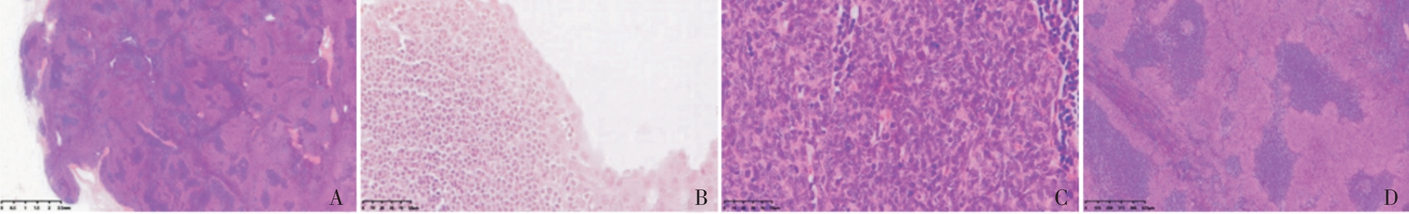
图2
MNT患者的免疫组化图片(Bench Mark)A:囊壁腔缘单层细胞EMA(+),基底部复层细胞及上皮性结节(-) (×200); B:囊壁腔缘单层细胞、基底部复层细胞及上皮性结节CK19(+) (×200);C:囊壁腔缘单层细胞P63(-),基底部复层细胞及上皮性结节P63(+)( ×100);D:囊壁腔缘单层细胞Bcl-2(-),基底部复层细胞、上皮性结节及间质淋巴细胞Bcl-2(+) (×400);E:B淋巴细胞CD20(+) (×20 );F:上皮性结节周边未成熟T细胞TdT(+)(×40);G:淋巴滤泡生发中心CD10(+)(×40) (右上角×400);H:上皮性结节内朗格汉斯细胞CD1α(+)(×100)。
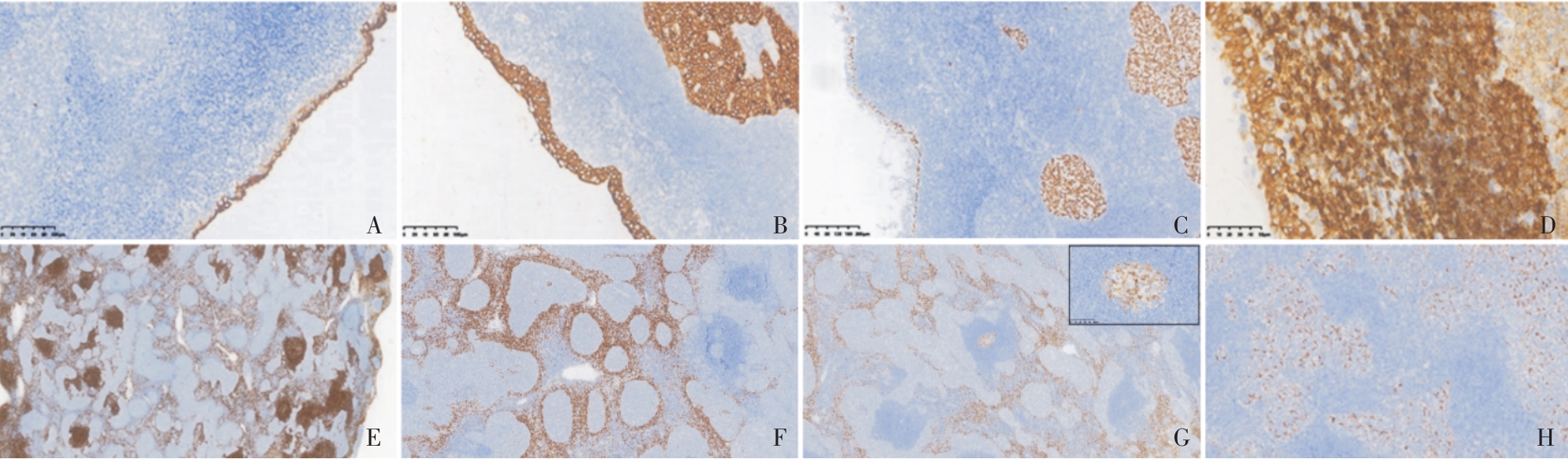
表2
文献及本文共211例MNT患者的临床资料
| Literature | Cases (male:female) | Ages (median,years) | Location | Maximum size(cm) | Clinical symptoms | Macroscopically | Follow-up(months) |
|---|---|---|---|---|---|---|---|
| Wang[ | 107(1.3∶1.0) | 45-83 (Not detailed) | 103 cases occurred in the thymus, 4 cases occurred in the neck | 1.0-10.7 | 6 cases of severe myasthenia gravis, 3 cases of ptosis, 6 cases of chest discomfort, 1 case ofsternal pain, and the rest were found during physical examination or incidentally | 12 cystic, 30 solid, 7 cystic and solid, others not specified | 68 cases (4-190 months), 1 case died of esophageal cancer, 1 case of rectal cancer, and the rest had no recurrence |
| Zhang[ | 4(2∶2) | 5-62(52) | mediastinum | 3.3-5.0 | 1 case of severe myasthenia gravis, 3 cases found during physical examination | Not specified | 4 cases (19-32 months), disease-free survival |
| Yagi[ | 8(1∶1) | 47-76(60.5) | thymus | 1.8-6.0 | Not detailed | Not specified | 8 cases (14-120 months), 1 case recurred 10 years after surgery, and the rest had no recurrence |
| Oramas[ | 25(13∶12) | 38-69(57) | anterior mediastinum | 2.5-8.0 | Most presented with non-specific symptoms such as cough, chest pain, and dyspnea, and 4 cases were asymptomatic | Multilocular cystic | 19 cases (12-24 months), no recurrence |
| Hulme[ | 5(1∶4) | 58-70(65) | anterior mediastinum | 2.2-6.0 | Incidentally found | 1 solid, 4 cystic and solid | 5 cases (1-96 months), no recurrence |
| Zhao[ | 5(3∶2) | 49-68(64) | anterior mediastinum | 0.5-6.0 | 1 case of thyroid cancer, 1 case of emphysema, 1 case of bullae, 1 case of cough, 1 case asymptomatic | 4 solid, 1 multilocular cystic | 4 cases (9-29 months), no recurrence |
| Jiang[ | 5(3∶2) | 49-64(53) | anterior mediastinum | 3.5-8.2 | 1 case of repeated cough and chest distress, 4 cases asymptomatic | 1 solid, 4 cystic and solid | 5 cases (8-35 months), no recurrence |
| Bakshi[ | 3(2∶1) | 70-76(71) | thymus | 4.5-7.5 | 1 case of chest discomfort, 1 case of intermittent dry cough, 1 case asymptomatic | 2 solid, 1 cystic and solid | 3 cases(19-34 months), no recurrence |
| Yu[ | 8(5∶3) | 36-74(64.5) | anterior mediastinum | 2.1-12.0 | Found during physical examination or when seeking medical attention for other diseases (lung adenocarcinoma, chest discomfort) | 3 solid, 5 with cystic areas (3 multilocular cystic) | 8 cases (4-77 months), no recurrence |
| He[ | 7(4∶3) | 18-75(62) | 6 cases were located in the anterior mediastinum, 1 case was located in the middle mediastinum | 1.2-6.5 | 1 case of right upper eyelid ptosis,6 cases found during physical examination | 3 solid, 1 slightly solid 2 cystic and solid, 1 cystic | 7 cases (10-56 months), no recurrence |
| Liu[ | 4(1∶1) | 40-70(55) | thymus | 4.0-10.0 | 1 case of severe myasthenia gravis, 1 case of anemia, 1 case of cough, 1 found during physical examination | Not specified | 4 cases (4-51 months), 1 case died of other reasons 4 months later, the rest had no recurrence |
| Qin[ | 15(8∶7) | 5-73(61) | mediastinum | 1.4-6.0 | 4 cases of severe myasthenia gravis, 3 cases of chest distress and shortness of breath, 8 cases found during examination | 10 solid, 5 cystic and solid | 15 cases (3-97 months), no recurrence |
| Hsieh[ | 10(3∶7) | 53-80(70) | thymus | 2.0-16.5 | Not detailed | Not specified | 10 cases (3-124 months), no recurrence |
| The group | 5(3∶2) | 55-68(58) | 4 cases were located in the anterior mediastinum, 1 case was located in the superior mediastinum | 1.3-4.5 | Found during physical examination | 3 solid, 2 cystic and solid | 5 cases (7-39 months), no recurrence |
| [1] |
SUSTER S, MORAN C A. Micronodular thymoma with lymphoid B-cell hyperplasia: clinicopathologic and immunohistochemical study of eighteen cases of a distinctive morphologic variant of thymic epithelial neoplasm[J]. Am J Surg Pathol, 1999, 23(8):955-962.
doi: 10.1097/00000478-199908000-00014 pmid: 10435566 |
| [2] |
ISHIKAWA Y, TATEYAMA H, YOSHIDA M,et al. Micronodular thymoma with lymphoid stroma: an immunohistochemical study of the distribution of Langerhans cells and mature dendritic cells in six patients[J]. Histopathology, 2015, 66(2):300-307.
doi: 10.1111/his.12428 pmid: 24702632 |
| [3] | SUSTER S. The WHO 2021 thymoma classification: a work in progress[J]. J Cancer Metastasis Treat, 2022, 8:7. |
| [4] | 倪亚平, 笪倩, 袁菲,等. 混合性A型胸腺瘤与伴有淋巴样间质的微结节型胸腺瘤1例临床病理分析及文献复习[J]. 诊断病理学杂志, 2021, 28(7):557-561. |
| NI Y P, DA Q, YUAN F,et al. Mixed type A thymoma and micronodular thymoma with lymphoid stroma: a clinicopathologic analysis of one case and review of literature[J]. J Diag Pathol, 2021, 28(7):557-561. | |
| [5] | 王立娟, 曹友德, 曾敏, 等. 伴有淋巴样间质的微结节型胸腺瘤临床病理特征及文献复习[J]. 重庆医学, 2019, 48(17):2983-2987. |
| WANG L J, CAO Y D, ZENG M,et al. Clinicopathological features and literature review of micronodular thymoma with lymphoid stroma[J]. Chongqing Med, 2019, 48(17):2983-2987. | |
| [6] | 张冬梅, 魏建国, 方三高, 等. 4例伴淋巴样间质的微结节性胸腺瘤的临床病理分析[J]. 诊断病理学杂志, 2019, 26(9):562-565. |
| ZHANG D M, WEI J G, FANG S G,et al. Micronodular thymoma with lymphoid stroma: a clinicopathological study of four cases[J]. J Diag Pathol, 2019, 26(9):562-565. | |
| [7] | YAGI H, NAKAGURO M, ITO M,et al. Difference in the distribution of tumor infiltrating CD8+ T cells and FOXP3+ T cells between micronodular thymoma with lym-phoid stroma and micronodular thymic carcinoma with lymphoid stroma[J]. Pathol Int, 2021, 71(7):453-462. |
| [8] |
ORAMAS D M, MORAN C A. Micronodular thymomas with prominent cystic changes: a clinicopathological and immunohistochemical study of 25 cases[J]. Int J Surg Pathol, 2021, 29(4):352-357.
doi: 10.1177/1066896920963803 pmid: 33026263 |
| [9] | HULME K R, MAHAR A, CAO C,et al. Micronodular thymoma with lymphoid stroma: a clinicopathological study of five cases[J]. Pathology, 2021, 53(7):930-933. |
| [10] | 赵丽娜, 袁静萍, 黄亚冰, 等. 伴淋巴间质的微结节型胸腺肿瘤5例临床病理特征[J]. 临床与实验病理学杂志, 2021, 37(7):855-858. |
| ZHAO L N, YUAN J P, HUANG Y B,et al. Micronodular thymoma with lymphoid stroma: a clinicopathologic features of five cases[J]. J Clin Exp Pathol, 2021, 37(7):855-858. | |
| [11] | 江美辰, 郑巧灵, 杨映红. 伴淋巴样间质的微结节型胸腺瘤5例临床病理分析[J]. 临床与实验病理学杂志, 2021, 37(10):1234-1236. |
| JIANG M C, ZHENG Q L, YANG Y H. Micronodular thymoma with lymphoid stroma: a clinicopathologic analysis of five cases[J]. J Clin Exp Pathol, 2021, 37(10):1234-1236. | |
| [12] | BAKSHI N, DHAWAN S, RAO S,et al. Micronodular thymoma with lymphoid stroma: a trio of cases, with diverse-associated histological features[J]. Int J Surg Pathol, 2021, 29(6):693-697. |
| [13] | 余昶, 孙文勇. 伴淋巴样间质的微结节型胸腺瘤8例临床病理分析[J]. 肿瘤学杂志, 2022, 28(7):602-606. |
| YU C, SUN W Y. Micronodular thymoma with lymphoid stroma: a clinicopathologic analysis of eight cases[J]. J Chin Oncol, 2022, 28(7):602-606. | |
| [14] | 何晓顺, 詹升华, 黄山, 等. 伴淋巴样间质的微结节型胸腺瘤7例临床病理分析[J]. 临床肿瘤学杂志, 2022, 27(7):638-642. |
| HE X S, ZHAN S H, HUANG S,et al. Micronodular thymoma with lymphoid stroma: a clinicopathologic analysis of seven cases [J]. Chin Clin Oncol, 2022, 27(7):638-642. | |
| [15] | LIU P P, SU Y C, NIU Y,et al. Comparative clinicopathological and immunohistochemical study of micronodular thymoma and micronodular thymic carcinoma with lymphoid stroma[J]. J Clin Pathol, 2021, 75(10):702-705. |
| [16] | 秦积龙, 何萍, 范蕾, 等. 伴淋巴样间质的微结节型胸腺肿瘤17例临床病理与形态学分化谱系分析[J]. 临床与实验病理学杂志, 2023, 1(1):23-28. |
| QIN J L, HE P, FAN L,et al. Spectrum of morphological differentiation of micronodular thymic neoplasms with lymphoid stroma: a clinical and pathological analysis of seventeen cases[J]. J Clin Exp Pathol, 2023, 1(1):23-28. | |
| [17] | HSIEH M S, KAO H L, HUANG W C,et al. L424H mutation in GTF2I in micronodular thymomas with lymphoid stroma: evidence supporting close relationship with type A and AB thymomas[J]. Mod Pathol, 2023, 36(2):100008. |
| [18] | YOON J C, HAN J, KIM J,et al. A rare case of mixed type A thymoma and micronodular thymoma with lymphoid stroma[J]. Pathol Transl Med, 2015, 49(1):75-77. |
| [19] |
RADOVICH M, PICKERING C R, FELAU I,et al. The integrated genomic landscape of thymic epithelial tumors[J]. Cancer Cell, 2018, 33(2):244-258.
doi: S1535-6108(18)30003-5 pmid: 29438696 |
| [20] | 陈骏, 陈亭亭, 吴鸿雁, 等. 伴淋巴样间质的微结节型胸腺瘤2例并文献复习[J]. 临床与实验病理学杂志, 2014, 30 (7):766-770. |
| CHEN J, CHEN T T, WU H Y,et al. Micronodular thymoma with lymphoid stroma:two cases of report and lite-rature review[J]. J Clin Exp Pathol, 2014, 30(7):766-770. | |
| [21] |
STRÖBEL P, MARINO M, FEUCHTENBERGER M,et al. Micronodular thymoma: an epithelial tumour with abnormal chemokine expression setting the stage for lymphoma development[J]. J Pathol, 2005, 207 (1):72-82.
pmid: 15965907 |
| [22] | WELLS K, LAMRCA A, PAPAXOINIS G,et al. Unique correlation between GTF2I mutation and spindle cell morphology in thymomas (type A and AB thymomas)[J]. J Clin Pathol, 2023, 76(7):463-466. |
| [1] | . 三维平衡稳态自由进动磁共振成像序列在儿童冠状动脉异常起源于肺动脉诊断中的应用价值[J]. 诊断学理论与实践, 2020, 19(02): 145-150. |
| [2] | 许海敏, 张培培. 三款自动免疫组织化学染色仪在乳腺癌病理诊断中的应用比较[J]. 诊断学理论与实践, 2017, 16(06): 645-649. |
| [3] | 糜坚青, 金诗炜. 多发性骨髓瘤细胞分子遗传学异常与预后分层、治疗[J]. 诊断学理论与实践, 2017, 16(05): 460-463. |
| [4] | 乔长婷, 李蕾, 邬安妮, 袁菲. 进展期胃癌人表皮生长因子受体2蛋白表达与临床病理学特征的关系[J]. 诊断学理论与实践, 2017, 16(02): 166-170. |
| [5] | 李佳明, 张苏江. Ph样急性淋巴细胞白血病的分子遗传学改变及其对治疗的意义[J]. 诊断学理论与实践, 2017, 16(01): 27-31. |
| [6] | 韦玮, 吴春, 石群立,. 卵巢浆液性癌发病机制的研究进展[J]. 诊断学理论与实践, 2013, 12(01): 114-118. |
| [7] | 张青霞, 李晓,. 骨髓增生异常综合征异常克隆起源的研究进展[J]. 诊断学理论与实践, 2011, 10(04): 367-370. |
| [8] | 郭倞, 张丽华,. 大肠锯齿状病变的分子遗传学研究进展[J]. 诊断学理论与实践, 2008, 7(04): 451-453. |
| [9] | 骆天红,. 2型糖尿病分子遗传学研究的现今认识[J]. 诊断学理论与实践, 2007, 6(02): 101-107. |
| 阅读次数 | ||||||
|
全文 |
|
|||||
|
摘要 |
|
|||||