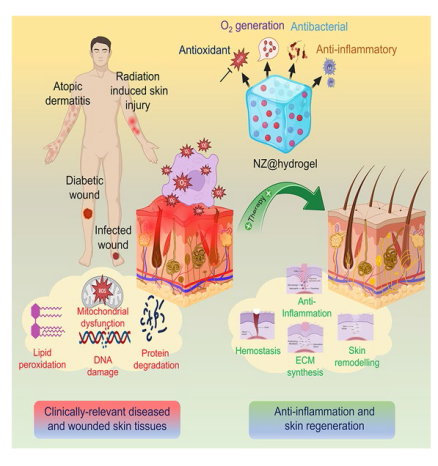
HIGHLIGHTS
1 Introduction
Fig. 1 Number of NZ-related articles published in the last 5 years according to Web of science (report acquired using the keyword ‘nanozymes’), indicating the scope and importance of this rapidly emerging platform |
2 Functional Roles of Hydrogel in NZ@Hydrogel System
3 Enzymatic Mechanisms and Multifunctional Roles of NZ@Hydrogels
3.1 Enzymatic Activities of NZ@Hydrogels
Fig. 2 Schematic showing the chemical design of NZ@hydrogels based on intrinsic enzymatic properties |
3.1.1 Peroxidase-like Activity
3.1.2 Catalase-like Activity
Fig. 3 Design of NZ@hydrogels with dominant enzymatic performance profiting skin regeneration. a Schematic diagram showcasing the synthesis process and the dual enzyme-mimic activity of MoS2@TA/Fe NSs aimed at enhancing wound repair [50]. Copyright 2021, Elsevier. b Synthesis of PFOB@PLGA@Pt/GelMA/ODex nanohybrid double network hydrogels with exceptional enzymatic and antibacterial attributes for advancing wound healing process [127]. Copyright 2023, American Chemical Society. c Synthesis of MoS2@Au@BSA NS and the preparation of an injectable hydrogel using an O2-supplying glucose-powered cascade reaction for the reconstruction of infected diabetic skin [133]. Copyright 2022, John Wiley and Sons, Inc |
3.1.3 Oxidase-like Activity
3.1.4 Superoxide Dismutase-like Activity
3.2 Tuning the Enzymatic Properties of NZ@Hydrogels
Table 1 List of various NZ platforms utilized to engineer hydrogels and their key catalytic properties analyzed |
| Classification | Nanozyme | Analyzed catalytic property | References |
|---|---|---|---|
| Ce-based | nCe | SOD, CAT | [49] |
| CeNZ | SOD, CAT | [77] | |
| Ce-MOF | SOD, CAT, GOx | [67] | |
| CeNZ | SOD, CAT | [45] | |
| Mn-based | MnO2 | CAT, POD | [158] |
| MnCoO | CAT | [159] | |
| GOx-MnO2 | POD, GOx | [160] | |
| EPL-MnO2 | SOD | [41] | |
| Fe-based | Fe@HCMS/GOx | POD, GOx, GSH | [114] |
| Mica-Fe3O4 | POD | [63] | |
| Fe3O4@GO | POD, SOD | [161] | |
| Fe-MIL-88NH2 | POD, OXD | [162] | |
| FePO4 | POD, SOD, CAT | [64] | |
| GA/Fe@AP (GAP) | CAT | [163] | |
| Fe2O3-GOx@ZIF-8 | GOx, CAT | [164] | |
| Mo-based | MoS2@TA/Fe | POD, CAT | [50] |
| CNT@MoS2 | POD, SOD, CAT | [83] | |
| MoS2 | CAT | [86] | |
| Ag/MoS2 | POD | [90] | |
| MoS2@Au@BSA | GOx, POD, CAT, SOD | [133] | |
| MoS2 | POD | [165] | |
| MoS2-PDA | CAT | [166] | |
| MoNP | POD, SOD, CAT | [167] | |
| MOF-based | MOF-818 | SOD, CAT | [75] |
| Ni3(HITP)2 MOF | SOD | [89] | |
| MOF(Fe-Cu)/GOx | POD, GOx | [168] | |
| Zr-Fc MOF | POD | [169] | |
| Cu-based | Au/Cu1.6O/P-C3N5/Arg NSs | SOD, CAT, POD, GOx, NOS | [88] |
| Cu5.4O | SOD, CAT, GPx | [170] | |
| Cu2O/Pt | POD, GOx, | [171] | |
| Cu2MoS4 | POD | [172] | |
| Ni4Cu2 | SOD, CAT | [51] | |
| Au-based | Au-Pt | POD | [80] |
| Au@ZIF-8 | POD | [115] | |
| Ag-based | PDA-AgNPs | POD | [56] |
| TA-Ag NZ | POD | [65] | |
| W-based | WS2/CL | CAT | [82] |
| PB-based | PB NZ | SOD | [42] |
| PB NZ/GOx | POD, GOx | [44] | |
| Pt-based | PtNZ | OXD, POD, GOx, NOx, SOD, CAT | [127] |
3.3 Multifunctional Roles of NZ@Hydrogels
Fig. 4 Schematic showing the multifunctional roles of NZ@hydrogels for skin therapy |
Table 2 Summary of various functional NZ@hydrogel platforms and their specific catalytic properties analyzed for skin therapy |
| Function | Hydrogel | Nanozyme | Analyzed catalytic property | Application | References |
|---|---|---|---|---|---|
| Antioxidant | PVA/SA | CNT@MoS2 | POD, SOD, CAT | Infected wound healing | [83] |
| Gelatin | MoNP | POD, SOD, CAT | Diabetic wound healing | [167] | |
| PLGA-PEG-PLGA | MOF-818 | SOD, CAT, GPx | Diabetic wound healing | [75] | |
| Antibacterial | HG | FePO4 | POD, SOD, CAT | Infected wound healing | [64] |
| GM-DC | Fe-MIL-88NH2 | POD, OXD | Infected wound healing | [162] | |
| Silk fibroin | Fe3O4 | POD | Infected wound healing | [63] | |
| Anti-inflammation | HMP | MnO2 | CAT, POD | Infected diabetic wound healing | [158] |
| Hep-PEG | Cu5.4O | SOD, CAT, GPx | Diabetic wound healing | [170] | |
| PAM | MOF(Fe-Cu)/GOx | POD, GOx | Infected wound healing | [168] | |
| Photothermal therapy | Carrageenan | Zr-Fc MOF | POD | Infected wound healing | [169] |
| PVA/Dex | MoS2@TA/Fe | POD, CAT | Infected wound healing | [50] | |
| MeHA | Fe3O4@GO | POD | Bacterial biofilm infection therapy | [161] | |
| Chemo-dynamic therapy | Chitosan thermogel | CuO2 nanodots | GSH, GPx | Melanoma therapy and infected wound healing | [207] |
| Pluronic F127 | TMB/Fe2+/GOx | GOx | Infected diabetic wound healing | [209] | |
| ALG-HA | Cu2O/Pt nanocubes | POD, GOx, | Infected wound healing | [171] | |
| Poloxamer F127 | Cu2MoS4 | POD | Infected burn wound healing | [172] | |
| Oxygen generation | HA-HYD | MnCoO | CAT | Diabetic wound healing | [159] |
| HA encapsulated L-arginine | Au/Cu1.6O/P-C3N5/Arg NSs | SOD, CAT, POD, GOx, NOS | Diabetic wound healing | [88] | |
| ODex/gC | MoS2 NSs | SOD, CAT, POD | Diabetic wound healing | [133] |
3.3.1 NZ@hydrogels as Antioxidants
3.3.2 NZ@hydrogels with Antibacterial Functions
3.3.3 NZ@hydrogels for Anti-inflammation
3.3.4 NZ@hydrogels for Photothermal Therapy
3.3.5 NZ@hydrogels for Chemo-dynamic Therapy
3.3.6 NZ@hydrogels for Oxygen Generation
4 Therapeutic Efficacy of NZ@Hydrogels in Clinically Relevant Diseased and Wounded Skin Tissues
4.1 Diabetic Skin Wound Healing
Fig. 5 Diabetic wound healing accelerated by NZ@hydrogels. a An illustration showing the fabrication process of PCN-miR/Col hydrogel for hastening diabetic skin regeneration. b Photographs of diabetic wounds at different periods after the hydrogel treatment. b Quantification of wound area obtained from photographs. d Masson’s trichrome staining of skin tissues after 28 days of treatment. Scale bars, 100 μm. e Fluorescent expression of VEGF and CD31 markers. Scale bars, 50 μm. f Quantification for VEGF expression and g Quantification of several blood vessels at 28 days [45]. Copyright 2019, American Chemical Society. h Representation of the accelerated wound healing coordinated by MnCoO@PLE/HA hydrogel. i An illustration showing the fabrication process of MnCoO@PLE/HA hydrogels for diabetic wound healing. j Photographs of diabetic wounds were evaluated up to 16 days after the hydrogel treatment. k A representation of relative wound healing at different time points. l Quantification of residual wound area from wound photographs. m Blood glucose levels in animals induced with diabetes for studying the wound healing process [159]. Copyright 2022, John Wiley and Sons, Inc |
4.2 Bacteria-Infected Skin Wound Healing
Fig. 6 Wound healing process accelerated by NZ@hydrogels with antibacterial properties. a Fabrication of a multifunctional anti-bacterial hydrogel based on PEGMA-GMA-Aam copolymer. b Photographs of infected wounds after hydrogel treatment. Scale bar: 1 cm. c Relative wound healing rates at different time points. d Number of days taken for complete wound healing. e Quantification of bacterial density in the infected wound [158]. Copyright 2022, Elsevier. f An illustration of the fabrication of FEMI hydrogel for infected diabetic wounds. g Photographs of the wound after hydrogel administration. h A representation of the wound closure at different time points. i Quantification of wound closure rates. j Blood glucose levels were examined in mice at different time points. k The fluorescent expression of ROS levels in wounds by dihydroethidium (DHE). Scale bar: 50 μm. l The fluorescence intensity of DHE for various hydrogel groups. m Histological examination of wounds following hydrogel treatment. Scale bar: 100 μm. n Epidermal thickness examined on the 7th day. o Blood vessel regeneration examined on the 7th and 14th days. p Hair follicle formation examined on the 14th day [41]. Copyright 2020, American Chemical Society |
4.3 Atopic Dermatitis Skin Regeneration
Fig. 7 Therapeutic properties of NZ@hydrogels in alleviating AD. a Illustration of the design of a nCe-based hydrogel patch to treat AD. b Schematic of in vivo experiments and timeline for animal experiments. c Photographs of dorsal skin of different groups following treatment. d Evaluation of dermatitis score. e Histology of mouse skin sections stained with H&E after treatment. Scale bars, 100 μm. f Comparison of epidermal thickness. g Histology of the samples revealed infiltrated mast cells. Scale bars, 100 μm. h Relative quantification of infiltrated mast cells. i Representative immunofluorescence images of oxidative DNA damage marker: 8-OHdG. Scale bars, 100 μm. j Relative 8-OHdG quantification [49]. Copyright 2022, American Chemical Society. k An illustration highlighting the capacity of Gel@ZIF-8 to suppress oxidative stress and alleviate inflammatory responses. l Photographs of mice dorsal skin following treatment. m Evaluation of dermatitis score. n Comparison of epidermal thickness between groups. Scale bars, 100 μm. o Relative quantification of epidermal thickness. p Histology of the samples showing infiltrated mast cells. Scale bars, 100 μm. q Relative quantification of mast cells [239]. Copyright 2023, Springer Nature |
4.4 Radiation-Induced Skin Wound Healing
Fig. 8 NZ@hydrogels act as radioprotectants for skin therapy. a Graphical representation showing the skin radioprotection capacity of the nano-GDY@SH hydrogel. b The photos of the skin wound changes of mice after different treatments. c The examination of skin tissues by H&E and Masson staining Scale bars, 100 μm [258]. Copyright 2021, Elsevier. d Schematic depicting the fabrication of IFI6-PDA@GO/SA hydrogel for RISI. e Images showing medical device used to induce the RISI model in mice. f Photographs of the dorsal skin of mice during the treatment period. g H&E staining of skin tissues on the 14th day. Scale bars, 200 μm. h Relative quantification of the wound area, granulation tissue thickness, and density of wound micro vessels [259]. Copyright 2022, Springer Nature |
5 Consideration of NZ@Hydrogels with Mechanobiological Functions
Fig. 9 NZ@hydrogels act mechano-chemically in alleviating symptoms of AD. a Graphical illustration displaying inflammatory reactions of AD due to oxidative harm and the outcome of mechanical scratching on AD. It also depicts the synergistic action of NZ@hydrogels in treating AD by ROS scavenging and FAK phosphorylation inhibition. b Representation of in vivo experiments. c Photographs of the dorsal skin of mice on the 24th day. d Evaluation of dermatitis score. e H&E staining of skin sections. Scale bars, 100 μm. F Epidermal thickness quantified from H&E staining. g TB staining of the skin section. Scale bars, 100 μm. h Measurement of the density of mast cells for each group after treatments [238]. Copyright 2023, Springer Nature |
6 Outlook and Challenges
Fig. 10 Design of advanced NZ@hydrogel platforms for skin therapeutics. a Making of MoS2-aided gelling hydrogel scaffolds through a microfluidic-assisted in situ printing approach to expedite the healing of infected chronic wounds [86]. Copyright 2023, Elsevier. b Ultrasmall TA-Ag NZ-catalyzed hydrogel platform is a sticky, antibacterial, and implantable bioelectrode that detects bio-signals and accelerates tissue regeneration while preventing infection [65]. Copyright 2021, Elsevier |


