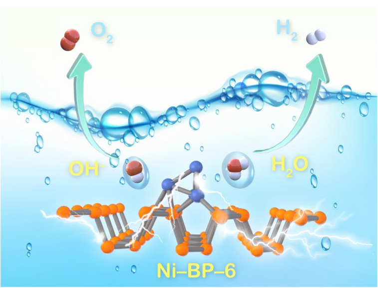
As illustrated in
Fig. 1a, the Ni-BP-x electrocatalysts were prepared via the one-step electro-exfoliation/covalent functionalized method from bulk BP and NiCl
2. Similarly, BP NSs were synthesized from bulk BP without the addition of metal ions (Fig. S1). The FE-SEM image revealed the ordered multilayer lamellar structure of bulk BP (
Fig. 1b), which were further exfoliated into few-layer nanosheets after the treatments (
Fig. 1c) [
29]. As revealed from AFM image in
Fig. 1d, the thickness of BP NSs is
ca. 3.75 nm (7 layers, the single layer thickness is 0.53 nm) [
30]. After the metal covalent functionalization, the BP NSs remained as a micron-sized nanosheet-like structure, suggesting that the macroscopic morphology of the structure was not damaged (Fig. S2). X-ray diffraction (PXRD) patterns proved that the Ni-BP-x samples were well-crystallized, with no obvious diffraction peaks of Ni metal phase (Fig. S3). All the diffraction peaks of Ni-BP-x can be indexed to the BP (JCPDS No. 01-073-1358) [
31,
32,
33]. A series of peaks located at 17.2°, 34.5°, and 52.6° can be assigned to the (020), (040), and (060) planes of BP NSs, respectively. The peak positions of Ni-BP-3, Ni-BP-6, and Ni-BP-9 materials slightly shifted to lower angles in comparison with those of BP NSs. According to the Bragg’s law, the stretched crystal plane spacing of BP NSs is caused by the covalent bonding of Ni. The few-layer BP NSs successfully incorporate Ni atoms as evidenced by the lattice expansion and lack of metal phase, which is consistent with the subsequent HRTEM data. TEM analysis revealed a comparatively thin view of BP NSs (
Fig. 1e) with a well-defined fringe spacing of 0.260 nm, corresponding to the (040) crystal planes in the high-resolution TEM (HRTEM) image (
Fig. 1f). The SAED pattern shows the rings corresponding to the (111), (112), and (002) crystal planes of BP NSs (Fig. S4). As demonstrated in
Fig. 1g, the Ni-BP-6 maintained the lamellar microstructure after the introduction of Ni
2+ ions. The SAED pattern of Ni-BP-6 material presents the (111), (221), and (022) crystal planes of BP NSs (Fig. S5) in the absence of any diffraction rings of Ni metal phase, indicating the Ni dispersion on the BP NSs. The HRTEM image exhibits the (040) crystal plane with a fringe spacing of 0.262 nm (
Fig. 1h). Such a lattice after Ni inserting/covalent binding is slightly larger than that of BP NSs (0.260 nm), which demonstrates that the Ni element was covalent functionalized with BP. In the Ni-BP-6 sample, the elemental mapping images have clearly illustrated the uniform distribution of P/Ni/O (
Fig. 1i). Ni-BP-3 and Ni-BP-9 exhibit the preserved lamellar structure of BP NSs, with slight lattice stretching and uniform component distribution (Figs. S6-S7). The Ni inserting/covalent amounts of Ni-BP-3, Ni-BP-6, and Ni-BP-9 were 0.7, 1.5, and 2.0 wt%, respectively, which were determined by inductively coupled Plasma mass spectrometer (ICP-MS) examinations (Table S1).


