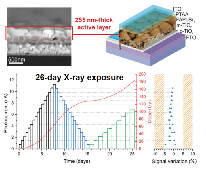
As can be inferred from the evaluation of the photoelectronic properties after the prolonged irradiation, our prototypal device demonstrated an excellent radiation hardness: this is quite unexpected for metal-halide perovskite thin films, which usually report structural damage after few hours of exposure to X-rays in air [
54]. In a subsequent phase of our study, aimed at a deeper investigation of the radiation hardness of the FAPbBr
3 film, a complete device stack selected from the same batch described in Sect. 3.2 was exposed to the same total ionizing dose (
TID) as the sample used for the 26-day long test (192 Gy, red curve of
Fig. 4b). Several XRD measurements (
Fig. 4g) were progressively performed during the experiment in order to monitor the structural integrity of the active layer: as can be seen, despite the high accumulated X-ray dose, the cubic structure of FAPbBr
3 is perfectly preserved, and no signs of structural degradation/modification is observed. It is worth highlighting here that a
TID value of 192 Gy exceeds of about two orders of magnitude the highest
TID reported up to now (2.2 Gy) on MHP thin films [
55]. In the literature on MHP-based X-ray detectors, comparable values of
TID have only been reported for millimeter-thick single crystals [
56,
91] or very thick (hundreds of micrometers) polycrystalline films [
67], but have been obtained by exposing the devices to high dose rates for short times, mostly aimed at evaluating the radiation hardness. However, a good radiation hardness is a necessary but not sufficient condition for a reliable X-ray detector, and this explains the necessity of a prolonged test. Monitoring the photocurrent signal under uninterrupted irradiation for a considerable amount of time (
e.g., weeks), so to take into account the possible effects of temperature and humidity on the performance of the detector, is indeed the most accurate method to assess the signal stability and reproducibility over time. And this is even more crucial for perovskite-based devices, the photoelectronic properties of which may be very sensitive to environmental conditions. In addition, the 26-day long test gave us the opportunity to assess the operational stability of the detector even at very low dose rates, as those used in the field of interventional radiology: in this case, signals may indeed be significantly weak, and the presence of instability over time (due, for instance, to noise drift) would be more obvious. It is also worth noting here that the signal stability and reproducibility, as well as the negligible degradation of the optoelectronic properties, are sufficient conditions to classify our detector at least as a rad-hard “D-level” (
i.e., compliant with a
TID > 100 Gy) device according to US MIL-PRF-38535F, which is the reference standard for electronic and optoelectronic devices suitable for military and space applications. Obviously, the upper
TID limit of the device (
i.e., the
TID corresponding to unacceptable performance degradation) is significantly higher, and depends on the “acceptable tolerance levels” (
ATL) defined for a specific dosimeter operating in a specific field of application. For instance, the
ATL established by
IEC (International Electrotechnical Commission) for X-ray dosimeters used in interventional radiology [
57] is ± 25%. This allows us to make a rough prediction of the operating lifetime of the device for this specific application. By considering that -1.3% is the average signal loss after exposure to a
TID = 192 Gy, and assuming a linear degradation model with an acceptable performance degradation of -25%, we can predict that our device can operate up to a
TID = 3.7 kGy. Of course, this is only a rough extrapolation, which does not take into account other possible degradation factors in addition to radiation-induced damage (
e.g., environmental factors), but highlights anyway the potentially excellent radiation hardness of the device: just to give an example, a
TID = 3.7 kGy corresponds to the total X-ray dose delivered in about 12,600 pulmonary angiographies [
58], that is one of the routine procedures in interventional radiology.


