HIGHLIGHTS
1 Introduction
Table 1 Representation of characteristic features of various forms of cell death |
| Type of cell death | Characteristic features |
|---|---|
| Apoptosis | It is a programmed cell death (PCD) wherein both the nuclear and cellular size decrease, leading to the fragmentation of nuclear bodies while maintaining an unchanged response to the mitochondria |
| Ferroptosis | A newly discovered regulated form of cell death that damages mitochondrial Crista and ruptures the nucleus and mitochondrial membrane |
| Necroptosis | This kind of PCD causes rupture of cell membrane, random DNA degradation, swelling of cell and its deformation and deformation of organelles |
| Cuproptosis | Aggregation of lipoylated mitochondrial proteins |
Table 2 Kinds of ferroptosis inducers |
| Inducers of ferroptosis | Function | References |
|---|---|---|
| Doxorubicin | HO-1 | [251] |
| RSL3 | GPX4 inhibition | [252] |
| Hemin | Iron accumulation | [253] |
| Sulfasalazine | Inhibition of System Xc− | [254] |
| Altretamine | GPX4 inhibition | [255] |
| Glutamate | Inhibition of System Xc− that affect cellular transport of cysteine | [256] |
| FINs | ROS generation | [10] |
| Bromelain | ROS generation in KRAS mutation | [257] |
| All-trans retinoic acid (ATRA) | ROS generation | [258] |
| Lapatinib | Improve oxidative stress inside the cells | [259] |
| Artesunate | Induces programmed cell death through ROS generation | [260] |
| Ferumoxytol | Lipid peroxidation | [261] |
| Cotylenin A | ROS generator | [262] |
2 Elucidating the Importance of Iron in Cancer Progression
3 Features of Ferroptosis
4 Understanding the Molecular Basis of the Ferroptosis Process
4.1 Suppression/Inhibition of GPX4
Fig. 1 Representation of the ferroptosis mechanism in cancer therapy. Three pathways are primarily associated; inhibition of GPX4, iron metabolism and lipid peroxidation. 1 Erastin, which is a prototype of ferroptosis inducer, blocks the uptake of cysteine (cys) by directly inhibiting system Xc-, which in turn promotes GPX4 degradation by encouraging chaperone-mediated autophagy, 2 increase in LIP downregulate DMT1 to promote oxidative stress causing ferroptosis, 3 recombinant lysophosphatidylcholine acyltransferase 3 (LPCAT3) and Acyl-CoA synthetase long-chain family member 4 (ACSL4) are the catalysts required for the synthesis of PUFA-containing phospholipids which under oxidative stress promotes ferroptosis |
4.2 Lipid Peroxidation
4.3 Iron Metabolism
5 Chemical Basis of Nanoparticle-Induced Ferroptosis in Cancer Management
Fig. 2 Representation of ferroptosis effect of endothelin-3 targeted nanoparticle in production of highly reactive hydroxyl radical [101] |
Fig. 3 The cascade of cell death inducing mechanism of nanomicelles. The theranostic nanomicelles produced highly reactive hydroxyl radical by releasing of AscH−, Fe2+ and the adjuvant Cy7QB [103] |
Fig. 4 a pH-responsive polyethylene glycolpoly(allylamine)-dimethylmaleic anhydride (PEG-PAH-DMMA)-coated tamoxifen-loaded copper peroxide nanoparticle-capped hollow mesoporous organosilica nanoparticle (HMON), b illustration of ROS storm generation by developed nanoparticle,c lscm images showing GPX4, ROS and lipoxygenase level, d estimation of ROS level by flow cytometry in 4T1 cells laden mice, e relative complex I activity measurement and f lactate level measurement in 4T1 cells laden mice, g measurement of GSH level [105] |
Fig. 5 a Representation of formation of Lapa-loaded HAS-Fe2O3 NP. b Following intravenous administration, the EPR effect caused HAS-Fe2O3 NP to concentrate in tumor tissues and be absorbed by tumor cells. Superoxide dismutase (SOD) then transformed the O2· into a mass of H2O2 as a result of the excessive O2· produced by the catalytic release of Lapa by the overexpressed NQO1 enzyme. Meanwhile, in the acidic endosomal environment, ferrous ions from self-sacrificing Fe3O4 combined with accumulating H2O2 via Fenton reactions to form extremely harmful ·OH leading to cell death. Additionally, consumption of NADPH in the Lapa futile cycle led to GSH depletion, impairing the antioxidant defense system and increasing the sensitivity of tumor cells to •OH produced in the Fe2+-mediated Fenton chemical reaction, intensifying the anticancer impact [111] |
Fig. 6 Representation of effect of molecularly engineered pH-responsive photothermal oxazine self-assembled nanoparticles (PTO-Biotin Nps) for spatiotemporal controlled tumor ferroptosis [112] |
6 Challenges of Ferroptosis
7 Unraveling the Scope of Nanoparticulate Systems in Ferrotherapy
Table 3 Illustration of nanoferrotherapeutics involved in treatment of various cancers |
| Type of nanocarrier system | Chemotherapeutics | Genes | Cancer type | In vitro | In vivo | Inference | References |
|---|---|---|---|---|---|---|---|
| MOF | Doxorubicin | - | Breast cancer | 4T1 cells | Balb/c mice | Doxorubicin-induced ferroptosis causing immunogenic cell death (ICD) | [251] |
| Gold mesoporousMOF | - | - | Breast cancer | 4T1 cells | Balb/c mice | Cu and Fe ion bridged using disulfide bond induces ferroptosis by depleting GSH, finally inhibiting GPX-4 level | [137] |
| MOF | Doxorubicin | - | Breast cancer | 4T1 and MCF-7 | Mice (name not mentioned) | The drug-loaded nanoparticle showed great potency and clinical translation by targeting CD44 over-expressed cells | [141] |
| Fe(III)- porphyrin-MOF | Oxaliplatin | - | - | Breast cancer | 4T1 cells | The therapy increased the level of H2O2 and (IFN-γ), which in turn caused ferroptosis of cells | [145] |
| Fe-MOF | - | - | Breast cancer | 4T1 cells | Aptamer PD-L1-attached glucose oxidase and PEG-modified iron-based MOF were an innovative strategy to disrupt iron homeostasis and hinder intracellular redox | [21] | |
| RBC membrane camouflaged MOF | Doxorubicin | - | Breast cancer | MCF-7 | Balb/c nude mice | Results illustrated that self-assembled RBC membrane camouflaged MOF could amplify the oxidative stress of ROS, reduce glutathione potentiating remarkable anticancer effects | [146] |
| MOF | Doxorubicin | - | Breast cancer | 4T1 cells | Balb/c mice | The nanoplatform downregulated GPX4 to induce ferroptosis | [251] |
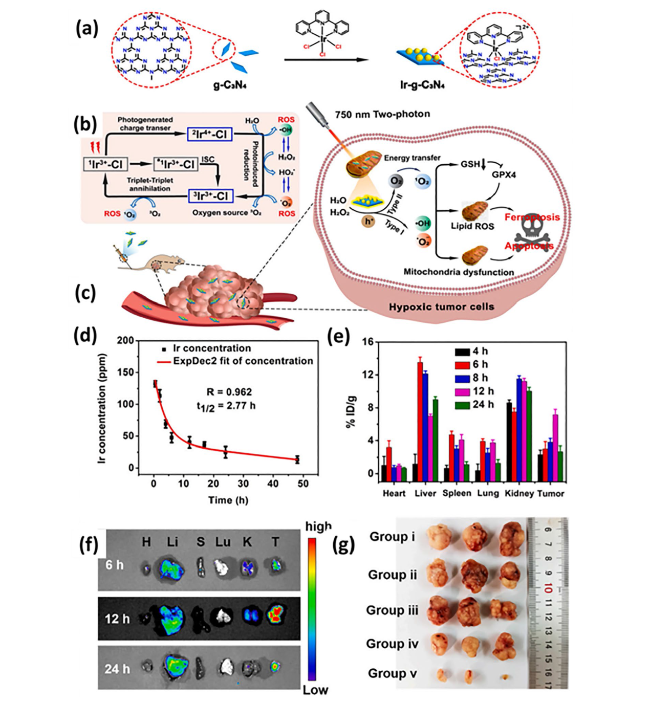
| Nanophoto-sensitizer | - | - | Human melanoma cancer | A375 | Nu/Nu female mice | A photodynamic therapy-based nanosheets of graphitic carbon nitride functionalized with mitochondria-targeting iridium (III) polypyridine complexes caused hypoxic environment resulting in cell death | [225] |
| Theranostic nanoparticle composed of iron ions, cinnamaldehyde prodrug and amphiphilic polymer skeletal (FCS/GCS) | - | - | Breast cancer | 4T1 cells | Balb/c mice | The preparation with the help of CA induces Fenton reaction, generated ·OH and accelerated lipid peroxides (LPO) accumulation and accordingly augments ferroptosis | [229] |
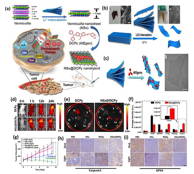
| Bonsai-inspired AIE nanohybrid photosensitizer | - | - | Colon cancer | MC38 cells | Balb/c | The Bonsa-inspired nanopreparation in presence of white light irradiation produced hydroxyl radical and depleted GSH to induce ferroptosis in cancer cells | [240] |
| Cell membrane decorated iron-siRNA nanohybrid | - | Anti- SLC7A11 siRNA | Human oral squamous cell carcinoma | CAL-27 | Male Balb/c | The nanohybrid system elevated the ROS level to show synergic anti-cancer effect in vivo | [96] |
| Magnetic lipid nanoparticle | - | siDECR1 | Castration-resistant prostate cancer | C4-2B or C4-2BEnz cells | Nude mice (name not mentioned) | The biomimetic nanoparticles were stable, safe and effective in showing remarked inhibition of distant organ metastasis | [218] |
| Graphene oxide-PEG-PEI nanoparticle | Sorafenib | PD-L1 siRNA | Hepatocellular carcinoma | MHCC97H cells | C57BL/6 mice | The developed preparation reduced the expression of GPX4 in the intrahepatic tumor regions in immunocompetent mice | [219] |
| Liposome | Artemisinin | - | Lung carcinoma | LLC cells | Balb/c nude mice | The remarkable autophagy-mediated ferroptosis-involved cancer-therapeutic efficacy is suggested by therapeutic outcomes both in vitro and in vivo which is further confirmed by transcriptome sequencing | [160] |
| Nanostructured lipid carrier | Doxorubicin, ferrocene | - | Breast cancer | 4T1 cells | Balb/c mice | TGF-β receptor inhibitor along with ferrocene and doxorubicin-loaded NLC inhibited mammary cancer metastasis by extracellular as well as intracellular hybrid mechanism | [164] |
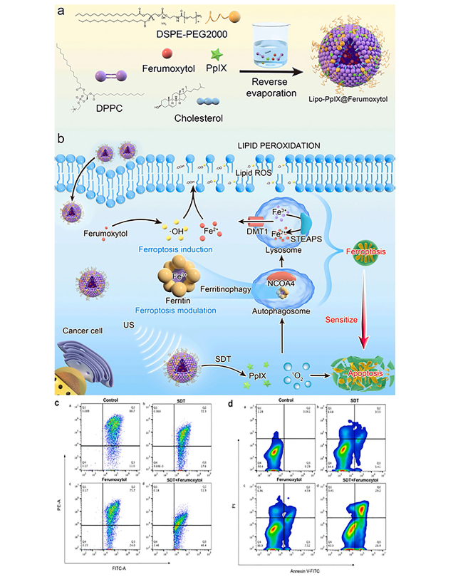
| Liposomes | - | - | Breast cancer | 4T1 cells | Balb/c mice | An amalgamation of sonosensitizing agent (PpIX) and ferumoxytol in liposomes induced apoptosis and ferroptosis overcoming the tumor resistance | [215] |
| Iron oxide nanoparticles | Paclitaxel | - | Glioblastoma | U251 and HMC3 cells | Balb/c-nu mice | PTX-IONP reduced the ability of cells to invade and migrate, elevated ROS, iron ions and lipid peroxidation, enhanced the expression of the autophagy-related proteins LC3II, and Beclin1 and suppressed the expression of the p62 and GPX4 | [173] |
| Nanozyme | Cisplatin | - | Ovarian cancer | SKOV3/DDP cells | BALB/cJGpt-Foxn1nu/Gpt mice | The formulation was able to induce both apoptosis and ferroptosis with the help of ultrasound treatment for cisplatin-resistant cancer cells | [193] |
| Nanozyme | - | - | Triple-negative breast cancer | MDA-MB-231 cells | Balb/c nude mice | The imaging-based nanosystem used SPIO and Avastin to induce tumor starvation and ferroptosis | [263] |
| Nanozyme | - | - | Breast cancer | 4T1 and MCF-7 cells | Balb/c mice | Tumor ablation was accomplished with Fenton reaction-independent ferroptosis driven by photothermal nanozyme | [197] |
| Nanozyme | Gemcitabine | - | Pancreatic cancer | PANC02 | Balb/c mice | GEM and MnFe2O4 can synergistically improve anti-cancer profile via ferroptosis and GEM-mediated chemotherapy | [198] |
| Human serum albumin nanoparticle (HAS-NP) | Pt (IV) | - | Ovarian cancer | SKOV3 | Mice (name not mentioned) | The prodrug of platinum in HAS nanoparticles was effective for ovarian cancer cell resistance to platinum | [241] |
| Iron-doped calcium carbonate nanoparticles | Pt(IV) | - | Breast cancer | 4T1 and CT26 cells | Balb/c | Iron-doped platinum-SA-based CaCO3 showed development of ROS and lipid peroxidation to mediate cancer cell death | [242] |
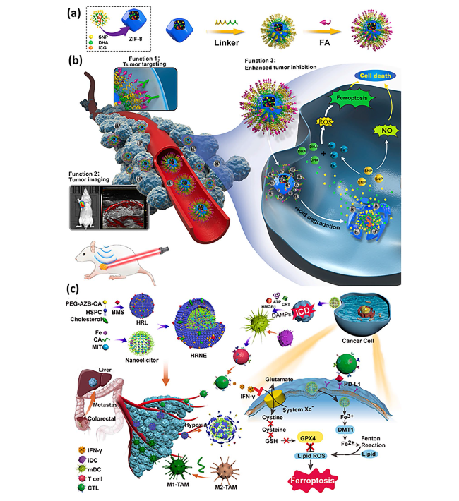
| Zeolite imidazolate framework (Zif-8) | - | - | Head and neck squamous cell carcinoma | HN6 | Male Balb/c | DHA and SNP enveloped nanoreactor system prompted Fe2+ and NO release to initiate ferroptosis and apoptosis synergistically | [247] |
| Hypoxia-responsive nanoelicitor | Mitoxantrone | - | Colorectal cancer | CT26 cells | Balb/c nude mice | The simultaneous co-stimulating effects of CA and MIT resulted in an elevated antiproliferative and anti-cancer immunity, which in turn aborted the system Xc− to GPX4 pathway and increased the iron-initiated tumor cell destruction | [248] |
7.1 Metal-Organic Framework Nanoparticles
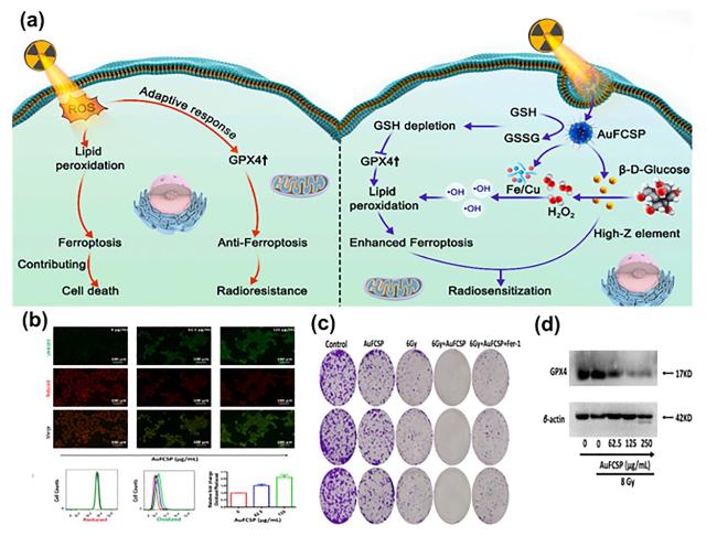
Fig. 7 a Schematic illustration of synergistic effect of radiotherapy and ferroptosis mediated by Au-FCP-MOF-NP. The left side represents the negative role of GPX4 stimulated by radiotherapy to resist ferroptosis and finally radio-resistance, while the right side shows induction of ferroptosis due to intratumoral release of Fe-Cu dual ion along with Au NP which catalyzed β-D-glucose oxidation to develop gluconic acid. Increased amount of H2O2 released toxic hydroxyl radicals which induced ferroptosis. Overall, radiosensitization increased due to synergic effect of high Z element (Au) and ferroptosis; b fluorescence images of cell (4T1) treated with Au-FCP-MOF-NP to evaluate peroxidation with respective flow cytometry analysis; c Au-FCP-MOF-NP and IR treated 4T1 cells showing extend of colon formation and d western blotting results showing expression of GPX4 with various concentration of Au-FCP-MOF-NP with or without exposure of IR. Reproduced with permission from Ref. [137] |
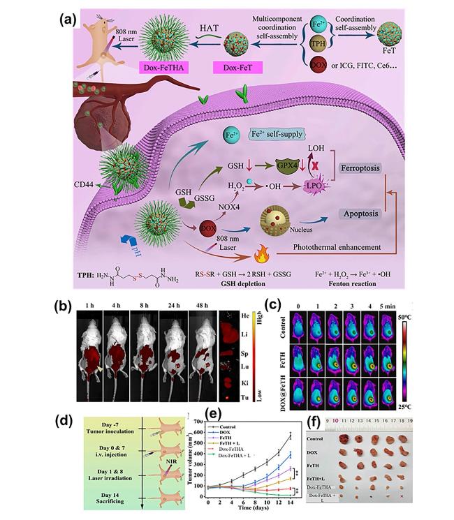
Fig. 8 a Schematic representation of development process of Dox-FeTHA-aMOF with its application in photothermal-assisted synergistic ferroptosis-apoptosis cancer therapy; b time-dependent fluorescence descriptions of 4T1 tumor-bearing mouse post-injection with dye (Cy5.5) labeled aMOF with detected fluorescence in major organs at 24 h post-treatment; c infrared thermal images under 808-nm laser irradiation; d representation of development of tumor model and respective treatment strategy; e illustration of tumor volume with f digital pictures of excised tumor lesions. Reproduced with permission from Ref. [141] |
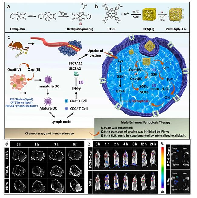
Fig. 9 The triple-hit effect of (PCN-Oxpt/PEG) for inducing chemotherapy, ferroptosis and immunotherapy, a representation of development of oxaliplatin prodrug and b PCN-Oxpt/PEG, c magnetic resonance images of mice after treatment with PBS, iron chloride and nanoparticles showing accumulation at tumor site [145] |
Fig. 10 a Schematic representation of development of PD-L1-targeted aptamer-grafted iron-based MOF, b the developed therapy based on ferroptosis-mediated immunotherapy disrupts iron homeostasis and redox balance through inactivation of GSH, ROS accumulation and transferrin 1 downregulation to finally suppress PD-L1 checkpoints, c illustration of blocking the targeted site (PD-L1), d illustration of therapy-induced in mouse models, e digital images of different treatment groups, f estimation of tumor volume (mm3) and g tumor weight of xenograft model of mouse bearing 4T1 cells, h tumor section analysis through H&E-stained images, analysis of Ki 67 and TUNEL test. Reproduced with permission from Ref. [21] |
7.2 Lipid Nanoparticles
Fig. 11 Representation of ferrocene (Fc) and Dox-loaded cationic NLCs developed by emulsion solvent evaporation method, which was then decorated with β-cyclodextrins grafted heparin (β-CD-HEP) by the help of electrostatic interaction. The preparation triggered Fenton reaction, reduced TGF-β secretion and induced immune responses for ferroptosis-mediated anti-cancer therapy |
7.3 Iron Nanoparticle
7.3.1 Nanozyme
Fig. 12 a Synthesis of SO2, •CF3 and •OH bearing nanoflower and its inhibiting mechanism for cisplatin resistance cancer under ultrasonic radiation, b ROS suppressing effect of nanoflower detected by 2,7-dichlorodihydrofluorescein diacetate (DCFH-DA), dihydroethidium (DHE) and hydroxyphenyl fluorescein (HPH), c determination of SO2 production capacity coumarin-hemicyanine dye, d cell cytotoxicity study of different nanopreparations under US irradiation. Reproduced with permission from Ref. [193] |
7.3.2 Magnetic Control of Iron-Based Nanoparticle
7.4 Mesoporous Silica Nanoparticle
7.5 Assisted Therapies in Amalgamation with Nanoferroptosis
7.5.1 Nanoparticle Mediated Sonodynamic-Ferrotherapy Against Cancer
Fig. 13 a Schematic representation of development method of ferumoxytol-loaded nanoliposome to induce synergistic anti-cancer response (by applying sonodynamic approach) using reverse evaporation method. Ferumoxytol successfully imported iron ions into the cell to stimulate ferroptosis, while photosensitizing agent induced apoptosis by making cancer cells responsive toward therapy, c, d apoptosis and destruction of 4T1 cells post-treatment with various formulations. Reproduced with permission from Ref. [215] |
7.5.2 Amalgamation of Nanoparticle-Based Gene Therapy and Ferroptosis
7.5.3 Nanoparticle-Based Photodynamic Therapy in Association with Ferroptosis
Fig. 14 a Representation of development of Ir-g-C3N4, b regeneration of different kinds of ROS using photosensitizer, c representation of mechanism of action of mitochondrial targeting nanosystem responsible for ferroptosis and apoptosis-mediated cell death, d blood circulation time post-treatment of Nu/Nu mice with Ir-g-C3N4, e biodistribution assay for evaluation of Ir in major organs, f luminescence intensity measurement of nanoplatforms at various time interval, g digital images of tumor after treatment. Reproduced with permission from Ref. [225] |
Fig. 15 a Representation of bonsai-inspired AIE nanohybrid photosensitizer as a key driver in ferroptosis-mediated self-reliable PDT, b preparation steps of ultrathin vermiculite nanosystem showing vermiculite in bulk (i), its SEM image (ii) and vermiculite NSs solution (iii), c preparation steps of vermiculite NSs@DCPy and its TEM image, d In vivo study in mice model showing tumor bio-images with white dotted lines, e images of major organs post 24 h of therapy showing spleen (S), heart (H), kidney (K), tumor (T), liver (Li) and lungs (Lu), f bio-distribution of NSs@DCPy and DCPy in Balb/c mice model, g tumor growth curves, h estimation of expression of Caspase 3 and i GPX4 by immunohistochemical analysis. Reproduced with permission from Ref. [240] |
7.6 Miscellaneous Nanoparticles with Combinatorial Therapy
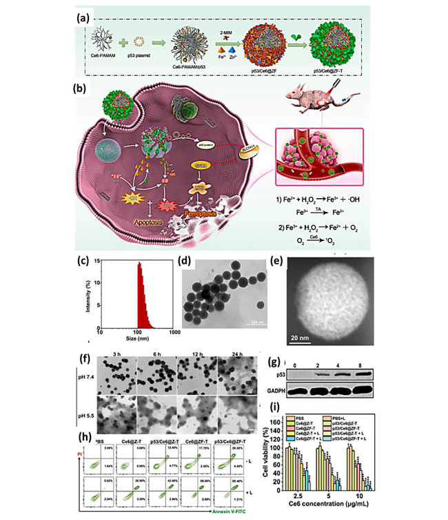
Fig. 16 a, b Preparatory steps of mesoporous zeolite framework comprising of C6-PAMAM and tannic acid, which is then laden with p53 to deplete GSH which is attributed to SLC7A11 inhibition, further causing prominent downregulation of GPX4, c size determination using DLS, d TEM image, e high-angle annular dark-field scanning TEM (HAADF-STEM) image, f TEM images of preparation in pH (5.5 or 7.4), g estimation of expression of p53 post-incubation of H1299 cells with developed therapy, h cell apoptosis study with various treatment groups, i cell viability assay. Reproduced with permission from Ref. [246] |
Fig. 17 a Development process of PEG-functionalized folic acid (FA)-modified zeolite imidazolate frameworks (Zifs) carrying dihydroartemisinin (DHA) as ferroptosis inducer and sodium nitroprusside (SNP) as apoptosis inducer for the treatment of HNSCC; b (1) folate receptor enhanced the cellular uptake after binding with FA-functionalized preparation, (2) location of tumor after treatment using imaging tool and (3) elevated anti-cancer effect, c representation of development process of MIT-based nanoelicitor to suppress GPX4 expression, enable lipid peroxidation to induce cell ferroptosis [247,248] |


