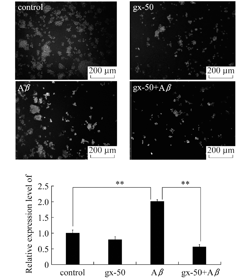
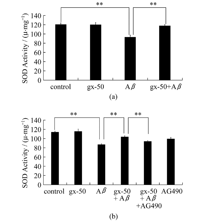
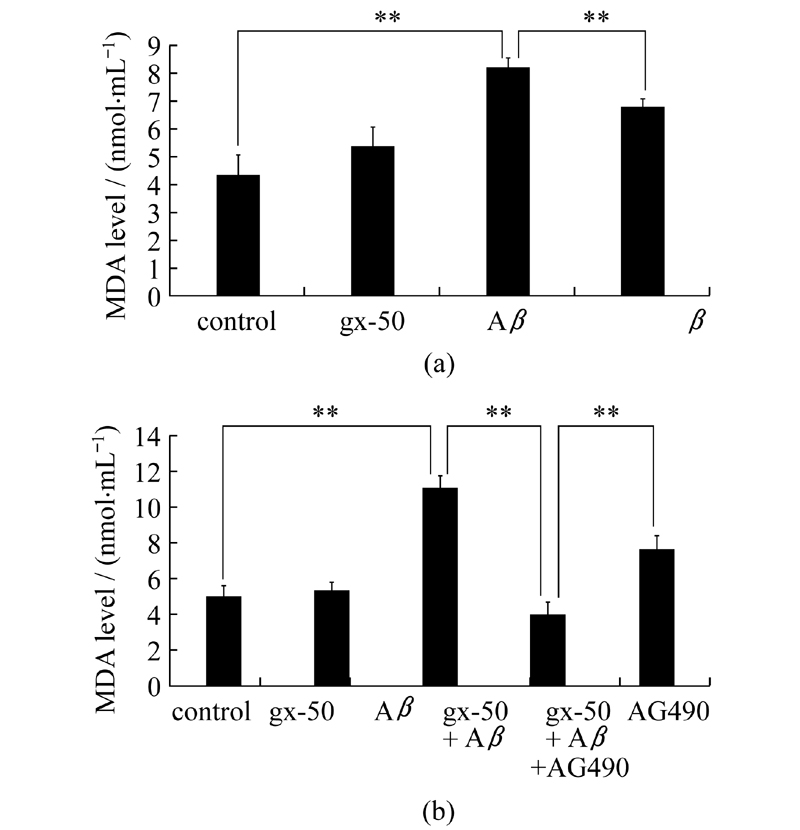
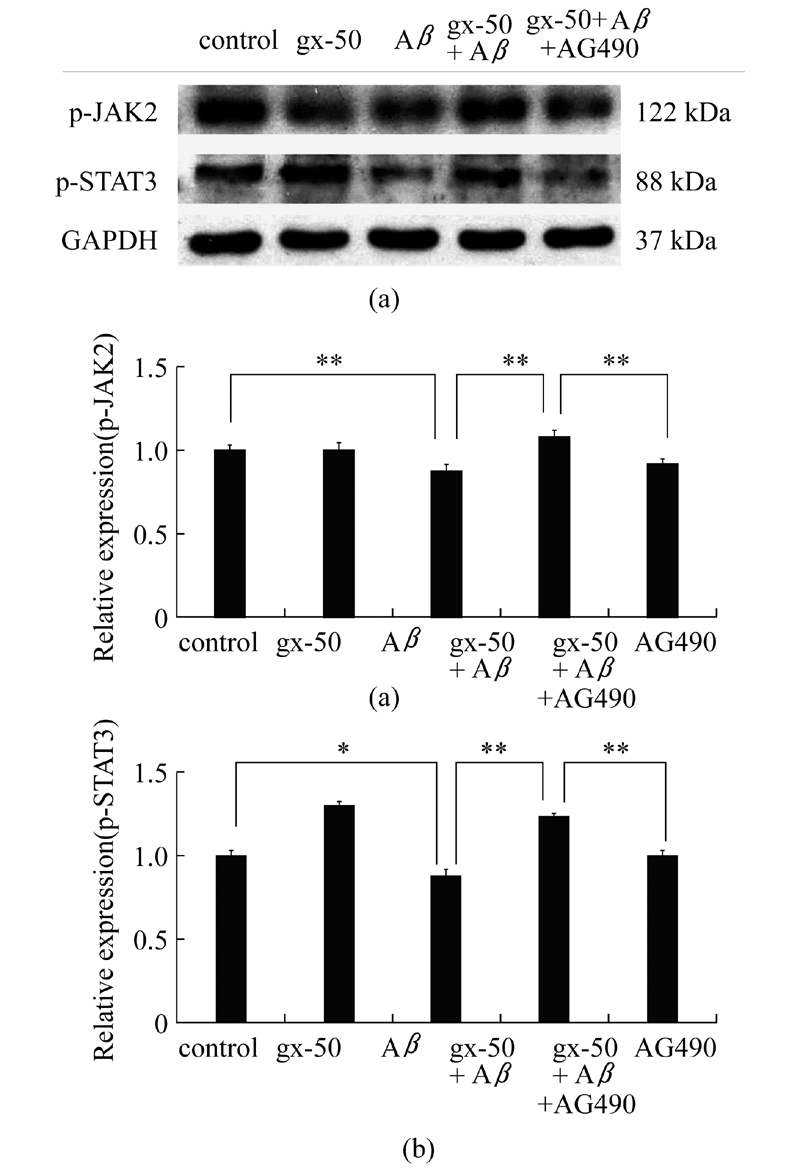
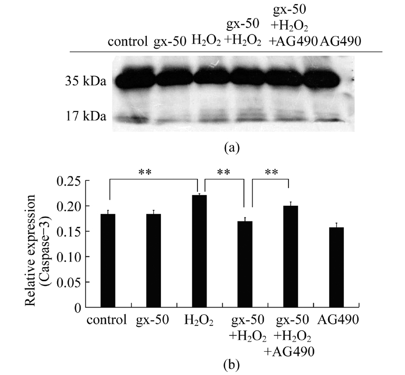
A novel compound derived from Zanthoxylum, gx-50(N-[2-(3, 4-dimethoxyphenyl)ethyl]-3-phenyl-acrylamide), has been demonstrated that it has neuroprotective effects against Alzheimer’s disease(AD)by our previous study.This study focuses on the mechanism of its antioxidant properties against AD.We measured the levels of intracellular reactive oxygen species(ROS), superoxide dismutase(SOD)activity and malondialdehyde(MDA)to determine the anti-oxidative ability of gx-50 at cellular level.We also measured gx-50’s effect on JAK-STAT signaling pathway and the relative expression levels of activated caspase-3.Data showed that gx-50 reduced the levels of reactive oxygen species(ROS)and malondialdehyde(MDA), and recovered the activity of total intracellular superoxide dismutase(SOD)in neuronal PC12 cells exposed to Aβ.After gx-50 pretreatment, the levels of p-JAK2 and p-STAT3 both increased in PC12 cells, while they were down-regulated in Aβ-treated group.In present study, we also found that gx-50 reduced the relative expression level of the activated caspase-3 in PC12 cells by activating JAK-STAT signaling pathway.Results demonstrated that gx-50 reduced amyloid-beta(Aβ)induced oxidative stress in neuron-like PC12 cells by enhancing the activation of JAK-STAT signaling pathway.It might help to protect neurons in Alzheimer's disease.
Praticò D.Evidence of oxidative stress in Alzheimer's disease brain and antioxidant therapy[J].Annals of the New York Academy of Sciences, 2008, 1147(1):70-78.
doi: 10.1196/annals.1427.010
pmid: 19076432
[2]
McLellan M E, Kajdasz S T, Hyman B T, et al.In vivo imaging of reactive oxygen species specifically associated with thioflavine S-positive amyloid plaques by multiphoton microscopy[J].The Journal of Neuroscience, 2003, 23(6):2212-2217.
pmid: 328613207063181838931172922232222126576808
[3]
Tang M, Shi S, Guo Y, et al.GSK-3/CREB pathway involved in the gx-50's effect on Alzheimer's disease[J].Neuropharmacology, 2014, 81:256-266.
doi: 10.1016/j.neuropharm.2014.02.008
pmid: 24565641
[4]
Manickam M, Tulsawani R.Survival response of hippocampal neurons under low oxygen conditions induced by hippophae rhamnoides is associated with JAK/STAT signaling[J].PloS One, 2014, 9(2):e87694.
doi: 10.1371/journal.pone.0087694
pmid: 24516559
[5]
Zeng H, Chen Q, Zhao B.Genistein ameliorates beta-amyloid peptide(25-35)-induced hippocampal neuronal apoptosis[J].Free Radical biology and Medicine, 2004, 36(2):180-188.
doi: 10.1016/j.freeradbiomed.2003.10.018
[6]
Finkel T, Holbrook N J.Oxidants, oxidative stress and the biology of ageing[J].Nature, 2000, 408(6809):239-247.
doi: 10.1038/35041687
pmid: 11089981
[7]
Vanfleteren J R.Oxidative stress and ageing in Caenorhabditis elegans[J].Biochem J, 1993, 292:605-608.
[8]
Larsen P L.Aging and resistance to oxidative damage in Caenorhabditis elegans[J].Proceedings of the National Academy of Sciences, 1993, 90(19):8905-8909.
doi: 10.1073/pnas.90.19.8905
pmid: 8415630
[9]
Martin G M, Austad S N, Johnson T E.Genetic analysis of ageing:role of oxidative damage and environmental stresses[J].Nature Genetics, 1996, 13(1):25-34.
doi: 10.1038/ng0596-25
pmid: 8673100
[10]
Yan L J, Levine R L, Sohal R S.Oxidative damage during aging targets mitochondrial aconitase[J].Proceedings of the National Academy of Sciences, 1997, 94(21):11168-11172.
doi: 10.1073/pnas.94.21.11168
pmid: 9326580
[11]
Reddy P H.Amyloid precursor protein-mediated free radicals and oxidative damage:Implications for the development and progression of Alzheimer's disease[J].Journal of Neurochemistry, 2006, 96(1):1-13.
[12]
Tang M, Wang Z, Zhou Y, et al.A novel drug candidate for Alzheimer's disease treatment:gx-50 derived from Zanthoxylum Bungeanum[J].Journal of Alzheimer's Disease, 2013, 34(1):203-213.
doi: 10.3233/JAD-121831
pmid: 23186988
[13]
Guo Y, Shi S, Tang M, et al.The suppressive effects of gx-50 on Aβ-induced chemotactic migration of microglia[J].International Immunopharmacology, 2014, 19(2):283-289.
[14]
Chiba T, Yamada M, Aiso S.Targeting the JAK2/STAT3 axis in Alzheimer's disease[J].Expert Opinion on Therapeutic Targets, 2009, 13:1155-1167.
doi: 10.1517/14728220903213426
pmid: 19663649
[15]
Chiba T, Yamada M, Sasabe J, et al.Amyloid-β causes memory impairment by disturbing the JAK2/STAT3 axis in hippocampal neurons[J].Molecular Psychiatry, 2008, 14(2):206-222.
doi: 10.1038/mp.2008.105
[16]
Zouein F A, Duhé R J, Arany I, et al.Loss of STAT3 in mouse embryonic fibroblasts reveals its Janus-like actions on mitochondrial function and cell viability[J].Cytokine, 2014, 66(1):7-16.
doi: 10.1016/j.cyto.2013.12.006
pmid: 24548419
[17]
Shaw S, Bencherif M, Marrero M B.Janus kinase 2, an early target of α7 nicotinic acetylcholine receptor-mediated neuroprotection against Aβ-(1-42)amyloid[J].Journal of Biological Chemistry, 2002, 277(47):44920-44924.
doi: 10.1074/jbc.M204610200
[18]
Szczepanek K, Lesnefsky E J, Larner A C.Multi-tasking:nuclear transcription factors with novel roles in the mitochondria[J].Trends in Cell Biology, 2012, 22(8):429-437.
doi: 10.1016/j.tcb.2012.05.001
pmid: 22705015
[19]
Szczepanek K, Chen Q, Larner A C, et al.Cytoprotection by the modulation of mitochondrial electron transport chain:the emerging role of mitochondrial STAT3[J].Mitochondrion, 2012, 12(2):180-189.
[20]
Andreyev A Y, Kushnareva Y E, Starkov A.Mitochondrial metabolism of reactive oxygen species[J].Biochemistry(Moscow), 2005, 70(2):200-214.
doi: 10.1007/s10541-005-0102-7
pmid: 15807660