Journal of Diagnostics Concepts & Practice ›› 2024, Vol. 23 ›› Issue (06): 602-611.doi: 10.16150/j.1671-2870.2024.06.007
Previous Articles Next Articles
ZHOU Henghuaa, LIN Lana, ZHU Guixianga, LIU Minb, HUANG Wentaoa( )
)
Received:2024-01-17
Online:2024-12-25
Published:2024-12-25
Contact:
HUANG Wentao
E-mail:wt.huang@hotmail.com
CLC Number:
ZHOU Henghua, LIN Lan, ZHU Guixiang, LIU Min, HUANG Wentao. Pure epithelial neuroendocrine neoplasms of the bladder: clinicopathological characteristics of 2 cases and literature review[J]. Journal of Diagnostics Concepts & Practice, 2024, 23(06): 602-611.
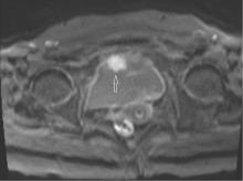
Figure 1
MRI showed thickening of the anterior wall of the bladder in Example 2, with a nodular shadow (indicated by the arrow) measuring 3.0 cm in size with clear boundaries
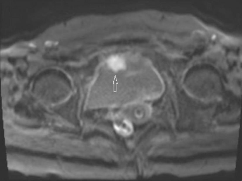
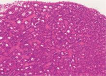
Figure 2
The tumor of case 1 was located within the lamina propria and presented a classic histological structure of NENs (beam like/pseudoglandular tube like/sieve like). The cell morphology was relatively consistent, and the surface was covered with normal mucosa (HE, × 200)
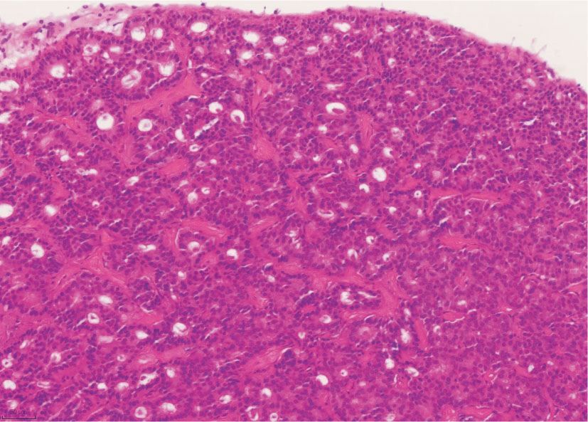
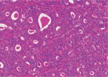
Figure 3
The tumor of case 2 was arranged in a pseudoglandular tube like/sieve like/solid sheet-like/nest like pattern, with fine thin-walled blood vessels abundant in the stroma. Concentrated eosinophilic secretions were observed in the pseudoglandular cavity (HE, × 200)
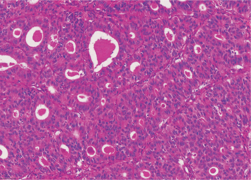
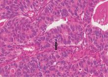
Figure 4
Tumor cells of case 2 were large with rich cytoplasm, eosinophilia, vacuolar nuclei, obvious nucleoli, and visible mitotic figures (indicated by arrows) (HE, × 400)
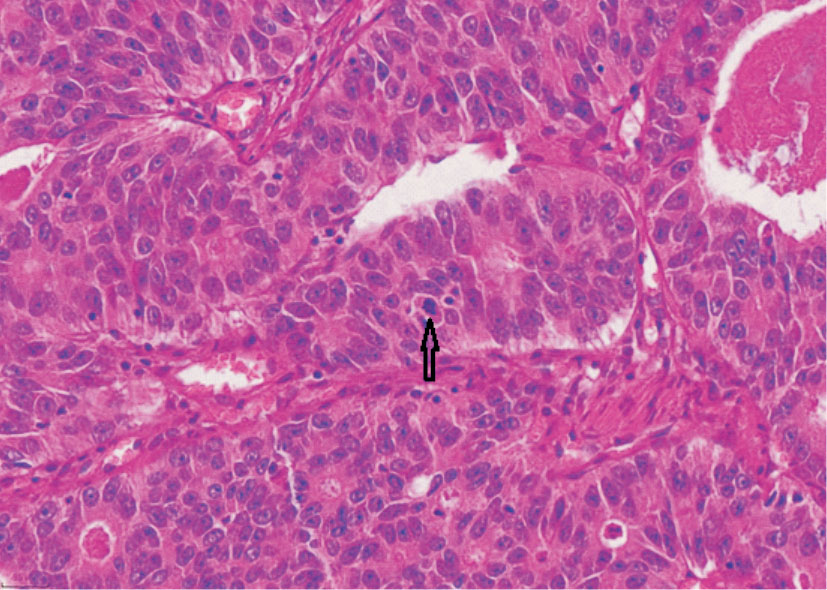
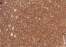
Figure 5
The cytoplasm of tumor cells in case 1 showing diffuse positive expression of Syn (EnVision two-step method, × 200)
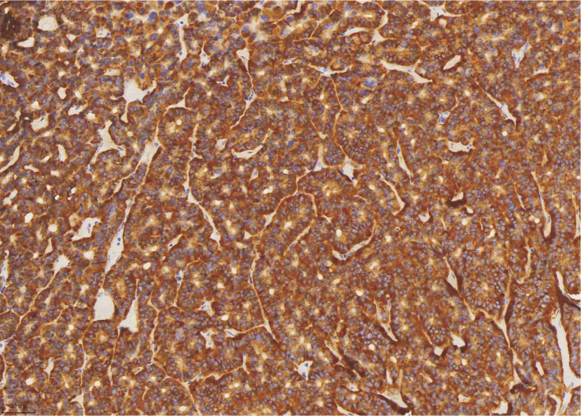
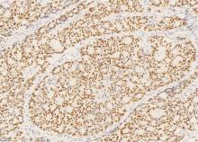
Figure 6
The nuclei of tumor cells in case 2 showing diffuse positive expression of INSM1.(EnVision two-step method, × 200)

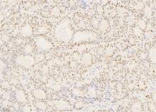
Figure 7
The nuclei of tumor cells in case 2 showing mottled wild-type expression of P53 (EnVision two-step method, × 200)
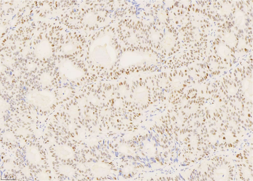
Figure 8
The Ki-67 proliferation index of in case 2 being as high as from 60% to 70% (EnVision two-step method, × 200)
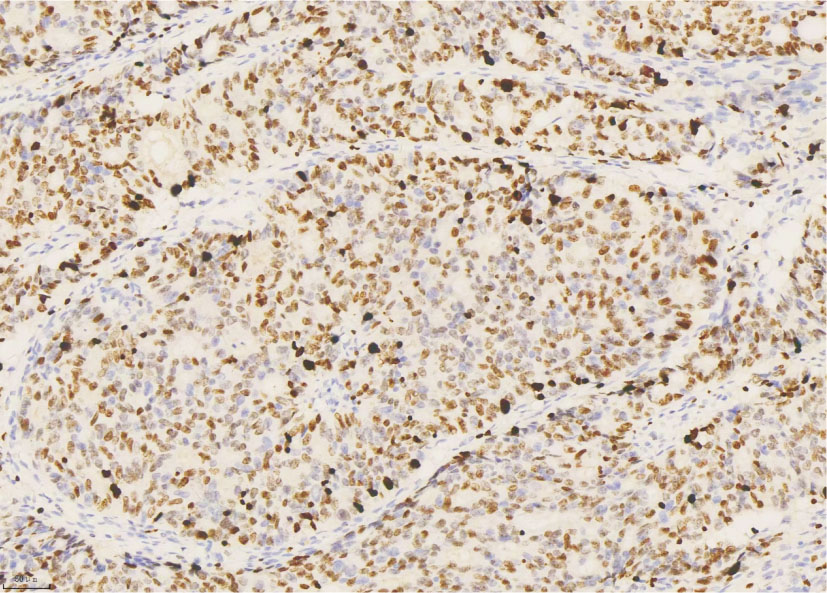
Table 1
Clinicopathological features and prognosis of 30 cases of bladder WD-NET
| Reference | Number | Age | Gender | Site and shape | Size (cm) | Clinical feature | Surgery | Staging | Associated urothelial alterations | Follow up and prognosis (mo) |
|---|---|---|---|---|---|---|---|---|---|---|
| [ | 1 | 30 | M | Neck, NA | 0.3 | Hematuria | Biopsy | NA | NA | 12,NED |
| [ | 1 | 61 | F | Trigone, pappilary | 0.3 | Hematuria | TURBT | pT1 | NA | NA |
| [ | 1 | 62 | F | Trigone, polypoid | 1.2 | Hematuria | TURBT | NA | CCG | NA |
| [ | 1 | 54 | F | Neck, polypoid | 0.9 | Hematuria and dysuria | TURBT | NA | Inverting papilloma | 6,NED |
| [ | 1 | 73 | M | Posterior wall, polypoid | 1.0 | Accidentally | TURBT | pT1 | NA | 22,NED |
| [ | 2 | 69/47 | M(2/2) | Neck(2/2),polypoid(2/2) | 0.3,0.7 | Hematuria(2/2) | TURBT(2/2) | pT1(2/2) | CCG/NA=1/1 | NA(2/2) |
| [ | 1 | 77 | M | Trigone, NA, | 1.5 | Accidentally | TURBT | pT1 | NA | NA |
| [ | 1 | 68 | M | Neck, polypoid | 0.4 | Hematuria | TURBT | pT1 | von Brunn’s nest | 14,NED |
| [ | 5 | Mean54.4 | M/F=4/1 | Neck/trigone=3/2, polypoid (5/5) | Mean0.34 | Hematuria/Accidentally=3/2 | TURBT(5/5) | pT1(5/5) | CCG(5/5) | Mean33.4,NED |
| [ | 1 | 49 | F | Trigone, NA | 3.0 | Hematuria | TURBT | pT2 | No | 6,NED |
| [ | 1 | 72 | M | Trigone, polypoid | 0.8 | Accidentally | TURBT | pT1 | CCG | 72,NED |
| [ | 1 | 71 | F | Trigone, polypoid | 2.8 | Vaginal pain and urinary incontinence | TURBT | pT1 | NA | NA |
| [ | 1 | 52 | M | Neck, polypoid | 0.7 | Urinary tract obstruction and hematuria | TURBT | pT1 | NA | 12,NED |
| [ | 1 | 44 | M | Left posterior lateral wall, nodule | 2.0 | Initial liver and peritoneal metastasis | Biopsy | pT2 | NA | 16,Died |
| [ | 1 | 83 | F | Neck, polypoid | 2.5 | Hematuria | TURBT | pT1 | CCG | NA |
| [ | 7 | Mean61.4 | M/F=5/2 | NA/neck=6/1,polypoid(7/7) | NA(7/7) | NA | Biopsy/TURBT=6/1 | pT1(7/7) | CCG/NA/papilloma=5/1/1 | Mean74.86,NED |
| [ | 1 | 51 | F | Neck, polypoid | 0.8 | Hematuria | TURBT | pT1 | CCG | 4,NED |
| [ | 1 | 90 | F | Bottom, polypoid | NA | Dysuria | TURBT | pT1 | CCG | 6,NED |
| Present case | 1 | 85 | F | Trigone, polypoid | 0.6 | Accidentally | TURBT | pT1 | CCG | 76,NED |
Table 2
Clinicopathological features and prognosis of 32 cases of bladder LCNEC
| Reference | Number | Age | Gender | Site | Size (cm) | Clinical feature | Staging | Treatment | Follow up and prognosis (mo) |
|---|---|---|---|---|---|---|---|---|---|
| [ | 1 | 73 | M | Posterior wall | 4.0 | Hematuria | pT3b | RC+LND | 2,died of RE/ME |
| [ | 1 | 32 | M | Anterior superior wall | 3.0 | Hematuria | pT3 | PC+CT | 12,alive with RE/ME |
| [ | 2 | 40/43 | M/F=1:1 | NA | NA | NA | pT2,pT4 | RC+CT,RC+RT | 13,NED;12,died of ME |
| [ | 1 | 37 | M | Posterior wall | 2.5 | Hematuria | pT3b | RC+LND+CT | 22,NED |
| [ | 1 | 19 | M | NA | NA | NA | NA | PC+CT | 14,died of ME |
| [ | 1 | 74 | M | Left wall | NA | Brain metastases | NA | PC+CT+RT | 5,died of pulmonary embolism |
| [ | 1 | 67 | M | Left posterior lateral wall | 5.0 | Hematuria | pT2 | TURBT | 0.5,died of heart failure |
| [ | 1 | 59 | M | Bottom | Huge | Dysuria and hematuria | pT4a | TURBT | NA |
| [ | 1 | 68 | M | Top wall | 2.8 | Hematuria | pT2b | RC+LND+CT | 30,died of RE/ME |
| [ | 1 | 84 | M | Posterior wall | NA | Hematuria | NA | TURBT+CT | NA |
| [ | 1 | 68 | M | Top wall | 2.8 | Hematuria | pT2b | RC+LND | NA |
| [ | 1 | 70 | M | Left wall | 3.5 | Hematuria | pT2b | TURBT | 7,died of ME |
| [ | 1 | 58 | M | Left and anterior walls | 6.5 | Hematuria | pT3b | RC | 5,died of ME |
| [ | 5 | Mean71.8 | M/F=4/1 | NA | NA | NA | 3,pT3a/2,pT4a | RC(5/5);1 NACT | 2.4-116.4(Mean14.4), 3 died of RE/2 died from other |
| [ | 1 | 72 | M | Bottom | NA | Back pain and acute renal failure | pT4a | RC+CT | 36,RE with regression |
| [ | 1 | 72 | M | Diffuse | NA | Hematuria | pT2 | RC+CT+IT | 11,died of ME |
| [ | 1 | 45 | M | Left wall | 4.0 | Acute renal failure | NA | TURBT+CT+RT | NA |
| [ | 1 | 39 | M | Right wall | 4.1 | Hematuria | pT2b | RC+LND+CT | 59,NED |
| [ | 1 | 30 | M | Anterior wall | 3.4 | Hematuria | pT3b | PC+CT | 24,NED |
| [ | 1 | 49 | M | Left wall | 7.0 | Vertebral metastasis | pT2 | TURBT+CT+RT | 12,Pain improvemen |
| [ | 1 | 66 | M | Left posterior wall | 4.3 | Hematuria | pT3b | NA | 10,NED |
| [ | 1 | 67 | M | Right posterior wall and right ureteral orifice | 4.4 | Hematuria | pT3a | RC+LND+CT | 39,NED |
| [ | 1 | 56 | F | Anterior wall, posterior wall, and trigone | 3.8 | Hematuria | pT2 | RC+CT | 6,Lost follow-up |
| [ | 1 | 79 | M | Left wall | 5.0 | Hematuria | pT3 | RC+IT | 24,RE |
| [ | 1 | 72 | M | Anterior wall | 2.0 | Hematuria | pT2b | PC | 10,NED |
| [ | 1 | 65 | M | Right anterior wall | 3.0 | Hematuria | pT2a | PC+CT | 7,NED |
| Present case | 1 | 84 | F | Anterior wall | 3.0 | Urinary frequency, urgency, and hematuria | pT3a | PC | 3, died of RE/ME |
| [1] |
KOUBA E, CHENG L. Neuroendocrine tumors of the urinary bladder according to the 2016 World Health Organization classification: molecular and clinical characteristics[J]. Endocr Pathol, 2016, 27(3):188-199.
doi: 10.1007/s12022-016-9444-5 pmid: 27334654 |
| [2] | MENON S, MOCH H. Neuroendocrine neoplasms[M]// WHO Classif. Tumours - Urin. Male Genit. Tumours, 5th ed. IARC,Lyon; 2022:386-391 |
| [3] |
WANG G, YUAN R, ZHOU C, et al. Urinary large cell neuroendocrine carcinoma: a clinicopathologic analysis of 22 cases[J]. Am J Surg Pathol, 2021, 45(10):1399-1408.
doi: 10.1097/PAS.0000000000001740 pmid: 34074810 |
| [4] |
COLBY T V. Carcinoid tumor of the bladder. A case report[J]. Arch Pathol Lab Med, 1980, 104(4):199-200.
pmid: 6892681 |
| [5] | HAILEMARIAM S, GASPERT A, KOMMINOTH P, et al. Primary, pure, large-cell neuroendocrine carcinoma of the urinary bladder[J]. Mod Pathol, 1998, 11(10):1016-1020. |
| [6] |
BURGESS N A, LEWIS D C, MATTHEWS P N. Primary carcinoid of the bladder[J]. Br J Urol, 1992, 69(2):213-214.
pmid: 1537038 |
| [7] |
WALKER B F, SOMEREN A, KENNEDY J C, et al. Primary carcinoid tumor of the urinary bladder[J]. Arch Pathol Lab Med, 1992, 116(11):1217-1220.
pmid: 1444752 |
| [8] |
STANFIELD B L, GRIMES M M, KAY S. Primary carcinoid tumor of the bladder arising beneath an inverted papilloma[J]. Arch Pathol Lab Med, 1994, 118(6):666-667.
pmid: 8204019 |
| [9] |
SUGIHARA A, KAJIO K, YOSHIMOTO T, et al. Primary carcinoid tumor of the urinary bladder[J]. Int Urol Nephrol, 2002, 33(1):53-57.
doi: 10.1023/a:1014400818905 pmid: 12090339 |
| [10] |
MARTIGNONI G, EBLE J N. Carcinoid tumors of the urinary bladder. Immunohistochemical study of 2 cases and review of the literature[J]. Arch Pathol Lab Med, 2003, 127(1):e22-24.
doi: 10.5858/2003-127-e22-CTOTU pmid: 12562289 |
| [11] |
MCCABE J E, DAS S, DOWLING P, et al. Oncocytic carcinoid tumour of the bladder[J]. J Clin Pathol, 2005, 58(4):446-447.
pmid: 15790719 |
| [12] |
MASCOLO M, ALTIERI V, MIGNOGNA C, et al. Calcitonin-producing well-differentiated neuroendocrine carcinoma (carcinoid tumor) of the urinary bladder: case report[J]. BMC Cancer, 2005, 5:88.
pmid: 16048646 |
| [13] | CHEN Y B, EPSTEIN J I. Primary carcinoid tumors of the urinary bladder and prostatic urethra: a clinicopathologic study of 6 cases[J]. Am J Surg Pathol, 2011, 35(3):442-446. |
| [14] | BAYDAR D E, TASAR C. Carcinoid tumor in the urinary bladder: unreported features[J]. Am J Surg Pathol, 2011, 35(11):1754-1757. |
| [15] |
ZOZUMI M, NAKAI M, MATSUDA I, et al. Primary carcinoid tumor of the urinary bladder with prominent subnuclear eosinophilic granules[J]. Pathol Res Pract, 2012, 208(2):109-112.
doi: 10.1016/j.prp.2011.10.008 pmid: 22115748 |
| [16] | KAPLAN A L, MARGOLIS D J, SAID J, et al. Primary carcinoid tumor of urinary bladder discovered on pelvic magnetic resonance imaging[J]. Urology, 2012, 80(5):e55-57. |
| [17] | MONDAL K, MANDAL R. A carcinoid tumor in the urinary bladder with uncommon clinicopathological presentation[J]. Iran J Pathol, 2017, 12(3):277-280. |
| [18] | DADHWAL R, JAIN S, SETH A, et al. Neuroendocrine tumour of urinary bladder: a rare case of aggressively behaving primary well-differentiated neuroendocrine tumour with review of literature[J]. BMJ Case Rep, 2019, 12(11):e231061. |
| [19] |
WARNCKE J, WHITE S, O'KEEFE M, et al. Primary carcinoid tumor of the bladder[J]. Can J Urol, 2018, 25(4):9421-9423.
pmid: 30125523 |
| [20] |
RODRIGUEZ PENA M D C, SALLES D C, EPSTEIN J I, et al. Well-differentiated neuroendocrine tumors of the lower urinary tract: biologic behavior of a rare entity[J]. Hum Pathol, 2021, 109:53-58.
doi: 10.1016/j.humpath.2020.11.014 pmid: 33301750 |
| [21] | MARLETTA S, MARTIGNONI G, GHIMENTON C, et al. Well-differentiated neuroendocrine tumor of the urinary bladder expressing GATA 3[J]. Virchows Arch, 2023, 482(4):783-788. |
| [22] | XU Q, WANG C, WANG Q, et al. Primary well-differentia-ted neuroendocrine tumor of the urinary bladder: report of a very rare case and literature review[J]. Asian J Surg, 2024, 47(12):5450-5451. |
| [23] |
LEE K H, RYU S B, LEE M C, et al. Primary large cell neuroendocrine carcinoma of the urinary bladder[J]. Pathol Int, 2006, 56(11):688-693.
doi: 10.1111/j.1440-1827.2006.02031.x pmid: 17040293 |
| [24] |
ALIJO SERRANO F, SÁNCHEZ-MORA N, ANGEL ARRANZ J, et al. Large cell and small cell neuroendocrine bladder carcinoma: immunohistochemical and outcome study in a single institution[J]. Am J Clin Pathol, 2007, 128(5):733-739.
doi: 10.1309/HTREM6QYQDYGNWYA pmid: 17951193 |
| [25] |
BERTACCINI A, MARCHIORI D, CRICCA A, et al. Neuroendocrine carcinoma of the urinary bladder: case report and review of the literature[J]. Anticancer Res, 2008, 28(2B):1369-1372.
pmid: 18505081 |
| [26] |
LEE W J, KIM C H, CHANG S E, et al. Cutaneous metastasis from large-cell neuroendocrine carcinoma of the urinary bladder expressing CK20 and TTF-1[J]. Am J Dermatopathol, 2009, 31(2):166-169.
doi: 10.1097/DAD.0b013e31818eba4c pmid: 19318803 |
| [27] |
TSUGU A, YOSHIYAMA M, MATSUMAE M. Brain metastasis from large cell neuroendocrine carcinoma of the urinary bladder[J]. Surg Neurol Int, 2011, 2:84.
doi: 10.4103/2152-7806.82250 pmid: 21748036 |
| [28] |
SARI A, ERMETE M, SADULLAHOĞLU C, et al. Large cell neuroendocrine carcinoma of urinary bladder; case presentation[J]. Turk Patoloji Derg, 2013, 29(2):138-142.
doi: 10.5146/tjpath.2013.01165 pmid: 23661352 |
| [29] | JAGGON J R, BROWN T A, MAYHEW R. Metastatic primary neuroendocrine carcinoma of the genitourinary tract: A case report of an uncommon entity[J]. Am J Case Rep, 2013, 14:147-149. |
| [30] | 杜元程, 孙美红, 杨栋嵛, 等. 长链非编码RNA作为竞争性内源RNA在膀胱癌中的研究进展[J]. 中国临床研究, 2022, 35(4):560-562,567. |
| DU YC, SUN MH, YANG DY, et al. Research progress of long chain non coding RNA as competitive endogenous RNA in bladder cancer[J]. Chin Clin Res, 2022, 35(4):560-562,567. | |
| [31] |
TREGLIA G, PAONE G, FLORES B, et al. A rare case of large cell neuroendocrine carcinoma of the urinary bladder evaluated by ¹⁸F-FDG-PET/CT[J]. Rev Esp Med Nucl Imagen Mol, 2014, 33(5):312-313.
doi: 10.1016/j.remn.2013.10.007 pmid: 24440201 |
| [32] |
PUSIOL T, MORICHETTI D, ZORZI M G. "Pure" primary large cell neuroendocrine carcinoma of the urinary bladder: case report, literature review and diagnostic criteria[J]. Pathologica, 2014, 106(2):82-85.
pmid: 25291874 |
| [33] | 蒋艳霞, 于文娟, 张伟, 等. 膀胱神经内分泌癌17例临床病理特征分析[J]. 中华病理学杂志, 2014(11):736-741. |
| JIANG Y X, YU W J, ZHANG W, et al. Clinical and pathological characteristics analysis of 17 cases of bladder neuroendocrine carcinoma[J]. Chin J Pathol, 2014(11):736-741. | |
| [34] | RADOVIĆ N, TURNER R, BACALJA J. Primary "pure" large cell neuroendocrine carcinoma of the urinary bladder: a case report and review of the literature[J]. Clin Genitourin Cancer, 2015, 13(5):e375-377. |
| [35] | GUPTA S, THOMPSON R H, BOORJIAN S A, et al. High grade neuroendocrine carcinoma of the urinary bladder treated by radical cystectomy: a series of small cell, mixed neuroendocrine and large cell neuroendocrine carcinoma[J]. Pathology, 2015, 47(6):533-542. |
| [36] | CHONG V, ZWI J, HANNING F, et al. A case of large cell neuroendocrine carcinoma of the bladder with prolonged spontaneous remission[J]. J Surg Case Rep, 2017, 2017(5):rjw179. |
| [37] | ZAKARIA A, AL SHARE B, KOLLEPARA S, et al. External beam radiation and brachytherapy for prostate cancer: is it a possible trigger of large cell neuroendocrine carcinoma of the urinary bladder?[J] Case Rep Oncol Med, 2017, 2017:1853985. |
| [38] |
AKDENIZ E, BAKIRTAS M, BOLAT M S, et al. Pure large cell neuroendocrine carcinoma of the bladder without urological symptoms[J]. Pan Afr Med J, 2018, 30:134.
doi: 10.11604/pamj.2018.30.134.13437 pmid: 30374380 |
| [39] |
XIA K, ZHONG W, CHEN J, et al. Clinical characteristics, treatment strategy, and outcomes of primary large cell neuroendocrine carcinoma of the bladder: a case report and systematic review of the literature[J]. Front Oncol, 2020, 10:1291.
doi: 10.3389/fonc.2020.01291 pmid: 32850401 |
| [40] |
LI W, SU Z Z, KANG J H, et al. Application of contrast-enhanced ultrasonography for large cell neuroendocrine carcinoma in the urinary bladder: a case report[J]. BMC Med Imaging, 2020, 20(1):46.
doi: 10.1186/s12880-020-00447-6 pmid: 32362278 |
| [41] | TLILI G, AMMAR H, MAJDOUB W, et al. Paraplegia due to medullary compression caused by a large cell neuroendocrine carcinoma of the urinary bladder: a case report[J]. Ann Med Surg (Lond), 2021, 67:102475. |
| [42] | XIAO P, LIU J, SUN W, et al. Large cell neuroendocrine carcinoma of the urinary bladder: A case report and literature review[J]. Asian J Surg, 2023, 6(12):6049-6050. |
| [43] | HE B, CHEN Y, HUI Z. Primary pure bladder large cell neuroendocrine carcinoma: a case report[J]. Asian J Surg, 202, 46(12):5454-5455. |
| [44] |
MOHANTY P, MOHAPATRA A S, SABAT D, et al. Unusual histomorphological spectrum of urinary bladder cancers and their treatment modalities revisited: Our experience with series of five cases[J]. J Cancer Res Ther, 2023, 19(3):617-623.
doi: 10.4103/jcrt.jcrt_134_21 pmid: 37470584 |
| [45] | SUN Z, LIANG X, ZHANG C, et al. Primary pure large cell neuroendocrine carcinoma of the urinary bladder: a case report and literature review[J]. Front Oncol, 2024, 14:1337997. |
| [46] | BAI L L, GUO Y X, SONG S Y, et al. Primary large cell neuroendocrine carcinoma of the bladder: a case report[J]. World J Clin Cases, 2024, 12(21):4783-4788. |
| [47] |
ZHOU Y, YANG L. Large-cell neuroendocrine carcinoma of the bladder: a case report[J]. World J Clin Oncol, 2024, 15(9):1239-1244.
doi: 10.5306/wjco.v15.i9.1239 pmid: 39351458 |
| [48] |
SANGUEDOLCE F, CALÒ B, CHIRICO M, et al. Urinary tract large cell neuroendocrine carcinoma: diagnostic, prognostic and therapeutic issues[J]. Anticancer Res, 2020, 40(5):2439-2447.
doi: 10.21873/anticanres.14213 pmid: 32366387 |
| [49] | REKHTMAN N. Lung neuroendocrine neoplasms: recent progress and persistent challenges[J]. Mod Pathol, 2022, 35(Suppl 1):36-50. |
| [50] | 中华医学会病理学分会消化疾病学组, 2020年中国胃肠胰神经内分泌肿瘤病理诊断共识专家组. 中国胃肠胰神经内分泌肿瘤病理诊断共识(2020版)[J]. 中华病理学杂志, 2021, 50(1):14-20. |
| Digestive Disease Group, Pathology Branch, Chinese Medical Association, 2020 Consensus Expert Group On Pathological Diagnosis of Gastrointestinal, Pancreatic, Neuroendocrine Tumors in China. Pancreatic, Neuroendocrine Tumors in China. Chinese consensus on pathological diagnosis of gastrointestinal pancreatic neuroendocrine tumors (2020 edition)[J]. Chin J Pathol, 2021, 50(1):14-20. |
| [1] | ZHOU Xiaodie, CHEN Weiwei, YU Bo, WANG Xuan, WANG Jianjun, SHI Qunli, RAO Qiu, BAO Wei. Clinicopathological features of urothelial carcinoma [J]. Journal of Diagnostics Concepts & Practice, 2023, 22(03): 292-299. |
| [2] | FENG Meijing, REN Xinping. Application of contrast-enhanced ultrasound in diagnosis of gallbladder protrusion lesions [J]. Journal of Diagnostics Concepts & Practice, 2023, 22(01): 68-74. |
| [3] | HE Yanyan, LI Fengzhu. Primary epithelioid angiosarcoma of the bladder: clinicopathological analysis of a case and review of literature [J]. Journal of Diagnostics Concepts & Practice, 2022, 21(06): 719-725. |
| [4] | XU Ling, WANG Genfa, ZHANG Liang. Diagnostic value of UroVysion FISH for detecting urological malignancy and its use in monitoring recurrence of bladder cancer [J]. Journal of Diagnostics Concepts & Practice, 2018, 17(02): 159-164. |
| [5] | LI Li, BIAN Bingxian, ZHANG Liang, SHEN Lisong. Establishment of urinary micro-RNA detection method and its application in diagnosis of bladder cancer [J]. Journal of Diagnostics Concepts & Practice, 2017, 16(01): 93-97. |
| [6] | . [J]. Journal of Diagnostics Concepts & Practice, 2013, 12(03): 334-338. |
| [7] | . [J]. Journal of Diagnostics Concepts & Practice, 2012, 11(01): 47-51. |
| [8] | . [J]. Journal of Diagnostics Concepts & Practice, 2011, 10(06): 535-539. |
| [9] | . [J]. Journal of Diagnostics Concepts & Practice, 2009, 8(06): 627-630. |
| [10] | . [J]. Journal of Diagnostics Concepts & Practice, 2008, 7(01): 73-76. |
| [11] | . [J]. Journal of Diagnostics Concepts & Practice, 2005, 4(03): 206-208. |
| Viewed | ||||||
|
Full text |
|
|||||
|
Abstract |
|
|||||