Journal of Diagnostics Concepts & Practice ›› 2025, Vol. 24 ›› Issue (02): 163-169.doi: 10.16150/j.1671-2870.2025.02.007
Previous Articles Next Articles
ZHOU Shanshui1,2, QIN Le1, CHANG Rui1, DU Lianjun1, YAN Fuhua1,2, LIU Fangtao1( )
)
Received:2024-12-16
Accepted:2025-02-28
Online:2025-04-25
Published:2025-07-11
CLC Number:
ZHOU Shanshui, QIN Le, CHANG Rui, DU Lianjun, YAN Fuhua, LIU Fangtao. Prospective study on quantitative evaluation of femoral neck bone mineral density using spectral localizer radiograph from photon-counting detector CT[J]. Journal of Diagnostics Concepts & Practice, 2025, 24(02): 163-169.

Figure 1
Segmentation of the left femoral neck and background soft tissue in a 57-year-old female patient with lumbar spinal stenosisA: Hydroxyapatite map; B: Water map.
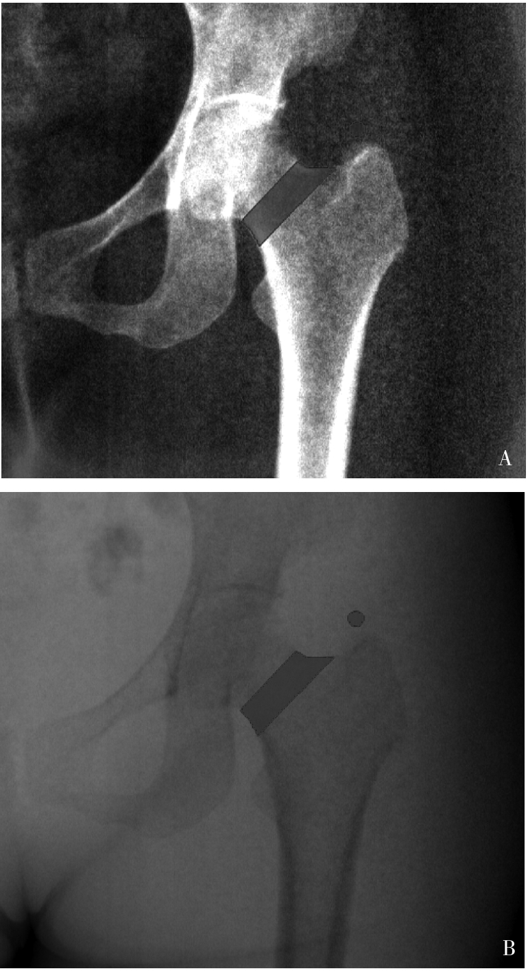

Figure 2
The linear regression result shows the high correlation between the SLR- and DXA-derived aBMD valuesSLR: spectral localizer radiography; DXA: dual-energy X-ray absorptio-metry; aBMD: areal bone mineral density.
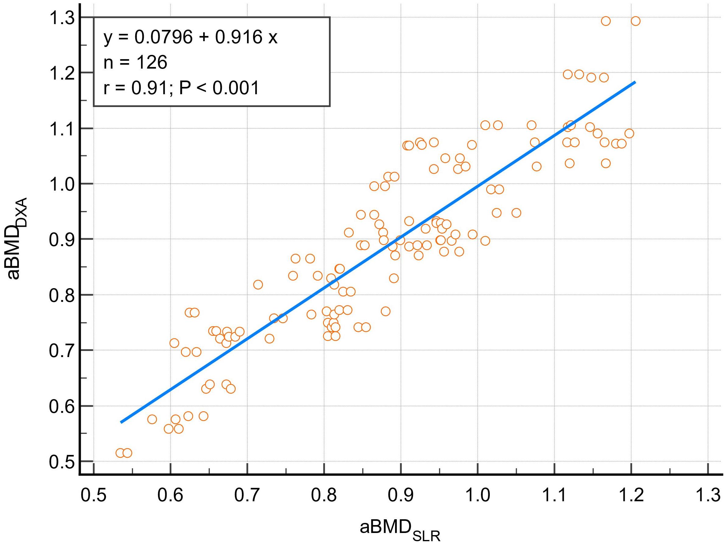

Figure 3
The Bland-Altman plot shows the high agreement between the SLR- and DXA-derived aBMD valuesSLR: spectral localizer radiography; DXA: dual-energy X-ray absorptio-metry; aBMD: areal bone mineral density.
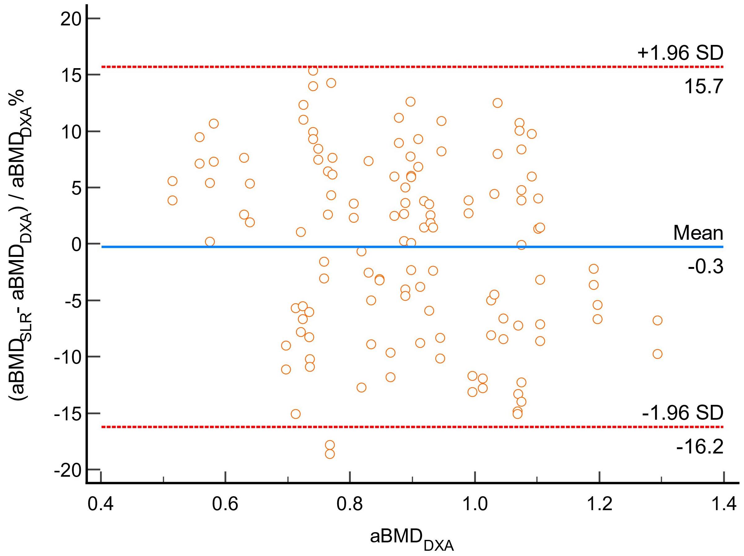

Figure 4
The Bland-Altman plot shows the high inter-o-bserver agreement of the aBMD value measured on SLRsSLR: spectral localizer radiography; aBMD: areal bone mineral density; 1 and 2: results measured by observer 1 and observer 2.
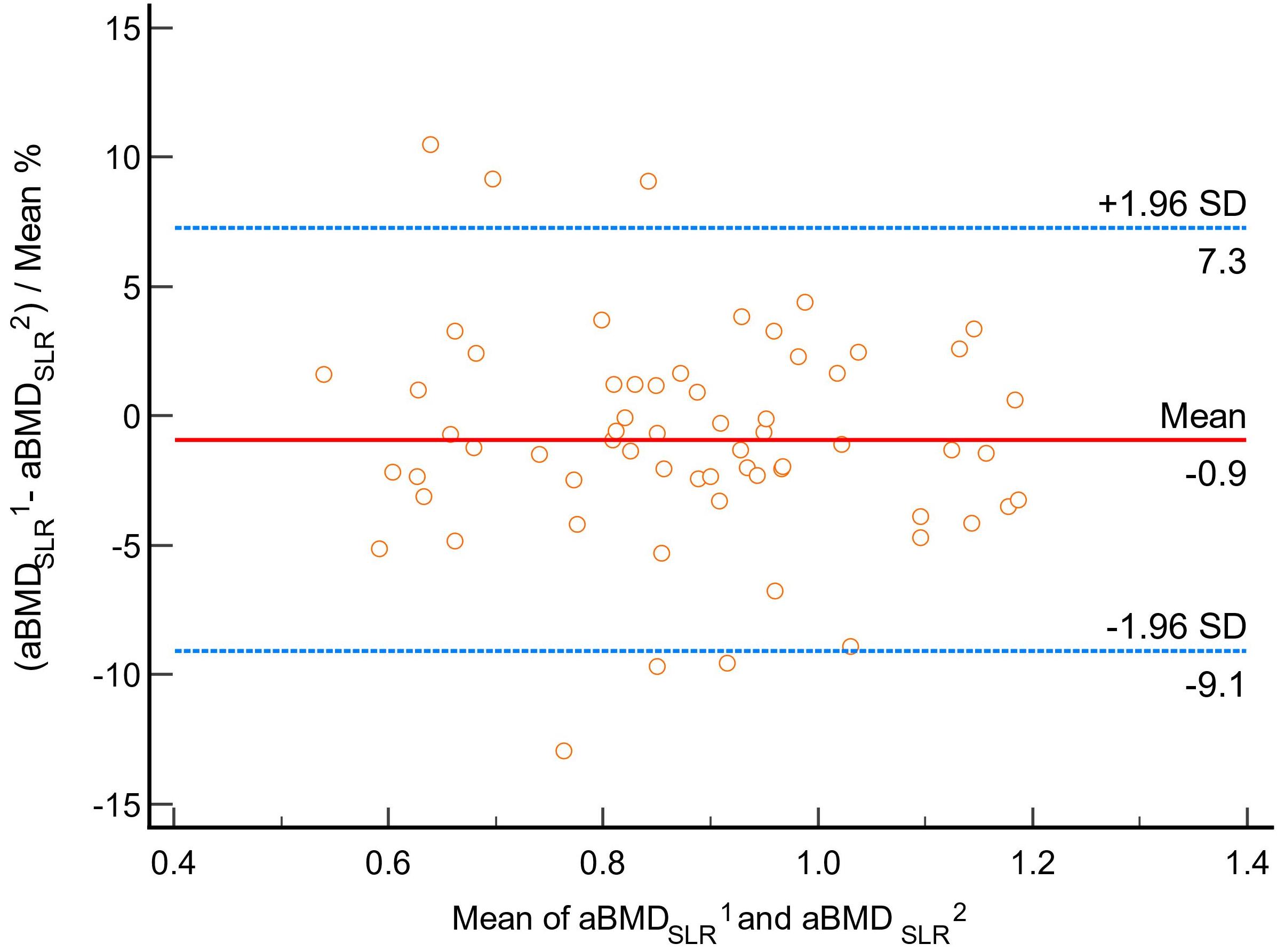
Table 1
The diagnostic performance for abnormal bona mass (T-score <-1.0) of SLR
| Item | Observer 1 | Observer 2 |
|---|---|---|
| Accuracy[%(N)] | 90.48% (57/63) | 95.24% (50/63) |
| Sensitivity[%(N)] | 86.96% (20/23) | 95.65% (22/23) |
| Specificity[%(N)] | 92.50% (37/40) | 95.00% (38/40) |
| PPV[%(N)] | 86.96% (20/23) | 91.67% (22/24) |
| NPV[%(N)] | 92.50% (37/40) | 97.44% (38/39) |
| [1] | XIA W B, HE S L, XU L, et al. Rapidly increasing rates of hip fracture in Beijing, China[J]. J Bone Miner Res,2012,27(1):125-129. |
| [2] |
SHEPSTONE L, LENAGHAN E, COOPER C, et al. Screening in the community to reduce fractures in older women (SCOOP): a randomised controlled trial[J]. Lancet,2018,391(10122):741-747.
doi: S0140-6736(17)32640-5 pmid: 29254858 |
| [3] |
US Preventive Services Task Force, CURRY S J, KRIST A H, et al. Screening for osteoporosis to prevent fractures: US preventive services task force recommendation statement[J]. JAMA,2018,319(24):2521-2531.
doi: 10.1001/jama.2018.7498 pmid: 29946735 |
| [4] |
KANIS J A. Assessment of fracture risk and its application to screening for postmenopausal osteoporosis: synopsis of a WHO report. WHO Study Group[J]. Osteoporos Int,1994,4(6):368-381.
doi: 10.1007/BF01622200 pmid: 7696835 |
| [5] |
中华医学会骨质疏松和骨矿盐疾病分会. 原发性骨质疏松症诊疗指南(2022)[J]. 中国全科医学,2023,26(14):1671-1691.
doi: 10.12114/j.issn.1007-9572.2023.0121 |
| Chinese Society of Osteoporosis and Bone Mineral Research. Guidelines for the Diagnosis and treatment of primary osteoporosis(2022)[J]. Chin Gen Pract,2023,26(14):1671-1691. | |
| [6] |
GILLESPIE C W, MORIN P E. Trends and disparities in osteoporosis screening among women in the United States, 2008-2014[J]. Am J Med,2017,130(3):306-316.
doi: S0002-9343(16)31191-3 pmid: 27884649 |
| [7] |
AMARNATH A L, FRANKS P, ROBBINS J A, et al. Underuse and overuse of osteoporosis screening in a regional health system: a retrospective cohort study[J]. J Gen Intern Med,2015,30(12):1733-1740.
doi: 10.1007/s11606-015-3349-8 pmid: 25986135 |
| [8] | CANN C E. Quantitative CT for determination of bone mine-ral density: a review[J]. Radiology,1988,166(2):509-522. |
| [9] |
ACU K, SCHEEL M, ISSEVER A S. Time dependency of bone density estimation from computed tomography with intravenous contrast agent administration[J]. Osteoporos Int,2014,25(2):535-542.
doi: 10.1007/s00198-013-2440-4 pmid: 23877871 |
| [10] |
KOCH V, HOKAMP N G, ALBRECHT M H, et al. Accuracy and precision of volumetric bone mineral density assessment using dual-source dual-energy versus quantitative CT: a phantom study[J]. Eur Radiol Exp,2021,5(1):43.
doi: 10.1186/s41747-021-00241-1 pmid: 34608576 |
| [11] | QIN L, HUANG J, YU P, et al. Accuracy, agreement, and reliability of DECT-derived vBMD measurements: an initial ex vivo study[J]. Eur Radiol,2021,31(1):191-199. |
| [12] |
Expert Panel on Musculoskeletal Imaging, YU J S, KRISHNA N G, et al. ACR appropriateness criteria® osteoporosis and bone mineral density: 2022 Update[J]. J Am Coll Radiol,2022,19(11S):S417-S432.
doi: 10.1016/j.jacr.2022.09.007 pmid: 36436967 |
| [13] |
KANIS J A. Diagnosis of osteoporosis and assessment of fracture risk[J]. Lancet,2002,359(9321):1929-1936.
doi: 10.1016/S0140-6736(02)08761-5 pmid: 12057569 |
| [14] | 陈海燕, 杨永波, 刘璐璐,等. 光子计数探测器CT初步临床应用的研究进展[J]. 中华放射学杂志,2022,56(2):213-216. |
| CHEN H Y, YANG Y B, LIU L L, et al. Research progress of clinical application of spectrum CT based on photon-counting detector[J]. Chin J Radiol,2022,56(2):213-216. | |
| [15] | 张挽时. 光子计数CT成像技术和临床价值[J]. 中华放射学杂志,2023,57(10):1133-1136. |
| ZHANG W S. Imaging technique and clinical value of photon counting CT[J]. Chin J Radiol,2023,57(10):1133-1136. | |
| [16] |
SYMONS R, KRAUSS B, SAHBAEE P, et al. Photon-counting CT for simultaneous imaging of multiple contrast agents in the abdomen: An in vivo study[J]. Med Phys,2017,44(10):5120-5127.
doi: 10.1002/mp.12301 pmid: 28444761 |
| [17] | CHRISTNER J A, KOFLER J M, MCCOLLOUGH C H. Estimating effective dose for CT using dose-length pro-duct compared with using organ doses: consequences of adopting International Commission on Radiological Protection publication 103 or dual-energy scanning[J]. Am J Roentgenol,2010,194(4):881-889. |
| [18] |
NOLDEN M, ZELZER S, SEITEL A, et al. The medical imaging interaction toolkit: challenges and advances : 10 years of open-source development[J]. Int J Comput Assist Radiol Surg,2013,8(4):607-620.
doi: 10.1007/s11548-013-0840-8 pmid: 23588509 |
| [19] |
DIMAI H P. Use of dual-energy X-ray absorptiometry (DXA) for diagnosis and fracture risk assessment; WHO-criteria, T- and Z-score, and reference databases[J]. Bone,2017,104:39-43.
doi: S8756-3282(16)30386-6 pmid: 28041872 |
| [20] |
KANIS J A, MELTON L J 3RD, CHRISTIANSEN C, et al. The diagnosis of osteoporosis[J]. J Bone Miner Res,1994, 9(8):1137-1141.
doi: 10.1002/jbmr.5650090802 pmid: 7976495 |
| [21] |
LAUGERETTE A, SCHWAIGER B J, BROWN K, et al. DXA-equivalent quantification of bone mineral density using dual-layer spectral CT scout scans[J]. Eur Radiol,2019,29(9):4624-4634.
doi: 10.1007/s00330-019-6005-6 pmid: 30758656 |
| [22] |
NOWAK T, EBERHARD M, SCHMIDT B, et al. Bone mineral density quantification from localizer radiographs: accuracy and precision of energy-integrating detector ct and photon-counting detector CT[J]. Radiology,2021,298(1):147-152.
doi: 10.1148/radiol.2020202767 pmid: 33141002 |
| [23] | EULER A, NOWAK T, BUCHER B, et al. Assessment of bone mineral density from a computed tomography topogram of photon-counting detector computed tomography-effect of phantom size and tube voltage[J]. Invest Radiol,2021,56(10):614-620. |
| [24] | SADANEY A O EL, FERRERO A, RAJENDRAN K, et al. Opportunistic bone mineral density measurement using photon-counting detector ct spectral localizer images: a prospective study[J]. Am J Roentgenol,2025,224(1):e2431909. |
| [25] | MOSER L J, KLAMBAUER K, DIAZ MACHICADO M C, et al. In vivo bone mineral density assessment with spectral localizer radiographs from photon-counting detector CT: Prospective comparison with DXA[J]. Invest Radiol,2025. |
| [26] |
MORI I, MACHIDA Y, OSANAI M, et al. Photon starvation artifacts of X-ray CT: their true cause and a solution[J]. Radiol Phys Technol,2013,6(1):130-141.
doi: 10.1007/s12194-012-0179-9 pmid: 23054905 |
| [27] |
LEMS W F, PACCOU J, ZHANG J, et al. Vertebral fracture: epidemiology, impact and use of DXA vertebral fracture assessment in fracture liaison services[J]. Osteoporos Int,2021,32(3):399-411.
doi: 10.1007/s00198-020-05804-3 pmid: 33475820 |
| [28] | MAZZIOTTI G, ANGELI A, BILEZIKIAN J P, et al. Glucocorticoid-induced osteoporosis: an update[J]. Trends Endocrinol Metab,2006,17(4):144-149. |
| [29] |
MARCUCCI G, BELTRAMI G, TAMBURINI A, et al. Bone health in childhood cancer: review of the literature and recommendations for the management of bone health in childhood cancer survivors[J]. Ann Oncol,2019,30(6):908-920.
doi: S0923-7534(19)31208-6 pmid: 31111878 |
| [1] | BAI Mengyao, KONG Bo, YANG Lihui, LI Lijuan, SHI Yanqing, SUN Lihao. Research progress on effects of berberine on bone metabolism and related mechanisms [J]. Journal of Diagnostics Concepts & Practice, 2024, 23(06): 634-640. |
| [2] | ZHANG Zhenlin, YUE Hua, LI Mei, XIA Weibo. Interpretation of guidelines for the diagnosis and treatment of primary osteoporosis (2022 version) in China: essential introduction [J]. Journal of Diagnostics Concepts & Practice, 2023, 22(03): 230-233. |
| [3] | LIU Xin, QI Caihui, WANG Zhenjing, LÜ Na, WANG Shaoting, WANG Shuping. Transcriptome study of glucagon like peptide-1 agonist Exendin-4 on mouse embryonic osteoblast precursor MC3T3-E1 in vitro [J]. Journal of Diagnostics Concepts & Practice, 2022, 21(03): 367-373. |
| [4] | Chinese Society of Endocrinology,Chinese Medical Association , et al . Expert suggestion for osteoporosis management during the recent COVID-19 pandemic [J]. Journal of Diagnostics Concepts & Practice, 2022, 21(02): 133-135. |
| [5] | CHANG Rui, XU Jiaxu, DONG Haipeng, WU Mengxiong, ZHAO Xuesong, MIAO Fei, YAN Fuhua. Value of CT spectral imaging in the evaluation of Crohn's disease activity [J]. Journal of Diagnostics Concepts & Practice, 2019, 18(04): 432-435. |
| [6] | . [J]. Journal of Diagnostics Concepts & Practice, 2014, 13(05): 476-480. |
| [7] | . [J]. Journal of Diagnostics Concepts & Practice, 2014, 13(01): 68-71. |
| [8] | . [J]. Journal of Diagnostics Concepts & Practice, 2014, 13(01): 82-85. |
| [9] | . [J]. Journal of Diagnostics Concepts & Practice, 2013, 12(02): 205-209. |
| [10] | . [J]. Journal of Diagnostics Concepts & Practice, 2011, 10(06): 517-522. |
| [11] | . [J]. Journal of Diagnostics Concepts & Practice, 2011, 10(06): 531-534. |
| [12] | . [J]. Journal of Diagnostics Concepts & Practice, 2011, 10(05): 428-433. |
| [13] | . [J]. Journal of Diagnostics Concepts & Practice, 2010, 9(02): 155-160. |
| [14] | . [J]. Journal of Diagnostics Concepts & Practice, 2003, 2(03): 44-46. |
| Viewed | ||||||
|
Full text |
|
|||||
|
Abstract |
|
|||||