Journal of Internal Medicine Concepts & Practice ›› 2025, Vol. 20 ›› Issue (04): 276-281.doi: 10.16138/j.1673-6087.2025.04.03
• Original article • Previous Articles Next Articles
SUN Jie1, XIE Jie1, MA Hongkun2, LIU Baolian1, CHEN Xueying1, HUANG Wenjie1, HE Shuilin1, CHEN Zijin2, ZHANG Wen2( )
)
Received:2024-07-31
Online:2025-07-31
Published:2025-10-27
SUN Jie, XIE Jie, MA Hongkun, LIU Baolian, CHEN Xueying, HUANG Wenjie, HE Shuilin, CHEN Zijin, ZHANG Wen. Clinical application of renal artery ultrasound monitoring hemodynamic parameters and non-contrast-enhanced magnetic resonance angiography in evaluation of renal artery stenosis and anatomic abnormalities[J]. Journal of Internal Medicine Concepts & Practice, 2025, 20(04): 276-281.
Table 1
Baseline characteristics and distribution of renal artery lesions in patients[n(%)/$\bar{x} \pm s$]
| 项目 | 整体人群 | RAS | 肾动脉变异 | ||
|---|---|---|---|---|---|
| 单侧 | 双侧 | 单侧 | 双侧 | ||
| 总例数[n(%)] | 149(100.0) | 32(21.5) | 11(7.4) | 52(34.9) | 11(7.4) |
| 男性[n(%)] | 108(72.5) | 29(19.5) | 5(3.4) | 42(28.1) | 9(6.0) |
| 年龄(岁) | 57.4±15.6 | 61.6±13.8 | 76.2±8.6 | 53.5±15.4 | 54.2±18.1 |
| 原发病[n(%)] | |||||
| 慢性肾炎 | 51(34.2) | 10(6.7) | 2(1.3) | 12(8.1) | 2(1.3) |
| 糖尿病肾病 | 31(20.8) | 7(4.7) | 3(2.0) | 17(11.4) | 3(2.0) |
| 高血压肾病 | 34(22.8) | 12(8.1) | 4(2.7) | 11(7.4) | 3(2.0) |
| 其他原发病 | 33(22.2) | 2(1.3) | 2(1.3) | 12(8.1) | 3(2.0) |
| 合并症[n(%)] | |||||
| 高血压 | 125(83.9) | 28(18.8) | 10(6.7) | 43(28.9) | 6(4.0) |
| 糖尿病 | 52(34.9) | 14(9.4) | 4(2.7) | 20(13.4) | 5(3.4) |
| eGFR [mL/(min·1.73 m2)] | 47.98±31.22 | 41.68±26.10 | 39.73±23.53 | 43.90±31.47 | 62.35±32.82 |
| GFR *(mL/min)(n=99) | 48.09±23.94 | 39.29±15.93 | 34.42±13.83 | 49.28±26.17 | 46.17±27.61 |

Figure 1
NCE-MRA assessment of renal arteries
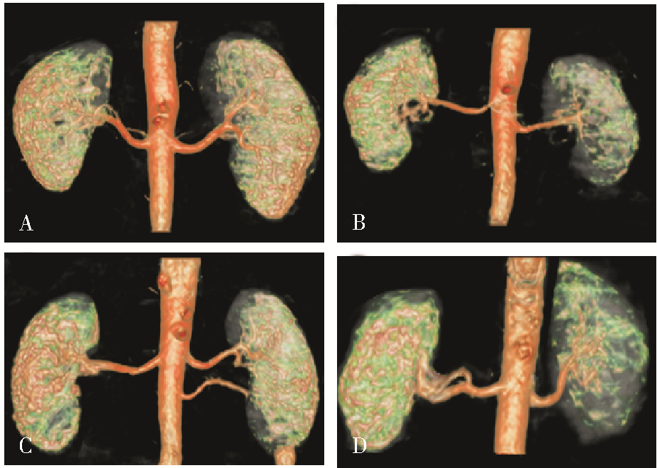
Table 2
Comparison of CDUS parameters in RAS patients with stenosis at different sites[M(Q1,Q3)/$\bar{x} \pm s$]
| 参数 | 肾动脉起始段无狭窄 (n=263) | 肾动脉起始段狭窄 (n=35) | t/Z | P | 肾动脉中段无狭窄 (n=277) | 肾动脉中段狭窄 (n=21) | t/Z | P |
|---|---|---|---|---|---|---|---|---|
| PSV(cm/s) | 74(54,90) | 53.5(42,78) | 0.767 | 0.443 | 41(33,50) | 40(33,44) | 1.310 | 0.190 |
| RI | 0.689±0.05 | 0.758±0.016 | -4.252 | 0.000 | 0.644±0.006 | 0.678±0.018 | -1.715 | 0.088 |
Table 3
Comparison of CDUS parameters between patients with renal artery anatomical variations and those without variations[M(Q1,Q3)/$\bar{x} \pm s$]
| 参数 | 无副肾动脉+提前分支 (n=224) | 有副肾动脉+提前分支 (n=74) | t/Z | P |
|---|---|---|---|---|
| 肾动脉起始段PSV(cm/s) | 71.5(53,90) | 71(48,90) | 0.585 | 0.559 |
| 肾动脉起始段RI | 0.701±0.006 | 0.687±0.009 | 1.232 | 0.219 |
| 肾动脉中段PSV(cm/s) | 40.5(33,50) | 41(31.5,49) | -0.616 | 0.538 |
| 肾动脉中段RI | 0.648±0.006 | 0.644±0.010 | 0.366 | 0.715 |
Table 4
Correlation between renal artery hemodynamic parameters (PSV and RI) and GFR
| 肾动脉 | r | P |
|---|---|---|
| 起始段RI | -0.446 | 0.000 |
| 起始段PSV | 0.302 | 0.000 |
| 中段RI | -0.294 | 0.000 |
| 中段PSV | 0.344 | 0.000 |
| [1] |
Chai JW, Lee W, Yin YH, et al. CT angiography for living kidney donors: accuracy, cause of misinterpretation and prevalence of variation[J]. Korean J Radiol, 2008, 9(4):333-339.
doi: 10.3348/kjr.2008.9.4.333 pmid: 18682671 |
| [2] | 郭震华, 那彦群. 实用泌尿外科学[M]. 2版. 北京: 人民卫生出版社,2014:112. |
| [3] |
AbuRahma AF, Yacoub M. Renal imaging: duplex ultrasound, computed tomography angiography, magnetic resonance angiography, and angiography[J]. Semin Vasc Surg, 2013, 26(4):134-143.
doi: 10.1053/j.semvascsurg.2014.06.001 pmid: 25220318 |
| [4] |
Herborn CU, Watkins DM, Runge VM, et al. Renal arteries: comparison of steady-state free precession MR angiography and contrast-enhanced MR angiography[J]. Radiology, 2006, 239(1):263-268.
doi: 10.1148/radiol.2383050058 pmid: 16493015 |
| [5] |
Holden A, Smith A, Dukes P, et al. Assessment of 100 live potential renal donors for laparoscopic nephrectomy with multi-detector row helical CT[J]. Radiology, 2005, 237(3):973-980.
pmid: 16304115 |
| [6] |
Bordei P, Sapte E, Iliescu D. Double renal arteries originating from the aorta[J]. Surg Radiol Anat, 2004, 26(6):474-479.
pmid: 15378279 |
| [7] |
Hänninen EL, Denecke T, Stelter L, et al. Preoperative evaluation of living kidney donors using multirow detector computed tomography: comparison with digital subtraction angiography and intraoperative findings[J]. Transpl Int, 2005, 18(10):1134-1141.
pmid: 16162099 |
| [8] | Gupta A, Tello R. Accessory renal arteries are not related to hypertension risk: a review of MR angiography data[J]. AJR Am J Roentgenol, 2004, 182(6):1521-1524. |
| [9] | Shen J, Lyu L, Wu X, et al. Correlation between renal artery anatomy and hypertension: a retrospective analysis of 3000 patients[J]. Evid Based Complement Alternat Med, 2021,2021:9957361. |
| [10] | Kang K, Ma Y, Jia C, et al. Relationship between accessory renal artery and clinical characteristics of middle-aged patients with primary hypertension[J]. Int J Hypertens, 2020,2020:7109502. |
| [11] |
刘方韬, 齐晓凤, 徐学勤, 等. 非增强磁共振血管成像在肾动脉狭窄评估方面的价值研究[J]. 诊断学理论与实践, 2019, 18(1):72-76.
doi: 10.16150/j.1671-2870.2019.01.014 |
| [12] |
Zhang LJ, Peng J, Wen J, et al. Non-contrast-enhanced magnetic resonance angiography: a reliable clinical tool for evaluating transplant renal artery stenosis[J]. Eur Radiol, 2018, 28(10):4195-4204.
doi: 10.1007/s00330-018-5413-3 pmid: 29666993 |
| [13] |
Bley TA, François CJ, Schiebler ML, et al. Non-contrast-enhanced MRA of renal artery stenosis: validation against DSA in a porcine model[J]. Eur Radiol, 2016, 26(2):547-555.
doi: 10.1007/s00330-015-3833-x pmid: 26017736 |
| [14] | Fananapazir G, McGahan JP, Corwin MT, et al. Screening for transplant renal artery stenosis: ultrasound-based stenosis probability stratification[J]. AJR Am J Roentgenol, 2017, 209(5):1064-1073. |
| [15] |
Granata A, Fiorini F, Andrulli S, et al. Doppler ultrasound and renal artery stenosis: an overview[J]. J Ultrasound, 2009, 12(4):133-143.
doi: 10.1016/j.jus.2009.09.006 pmid: 23397022 |
| [16] |
van Twist DJ, Houben AJ, de Haan MW, et al. Pathophysiological differences between multifocal fibromuscular dysplasia and atherosclerotic renal artery stenosis[J]. J Hypertens, 2017, 35(4):845-852.
doi: 10.1097/HJH.0000000000001243 pmid: 28060190 |
| [17] |
Lo R, Donaldson C. Vessel tortuosity causing false positives in detecting renal artery stenosis on doppler ultrasound[J]. Ultrasound Q, 2013, 29(1):47-50.
doi: 10.1097/RUQ.0b013e3182817b57 pmid: 23370780 |
| [18] | Kendrick J, Chonchol M. Renal artery stenosis and chronic ischemic nephropathy: epidemiology and diagnosis[J]. Adv Chronic Kidney Dis, 2008, 15(4):355-362. |
| [1] | SHEN Lianjun, WU Wei, JI Wei, WANG Hong, SUN Xing, SHI Qingqing, SUN Mei, GU Jian, NI Jun. A report on monitoring of coagulation indicators and treatment of microthrombus formation in AML (M4 type) after hematopoietic stem cell transplantation: a case report [J]. Journal of Diagnostics Concepts & Practice, 2024, 23(02): 180-183. |
| [2] | SUN Tiantian, YE Baoying, YANG Yu, NIU Jianmei. Color Doppler ultrasound and magnetic resonance imaging in prenatal diagnosis of pernicious placenta previa and pernicious placenta previa with placenta accreta: clinic value and analysis of missed diagnosis [J]. Journal of Diagnostics Concepts & Practice, 2021, 20(02): 173-177. |
| [3] | YUAN Yuwen,ZAN Tao,LI Qingfeng. Preoperative Vascular Imaging Techniques for Perforator Selections in Reconstructive Surgery [J]. Journal of Tissue Engineering and Reconstructive Surgery, 2014, 10(5): 285-288. |
| [4] | . [J]. Journal of Diagnostics Concepts & Practice, 2014, 13(05): 515-518. |
| [5] | . [J]. Journal of Surgery Concepts & Practice, 2011, 16(02): 151-154. |
| Viewed | ||||||
|
Full text |
|
|||||
|
Abstract |
|
|||||