外科理论与实践 ›› 2024, Vol. 29 ›› Issue (06): 529-532.doi: 10.16139/j.1007-9610.2024.06.12
丁勇, 周旻, 李旭 综述, 郭大乔, 王利新, 史振宇 审校
收稿日期:2024-10-22
出版日期:2024-11-25
发布日期:2025-03-17
基金资助:DING Yong, ZHOU Min, LI Xu, GUO Daqiao, WANG Lixin, SHI Zhenyu
Received:2024-10-22
Online:2024-11-25
Published:2025-03-17
摘要:
下肢静脉疾病的发病率高,严重危害病人的生活质量。腔内手术已成为下肢静脉疾病的重要治疗手段。术中常规使用的静脉造影无法提供静脉管腔内及周围结构的详细信息,而腔内成像技术如血管腔内超声和光学相干断层成像不仅能减少造影剂的使用、评估静脉管腔内及周围结构,而且精准测量并为后续治疗提供信息。本文梳理相关经验并总结腔内成像技术的成像原理、成像特点以及临床应用等,供临床实践参考。
中图分类号:
丁勇, 周旻, 李旭 综述, 郭大乔, 王利新, 史振宇 审校. 腔内成像技术在下肢静脉疾病中的应用[J]. 外科理论与实践, 2024, 29(06): 529-532.
DING Yong, ZHOU Min, LI Xu, GUO Daqiao, WANG Lixin, SHI Zhenyu. Application of intravascular imaging in lower extremities venous disorders[J]. Journal of Surgery Concepts & Practice, 2024, 29(06): 529-532.

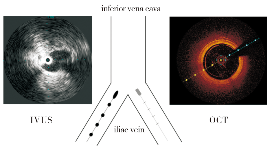
图1
腔内成像技术示意图
| [1] | 中华医学会外科学分会血管外科学组, 中国医师协会血管外科医师分会, 中国医疗保健国际交流促进会血管外科分会, 等. 中国慢性静脉疾病诊断与治疗指南[J]. 中华医学杂志, 2019, 99(39):3047-3061. |
| Vascular Surgery Group, Chinese Medical Association, Chinese Society of Surgery, China Association of Physicians in Vascular Surgery, China Society for the Promotion of International Exchange in Health Care, Vascular Surgery Branch, China Medical Association, et al. Chinese guidelines for the diagnosis and treatment of chronic venous diseases[J]. Natl Med J China, 2019, 99(39):3047-3061. | |
| [2] | 中国医师协会血管外科医师分会静脉学组. 常见静脉疾病诊治规范(2022年版)[J]. 中华血管外科杂志, 2022, 7(1):12-29. |
| Phlebology Group of Vascular Surgery Physicians Branch, Chinese Physicians Association. Diagnosis and treatment standard of common venous diseases (2022 version)[J]. Chin J Vasc Surg, 2022, 7(1):12-29. | |
| [3] | 中华医学会外科分会血管外科学组. 慢性下肢静脉疾病诊断与治疗中国专家共识[J]. 中华普通外科杂志, 2014, 29(4):246-252. |
| Vascular Surgery Group, Chinese Medical Association, Chinese Society of Surgery. Chinese expert consensus on the diagnosis and treatment of chronic lower extremity venous diseases[J]. Chin J Gen Surg, 2014, 29(4):246-252. | |
| [4] | PENG C, WU H, KIM S, et al. Recent advances in transducers for intravascular ultrasound (IVUS) imaging[J]. Sensors (Basel), 2021, 21(10):3540. |
| [5] | WANG G, QIAO W, XING C, et al. Diagnostic performance of 60 MHz high-definition intravascular ultrasound versus fourier domain optical coherence tomography for identifying plaque rupture, plaque erosion, and thrombosis in a rabbit model[J]. Rev Cardiovasc Med, 2023, 24(3):76. |
| [6] | KASHANI A H, CHEN C L, GAHM J K, et al. Optical coherence tomography angiography: a comprehensive review of current methods and clinical applications[J]. Prog Retin Eye Res, 2017,60:66-100. |
| [7] |
POTSAID B, GORCZYNSKA I, SRINIVASAN V J, et al. Ultrahigh speed spectral / Fourier domain OCT ophthalmic imaging at 70,000 to 312,500 axial scans per second[J]. Opt Express, 2008, 16(19):15149-15169.
doi: 10.1364/oe.16.015149 pmid: 18795054 |
| [8] | GARCÌA-GARCÌA H M, GOGAS B D, SERRUYS P W, et al. IVUS-based imaging modalities for tissue characte-rization: similarities and differences[J]. Int J Cardiovasc Imaging, 2011, 27(2):215-224. |
| [9] | DIETHRICH E B, IRSHAD K, REID D B. Virtual histo-logy and color flow intravascular ultrasound in peripheral interventions[J]. Semin Vasc Surg, 2006, 19(3):155-162. |
| [10] | 袁贤琳, 李百灵. 光学相干断层成像技术在医学应用中的研究进展[J]. 中国处方药, 2021, 19(11):71-73. |
| YUAN X L, LI B L. Research progress of medical application of optical coherence tomography imaging techno-logy[J]. J China Prescr Drug, 2021, 19(11):71-73. | |
| [11] | 罗明华, 关怀敏, 解金红, 等. 光学相干断层成像对支架内再狭窄冠脉内膜病理特点的观察[J]. 实用医学杂志, 2016, 32(1):94-97. |
| LUO M H, GUAN H M, XIE J H, et al. Optical coherence tomography on the pathological characteristics of the endothelium of in-stent restenotic coronary arteries[J]. J Pract Med, 2016, 32(1):94-97. | |
| [12] |
RAJU S, MARTIN A, DAVIS M. The importance of IVUS assessment in venous thrombolytic regimens[J]. J Vasc Surg Venous Lymphat Disord, 2013, 1(1):108.
doi: 10.1016/j.jvsv.2012.10.030 pmid: 26993928 |
| [13] | CHENG L, ZHAO H, ZHANG F X. Iliac vein compression syndrome in an asymptomatic patient population: a prospective study[J]. Chin Med J, 2017, 130(11):1269-1275. |
| [14] | 丁勇, 赵格非, 周旻, 等. 血管腔内超声与静脉造影在髂静脉压迫综合征术中诊断价值的比较[J]. 中华普通外科杂志, 2019, 34(9):753-756. |
| DING Y, ZHAO G F, ZHOU M, et al. Comparison of intravascular ultrasound and venography in the intraoperative evaluation of iliac vein compression syndrome[J]. Chin J Gen Surg, 2019, 34(9):753-756. | |
| [15] | GAGNE P J, TAHARA R W, FASTABEND C P, et al. Venography versus intravascular ultrasound for diagno-sing and treating iliofemoral vein obstruction[J]. J Vasc Surg Venous Lymphat Disord, 2017, 5(5):678-687. |
| [16] | 丁勇, 王永刚, 周旻, 等. 血管腔内超声在髂静脉疾病诊疗中的应用价值[J]. 中华普通外科杂志, 2023, 38(11):859-861. |
| DING Y, WANG Y G, ZHOU M, et al. The value of vascular endoluminal ultrasound in the diagnosis and treatment of iliac vein disease[J]. Chin J Gen Surg, 2023, 38(11):859-861. | |
| [17] |
MONTMINY M L, THOMASSON J D, TANAKA G J, et al. A comparison between intravascular ultrasound and venography in identifying key parameters essential for iliac vein stenting[J]. J Vasc Surg Venous Lymphat Disord, 2019, 7(6):801-807.
doi: S2213-333X(19)30253-7 pmid: 31196766 |
| [18] | GANGULI S, HAWKINS B M, ABTAHIAN F, et al. Comparison of inferior vena cava filters placed at the bedside via intravenous ultrasound guidance versus fluoroscopic guidance[J]. Ann Vasc Surg, 2017,39:250-255. |
| [19] | KITROU P M, SPILIOPOULOS S, KATSANOS K, et al. Venous drug-eluting vs. bare-metal stenting: an experimental animal study using frequency domain optical coherence tomography[J]. Hellenic J Cardiol, 2014, 55(5):386-392. |
| [20] |
LI G, HU B, SUN Y, et al. Histological features of in-stent restenosis after iliac vein thrombus removal and stent placement in a goat model[J]. J Vasc Interv Radiol, 2024, 35(4):611-617.
doi: 10.1016/j.jvir.2023.12.567 pmid: 38171414 |
| [21] | JONES G L, ALBADAWI H, HARIRI L P, et al. Aging of deep venous thrombosis in-vivo using polarization sensitive optical coherence tomography[J]. Biomed Opt Express, 2024, 15(6):3627-3638. |
| [22] | ROTUNNO G, DEINSBERGER J, MEIBURGER K M, et al. Optical coherence tomography angiography enables visualization of microvascular patterns in chronic venous insufficiency[J]. iScience, 2024, 27(11):110998. |
| [1] | 蒋金法,. 光学相干断层成像在诊断冠心病中的意义[J]. 诊断学理论与实践, 2011, 10(01): 10-14. |
| [2] | 邓钰蕾, 陈燕, 谢列琴, 王刚, 徐玮, 汤荟冬, 王瑛, 陈生弟,. 轻度阿尔茨海默病患者视网膜形态改变的观察[J]. 诊断学理论与实践, 2009, 8(04): 397-400. |
| 阅读次数 | ||||||
|
全文 |
|
|||||
|
摘要 |
|
|||||