外科理论与实践 ›› 2021, Vol. 26 ›› Issue (06): 550-554.doi: 10.16139/j.1007-9610.2021.06.018
肖莉, 王彦林, 董长宪( ), 孙斌
), 孙斌
收稿日期:2021-01-26
出版日期:2021-11-25
发布日期:2022-07-27
通讯作者:
董长宪
E-mail:zzchangxiand@126.com
基金资助:
XIAO Li, WANG Yanlin, DONG Changxian(), SUN Bin
Received:2021-01-26
Online:2021-11-25
Published:2022-07-27
Contact:
DONG Changxian
E-mail:zzchangxiand@126.com
摘要:
目的:根据静脉畸形(venous malformation,VM)组织结构、组织病理特点将病人分成3组,分析治疗方案及对应治疗结果,根据VM组织结构指导治疗方案选择。方法:回顾性分析本院血管瘤科2015年1月至2019年12月170例VM,男73例,女97例,年龄(23.4±9.9)岁。根据标本组织结构将VM分成包膜组141例,非包膜组22例,骨化组7例。超声、磁共振、X线和CT血管造影检查病人。手术或微创治疗VM。术中大体查看和术后VM组织病理检查。术前和出院后行疼痛视觉模拟评分、Oswestry功能障碍指数检测。术后1年随访163例,7例失访。结果:得到3组VM的影像、组织结构与病理表现、治疗方案及疗效评价的数据。包膜组手术101例,治愈78例(77.2%),术后复发2例(2.0%);微创治疗余40例,包括硬化治疗和射频消融,治愈23例(57.5%),复发2例(5.0%),是预后最佳的组织结构类型。非包膜组手术15例,治愈3例(20.0%),复发4例(26.7%);微创治疗余7例,治愈1例,复发3例,复发率高(42.9%)。骨化组均手术治疗,7例治愈,无复发。包膜组和非包膜组出院时疼痛视觉模拟评分较治疗前明显降低。所有病例出院时Oswestry功能障碍指数均较治疗前降低。术后6例发生切口延迟愈合等并发症。结论:组织结构可成为VM治疗方案选择及预后判断的有效依据,包膜组疗效最佳,非包膜组疗效差,骨化组只适用手术治疗。
中图分类号:
肖莉, 王彦林, 董长宪, 孙斌. 静脉畸形组织结构与治疗选择的临床分析[J]. 外科理论与实践, 2021, 26(06): 550-554.
XIAO Li, WANG Yanlin, DONG Changxian, SUN Bin. Clinical analysis on tissue structure and treatment option of venous malformation[J]. Journal of Surgery Concepts & Practice, 2021, 26(06): 550-554.
表1
3组VM的组织分布情况[n(%)]
| 组别 | 部位(n) | 皮肤脂肪 | 肌肉 | 骨骼 |
|---|---|---|---|---|
| 包膜组(n=141) | 头部(35),四肢(67),躯干(39) | 68(48) | 61(43) | 12(9) |
| 非包膜组(n=22) | 头部(4),四肢(15),躯干(3) | 5(24) | 17(76) | 0 |
| 骨化组(n=7) | 四肢(7) | 0 | 6(86) | 1(14) |
表2
3组VM的临床特点[n(%)]
| 组别 | 疼痛 | 功能障碍 | 血管内凝血 |
|---|---|---|---|
| 包膜组(n=141) | 118(83.7) | 43(30.5) | 46(32.6) |
| 非包膜组(n=22) | 6(27.3) | 1(4.5) | 1(4.5) |
| 骨化组(n=7) | 3(42.9) | 7(100) | 0 |

图1
颌面部包膜组VM的MRI检查表现 T2WI显示高信号,分叶状团块,有明显静脉石形成,伴纤维增生。
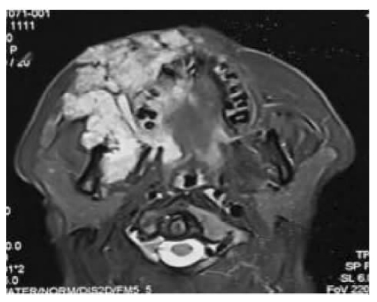
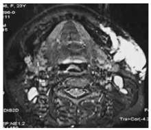
图2
颌面部非包膜组VM的MRI检查表现 T2WI显示高信号,均质,少见静脉石及纤维增生表现。
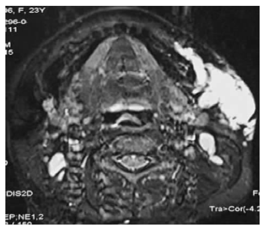

图3
骨化组VM的X线检查表现 瘤体组织内有类骨型增生。
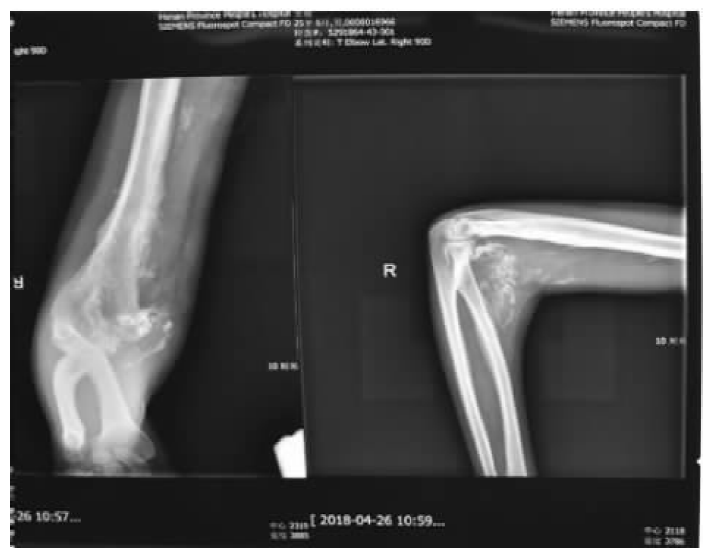
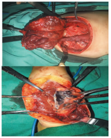
图4
包膜组VM大体观 瘤体边界清楚,外周有包膜覆盖,与周围正常组织界限清晰。
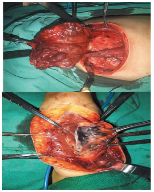

图5
非包膜组VM大体观 瘤体呈现立体网格状,对周围组织具侵袭性,血窦丰富。
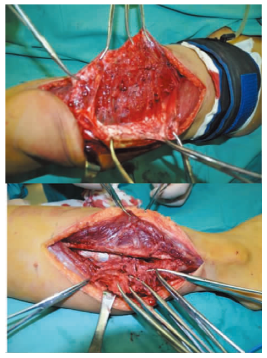

图6
骨化组VM大体观 肌肉内瘤体骨化,质地坚硬,失去原有肌肉功能。
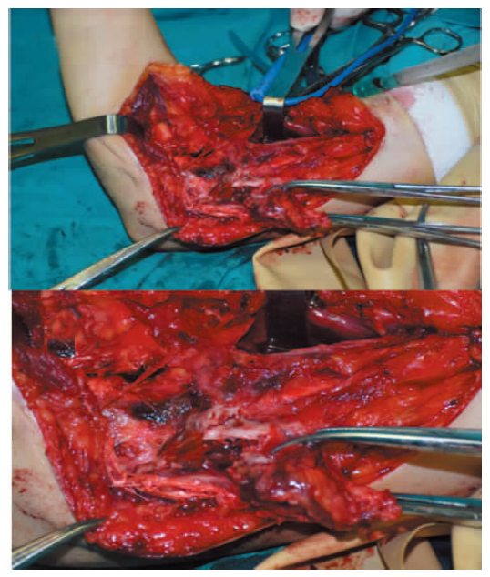
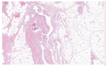
图7
包膜组VM病理表现 不规则扩张的畸形血管,管腔较大,有细小壁薄分枝状畸形血管(HE染色,×100)。
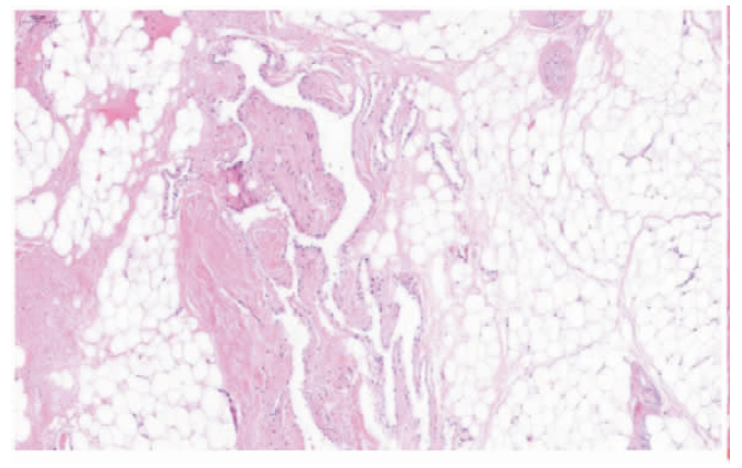

图8
非包膜组VM病理表现 海绵状、网状畸形血管,管腔淤血(HE染色,×200)。
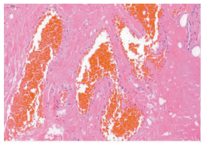
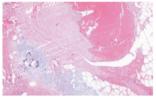
图9
骨化组VM病理表现 畸形血管及化生软骨组织(HE染色,×200)。

表3
3组 VM治疗效果评价[n(%)]
| 治疗 170 | 包膜组 141(82.9) | 非包膜组 22(12.9) | 骨化组 7(4.1) |
|---|---|---|---|
| 手术123(72.4) | 101 | 15 | 7 |
| 治愈 | 78(77.2) | 3(20.0) | 5(71.4) |
| 临床治愈 | 21(20.8) | 8(53.3) | 2(28.6) |
| 复发 | 2(2.0) | 4(26.7) | |
| 微创47(27.6) | 40 | 7 | 0 |
| 治愈 | 23(57.5) | 1(14.2) | |
| 临床治愈 | 15(37.5) | 3(42.9) | |
| 复发 | 2(5.0) | 3(42.9) |
表4
治疗前、后疼痛VAS与ODI比较
| 组别 | VAS | t值 | P值 | ODI | t值 | P值 | |||
|---|---|---|---|---|---|---|---|---|---|
| 术前 | 出院 | 术前 | 出院 | ||||||
| 包膜组(n=141) | 2.38±1.355 | 0.04±0.221 | 21.604 | <0.01 | 5.60±6.528 | 0.04±0.251 | 10.231 | <0.01 | |
| 非包膜组(n=22) | 0.45±0.858 | 0 | 2.485 | 0.02 | 0.55±0.858 | 0 | 2.982 | 0.01 | |
| 骨化组(n=7) | 0.714±0.951 | 0 | 1.987 | 0.09 | 17.71±3.817 | 1.14±1.069 | 14.388 | <0.01 | |
| [1] | Wieck MM, Nowicki D, Schall KA, et al. Management of pediatric intramuscular venous malformations[J]. J Pediatr Surg, 2017, 52(4):598-601. |
| [2] | Carqueja IM, Sousa J, Mansilha A. Vascular malformations: classification, diagnosis and treatment[J]. Int An-giol, 2018, 37(2):127-142. |
| [3] | Hage AN, Chick JFB, Srinivasa RN, et al. Treatment of venous malformations: the data, where we are, and how it is done[J]. Tech Vasc Interv Radiol, 2018, 21(2):45-54. |
| [4] | Zhong LP, Ow A, Yang WJ, et al. Surgical management of solitary venous malformation in the midcheek region[J]. Oral Surg Oral Med Oral Pathol Oral Radiol, 2012, 114(2):160-166. |
| [5] | Calandriello L, Grimaldi G, Petrone G, et al. Cavernous venous malformation (cavernous hemangioma) of the orbit: current concepts and a review of the literature[J]. Surv Ophthalmol, 2017, 62(4):393-403. |
| [6] | Jamshidi K, Jafari D, Ramezan Shirazi M, et al. An unusual presentation of ossified intramuscular hemangioma: a case report[J]. Acta Med Iran, 2014, 52(4):319-322. |
| [7] | Engelstad BL, Gilula LA, Kyriakos M. Ossified skeletal muscle hemangioma: radiologic and pathologic features[J]. Skeletal Radiol, 1980, 5(1):35-40. |
| [8] | Panda R, Mangal M, Reddy S, et al. Osseous metaplasia mimicking long bone in intramuscular vascualr malformation[J]. World J Plast Surg, 2018, 7(2):243-248. |
| [9] | Sadick M, Müller-Wille R, Wildgruber M, et al. Vascular anomalies(Part I): classification and diagnostics of vascular anomalies[J]. Rofo, 2018, 190(9):825-835. |
| [10] | 中华医学会整形外科分会血管瘤和脉管畸形学组. 血管瘤和脉管畸形的诊断及治疗指南(2019版)[J]. 组织工程与重建外科杂志, 2019, 15(5):277-317. |
| [11] | Clemens RK, Baumann F, Husmann M, et al. Percutaneous sclerotherapy for spongiform venous malformations-analysis of patient-evaluated outcome and satisfaction[J]. Vasa, 2017, 46(6):477-483. |
| [12] | Puig S, Aref H, Chigot V, et a1. Classification of venous malformations in children and implications for sclerothe-rapy[J]. Pediatr Radiol, 2003, 33(2):99-103. |
| [13] | Han YY, Sun LM, Yuan SM. Localized intravascular coa-gulation in venous malformations: a system review[J]. Phlebology, 2021, 36(1):38-42. |
| [14] | Dompmartin A, Acher A, Thibon P. Association of loca-lized intravascular coagulophathy with venous malformations[J]. Arch Dermatol, 2008, 144(7):873-877. |
| [15] | Binet Q, Lambert C, Hermans C. Dabigatran etexilate in the treatment of localized intravascular coagulopathy associated with venous malformations[J]. Thromb Res, 2018, 168:114-120. |
| [16] | Aronniemi J, Castren E, Lappalainen K, et al. Sclerothe-rapy complications of peripheral venous malformations[J]. Phlebology, 2016, 31(10):712-722. |
| [17] | Legiehn GM. Sclerotherapy with adjunctive stasis of efflux(STASE) in venous malformations: techniques and strategies[J]. Tech Vasc Interv Radiol, 2019, 22(4):100630. |
| [1] | 高定辉, 陈勇, 王倩, 等. 肌内静脉畸形的影像学观察 :单中心回顾性分析 [J]. 组织工程与重建外科杂志, 2023, 19(1): 37-. |
| [2] | 汤莹, 周燕春, 曹璇君, 等. 颅外动静脉畸形患者焦虑抑郁状态及相关因素分析[J]. 组织工程与重建外科杂志, 2023, 19(1): 54-. |
| [3] | 罗方秀, 马乾宸, 袁菲. 第5版WHO消化系统肿瘤分类解读:胆道系统肿瘤的更新及进展[J]. 外科理论与实践, 2023, 28(02): 124-131. |
| [4] | 郑亚民, 顾利国, 许臣. 胆囊结石病理生理进展分期和个性化诊治[J]. 外科理论与实践, 2023, 28(02): 94-99. |
| [5] | 高定辉, 陈勇, 王倩, 等. Bockenheimer综合征的诊断和治疗:7例报道与文献回顾 [J]. 组织工程与重建外科杂志, 2022, 18(6): 480-. |
| [6] | 戴梦婷 崔杰. 动静脉畸形动物模型的研究及应用进展[J]. 组织工程与重建外科杂志, 2022, 18(4): 355-. |
| [7] | 韩钰钰 余明薇 王慜 徐媛 陈勇 袁斯明. 体表软组织静脉畸形中局限性血管内凝血的临床特征分析[J]. 组织工程与重建外科杂志, 2022, 18(3): 214-. |
| [8] | 李函育 张世仁 仇雅璟 林晓曦. 病灶内注射博来霉素治疗局限型疣状静脉畸形[J]. 组织工程与重建外科杂志, 2022, 18(2): 99-. |
| [9] | 张燕 杨雅骊 刘洋 费谢婧 徐慧. 汉族人群获得性黑素细胞痣皮肤镜模式特点分析[J]. 组织工程与重建外科杂志, 2022, 18(2): 127-. |
| [10] | 谢吻, 梁怀予, 董磊, 袁菲, 王朝夫, 郭滟. 胰腺导管腺癌重要驱动基因突变与临床病理特征、预后间相关性的分析[J]. 诊断学理论与实践, 2022, 21(05): 581-587. |
| [11] | 刘庆华, 李真, 张晓伟, 张新焕, 张红. 胃镜下病理改变与血清幽门螺杆菌抗体分型的关系分析[J]. 内科理论与实践, 2022, 17(04): 313-316. |
| [12] | 车稳, 柳蒋书, 陈晓炎, 王朝夫, 袁菲, 王璇. 肺混合性鳞状细胞和腺性乳头状瘤2例临床病理特征及冷冻切片病理诊断误诊分析[J]. 诊断学理论与实践, 2022, 21(04): 476-481. |
| [13] | 宋洛卿, 戴廷军. 原发性抗磷脂综合征合并烟雾综合征一例并文献复习[J]. 诊断学理论与实践, 2022, 21(04): 497-503. |
| [14] | 李蕾, 袁菲, 王朝夫, 许海敏, 王婷. 101例壶腹部腺癌临床病理及预后因素分析[J]. 诊断学理论与实践, 2022, 21(03): 355-361. |
| [15] | 郭业兵, 郑金峰. 阴道壁胃肠道外间质瘤一例报道并文献复习[J]. 诊断学理论与实践, 2022, 21(03): 405-407. |
| 阅读次数 | ||||||
|
全文 |
|
|||||
|
摘要 |
|
|||||