Journal of Surgery Concepts & Practice ›› 2022, Vol. 27 ›› Issue (03): 229-233.doi: 10.16139/j.1007-9610.2022.03.009
• Original article • Previous Articles Next Articles
LIU Miao1,2, SHEN Yan2, FU Xiaohong2, HU Jiaojiao2, CHEN Qingqing2, YING Tao3( )
)
Received:2021-08-16
Online:2022-06-25
Published:2022-08-03
Contact:
YING Tao
E-mail:yingtaomail@yeah.net
CLC Number:
LIU Miao, SHEN Yan, FU Xiaohong, HU Jiaojiao, CHEN Qingqing, YING Tao. A comparative study on breast cancer between smaller and larger diameters using conventional ultrasound and contrast-enhanced ultrasound[J]. Journal of Surgery Concepts & Practice, 2022, 27(03): 229-233.
| 二维超声及 血流特征 | 最大直径≤ 2.0 cm(n=54) | 最大直径> 2.0 cm(n=53) | χ2值 | P值 |
|---|---|---|---|---|
| 形状 | 0.641 | 0.423 | ||
| 圆/椭圆 | 17(31.5) | 13(24.5) | ||
| 不规则 | 37(68.5) | 40(75.5) | ||
| 方位 | 8.507 | 0.004 | ||
| 平行 | 35(64.8) | 47(88.7) | ||
| 不平行 | 19(35.2) | 6(11.3) | ||
| 回声 | 0.725 | 0.867 | ||
| 低回声 | 41(75.9) | 37(69.8) | ||
| 高/等回声 | 2(3.7) | 2(3.8) | ||
| 不均质回声 | 7(13.0) | 10(18.9) | ||
| 囊实性复合回声 | 4(7.4) | 4(7.5) | ||
| 钙化 | 1.264 | 0.261 | ||
| 无 | 31(57.4) | 36(67.9) | ||
| 有 | 23(42.6) | 17(32.1) | ||
| 边缘 | 8.479 | 0.076 | ||
| 光整 | 5(9.3) | 9(17.0) | ||
| 成角 | 31(57.4) | 16(30.2) | ||
| 模糊 | 10(18.5) | 13(24.5) | ||
| 微分叶 | 5(9.3) | 10(18.9) | ||
| 毛刺 | 3(5.6) | 5(9.4) | ||
| 后方回声 | 0.421 | 0.810 | ||
| 无改变 | 48(88.9) | 45(84.9) | ||
| 回声增强 | 1(1.9) | 1(1.9) | ||
| 后方声影 | 5(9.3) | 7(13.2) | ||
| Adler分级 | 9.358 | 0.002 | ||
| 0~Ⅰ级 | 41(75.9) | 25(47.2) | ||
| Ⅱ~Ⅲ级 | 13(24.1) | 28(52.8) |
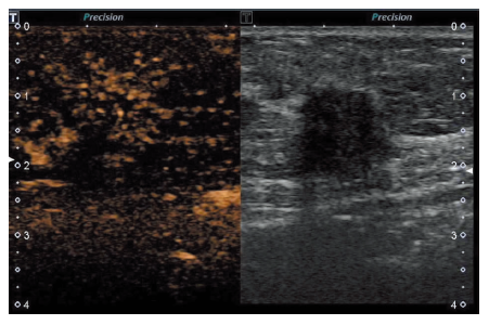
| 超声造影特征 | 最大直径≤ 2.0 cm(n=54) | 最大直径> 2.0 cm(n=53) | χ2值 | P值 |
|---|---|---|---|---|
| 增强时间 | 2.348 | 0.309 | ||
| 快进 | 33(61.1) | 39(73.6) | ||
| 等进 | 16(29.6) | 12(22.6) | ||
| 慢进 | 5(9.3) | 2(3.8) | ||
| 增强强度 | 15.222 | <0.001 | ||
| 低或等增强 | 23(42.6) | 5(9.4) | ||
| 高增强 | 31(57.4) | 48(90.6) | ||
| 对比剂分布 | 1.43 | 0.232 | ||
| 均匀 | 10(18.5) | 15(28.3) | ||
| 不均匀 | 44(81.5) | 38(71.7) | ||
| 增强后肿块形态 | 0.611 | 0.435 | ||
| 规则 | 18(33.3) | 14(26.4) | ||
| 不规则 | 36(66.7) | 39(73.6) | ||
| 增强后肿块边缘 | 0.202 | 0.653 | ||
| 清楚 | 11(20.4) | 9(17.0) | ||
| 模糊 | 43(79.6) | 44(83.0) | ||
| 增强顺序 | 3.302 | 0.069 | ||
| 向心性 | 44(81.5) | 35(66.0) | ||
| 弥漫性 | 10(18.5) | 18(34.0) | ||
| 增强后“蟹足征” | 0.082 | 0.775 | ||
| 无 | 29(53.7) | 27(50.9) | ||
| 有 | 25(46.3) | 26(49.1) | ||
| 增强后肿块周围穿支血管 | 6.81 | 0.009 | ||
| 无 | 34(63.0) | 20(37.7) | ||
| 有 | 20(37.0) | 33(62.3) | ||
| 增强后肿块内充盈缺损 | 16.133 | <0.001 | ||
| 无 | 49(90.7) | 30(56.6) | ||
| 有 | 5(9.3) | 23(43.4) | ||
| 增强后肿块增大 | 2.113 | 0.146 | ||
| 无 | 30(55.6) | 22(41.5) | ||
| 有 | 24(44.4) | 31(58.5) | ||
| 廓清时间 | 6.52 | 0.038 | ||
| 快出 | 12(22.2) | 12(22.6) | ||
| 等出 | 36(66.7) | 25(47.2) | ||
| 慢出 | 6(11.1) | 16(30.2) | ||


| [1] |
Siegel RL, Miller KD, Jemal A. Cancer statistics, 2020[J]. CA Cancer J Clin, 2020, 70(1):7-30.
doi: 10.3322/caac.21590 URL |
| [2] |
DeSantis CE, Ma J, Goding Sauer A, et al. Breast cancer statistics,2017, racial disparity in mortality by state[J]. CA Cancer J Clin, 2017, 67(6):439-448.
doi: 10.3322/caac.21412 URL |
| [3] | 余小琴, 姚兰辉, 于岚. 小乳腺癌超声直接及间接征象的诊断价值[J]. 中华超声影像学杂志, 2008, 17(10):879-882. |
| [4] | 汤兵辉, 肖秋金, 程淑珍. 二维超声联合弹性成像及三维超声对T1期乳腺癌的诊断价值[J]. 中国超声医学杂志, 2016, 32(11):973-976. |
| [5] |
Luo J, Chen JD, Chen Q, et al. Predictive model for contrast-enhanced ultrasound of the breast: is it feasible in malignant risk assessment of breast imaging reporting and data system 4 lesions?[J]. World J Radiol, 2016, 8(6):600-609.
doi: 10.4329/wjr.v8.i6.600 URL |
| [6] |
Janu E, Krikavova L, Little J, et al. Prospective evaluation of contrast-enhanced ultrasound of breast BI-RADS 3-5 lesions[J]. BMC Med Imaging, 2020, 20(1):66.
doi: 10.1186/s12880-020-00467-2 pmid: 32552678 |
| [7] | 沈若霞, 杨丽春, 罗晓茂, 等. 基于中国多中心研究数据的乳腺良恶性病灶超声造影定性特征的回顾性研究[J]. 中国医学影像学杂志, 2018, 26(12):885-889. |
| [8] |
Xiao X, Jiang Q, Wu H, et al. Diagnosis of sub-centimetre breast lesions: combining BI-RADS-US with strain elastography and contrast-enhanced ultrasound-a preliminary study in China[J]. Eur Radiol, 2017, 27(6):2443-2450.
doi: 10.1007/s00330-016-4628-4 URL |
| [9] |
Spak DA, Plaxco JS, Santiago L, et al. BI-RADS® fifth edition: a summary of changes[J]. Diagn Interv Imaging, 2017, 98(3):179-190.
doi: 10.1016/j.diii.2017.01.001 URL |
| [10] |
Adler DD, Carson PL, Rubin JM, et al. Doppler ultrasound color flow imaging in the study of breast cancer: preliminary findings[J]. Ultrasound Med Biol, 1990, 16(6):553-559.
pmid: 2238263 |
| [11] |
Gradishar WJ, Anderson BO, Abraham J, et al. Breast Cancer, Version 3.2020, NCCN Clinical Practice Guidelines in Oncology[J]. J Natl Compr Canc Netw, 2020, 18(4):452-478.
doi: 10.6004/jnccn.2020.0016 URL |
| [12] | 沈松杰, 孙强. 中国女性乳腺癌筛查现状及适宜模式探索[J]. 协和医学杂志, 2018, 9(4):298-302. |
| [13] |
Leng X, Huang G, Ma F, et al. Regional contrast-enhanced ultrasonography(CEUS) characteristics of breast cancer and correlation with microvessel density(MVD)[J]. Med Sci Monit, 2017, 23:3428-3436.
doi: 10.12659/MSM.901734 URL |
| [14] | 李静, 郭丽苹. 超声造影在乳腺癌中的临床应用进展[J]. 医学综述, 2018, 24(9):1817-1821. |
| [15] | 轩维锋, 徐晓红, 张建兴, 等. 乳腺超声与病理诊断[M]. 北京: 科学技术文献出版社, 2019:9-11. |
| [16] | 冷晓玲, 黄国福, 马富成. 乳腺癌病灶大小与超声造影表现的相关性[J]. 中华超声影像学杂志, 2015, 24(4):324-327. |
| [17] |
Golbabapour S, Pang WW, George J, et al. Chemically induced breast tumors in rats are detectable in early stages by contrast enhanced magnetic resonance imaging but not by changes in the acute-phase reactants in serum[J]. Int J Mol Sci, 2011, 12(2):1030-1040.
doi: 10.3390/ijms12021030 pmid: 21541040 |
| [18] | Cichon MA, Degnim AC, Visscher DW, et al. Microenvironmental influences that drive progression from benign breast disease to invasive breast cancer[J]. J Mammary Gland Boil Neoplasia, 2010, 15(4):389-397. |
| [19] |
Suzuki N, Shiota T, Watanabe F, et al. Discovery of novel 5-alkynyl-4-anilinopyrimidines as potent, orally active dual inhibitors of EGFR and Her-2 tyrosine kinases[J]. Bioorg Med Chem Lett, 2012, 22(1):456-460.
doi: 10.1016/j.bmcl.2011.10.103 URL |
| [20] | 高军喜, 王雅婷, 杨磊, 等. 乳腺癌超声造影特征及边缘带定量参数与生物学预后因子相关性研究[J]. 中国超声医学杂志, 2019, 35(4):306-309. |
| [21] | 赵璐, 张莹, 程颢, 等. 乳腺超声造影预测模型的建立及其对乳腺良恶性病变诊断效能的分析[J]. 中华医学超声杂志(电子版), 2019, 16(6):419-425. |
| [1] | FENG Meijing, REN Xinping. Application of contrast-enhanced ultrasound in diagnosis of gallbladder protrusion lesions [J]. Journal of Diagnostics Concepts & Practice, 2023, 22(01): 68-74. |
| [2] |
ZHU Fang, XU Zhe, WANG Xianming , et al.
Analysis of 50 cases of breast reconstruction with expanded latissimus dorsi musculocutaneous flap immediately after radical mastectomy [J]. Journal of Tissue Engineering and Reconstructive Surgery, 2022, 18(5): 377-. |
| [3] | REN Xinping, LI Junjian, ZHANG Jie, ZHAN Weiwei. Advances in the application of contrast-enhanced ultrasound in the diagnosis and treatment of focal liver lesions [J]. Journal of Diagnostics Concepts & Practice, 2022, 21(06): 684-690. |
| [4] | ZHANG Daojian, ZHANG Dexiang, WANG Jiwen, LU Pinxiang, LIU Houbao, LIU Han. Contrast-enhanced ultrasound in differential diagnosis of gallbladder cancer from xanthogranulomatous cholecystitis [J]. Journal of Surgery Concepts & Practice, 2020, 25(04): 322-325. |
| [5] | JI Ri, ZHOU Chun, ZHAN Weiwei, YANG Zhifang, GUO Wenjia. Evaluation of foot microcirculation by contrast-enhanced ultrasound in elderly male type 2 DM and IGT patients [J]. Journal of Diagnostics Concepts & Practice, 2017, 16(03): 287-291. |
| [6] | . [J]. Journal of Surgery Concepts & Practice, 2015, 20(02): 166-169. |
| [7] | MA Wen-jun1 (马文军), HONG Rong-rong2 (洪荣荣), YE Shao-zhen2 (叶少珍), YANG Yue3 (杨月),LI Yue-hua3 (李跃华), CHEN Li4, ZHANG Su1* (张素). Lesion Segmentation and Identification of Breast Tumor on Dynamic Contrast-Enhanced Magnetic Resonance Imaging [J]. Journal of shanghai Jiaotong University (Science), 2014, 19(5): 630-635. |
| [8] | . [J]. Journal of Diagnostics Concepts & Practice, 2009, 8(05): 515-519. |
| [9] | . [J]. Journal of Diagnostics Concepts & Practice, 2008, 7(03): 278-282. |
| [10] | . [J]. Journal of Diagnostics Concepts & Practice, 2007, 6(06): 533-535. |
| [11] | . [J]. Journal of Surgery Concepts & Practice, 2006, 11(06): 507-509. |
| [12] | . [J]. Journal of Surgery Concepts & Practice, 2006, 11(02): 139-141. |
| [13] | . [J]. Journal of Surgery Concepts & Practice, 2004, 9(04): 314-317. |
| Viewed | ||||||
|
Full text |
|
|||||
|
Abstract |
|
|||||