诊断学理论与实践 ›› 2019, Vol. 18 ›› Issue (03): 340-343.doi: 10.16150/j.1671-2870.2019.03.018

• 论著 • 上一篇    下一篇

双能量CT评价痛风性关节炎骨髓水肿的可行性研究

朱思奇, 吴梦雄(), 秦乐, 董海鹏   

  1. 上海交通大学医学院附属瑞金医院放射科,上海 200025
  • 收稿日期:2019-03-20 出版日期:2019-06-25 发布日期:2019-06-25
  • 通讯作者: 吴梦雄 E-mail:mengxiongwu@live.cn

Feasibility of dual-energy CT in evaluation of marrow edema of gouty arthritis

ZHU Siqi, WU Mengxiong(), QIN Le, DONG Haipeng   

  1. Department of Radiology, Ruijin Hospital, Shanghai Jiao Tong University School of Medicine, Shanghai 200025, China
  • Received:2019-03-20 Online:2019-06-25 Published:2019-06-25
  • Contact: WU Mengxiong E-mail:mengxiongwu@live.cn

摘要:

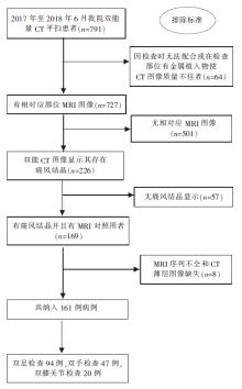
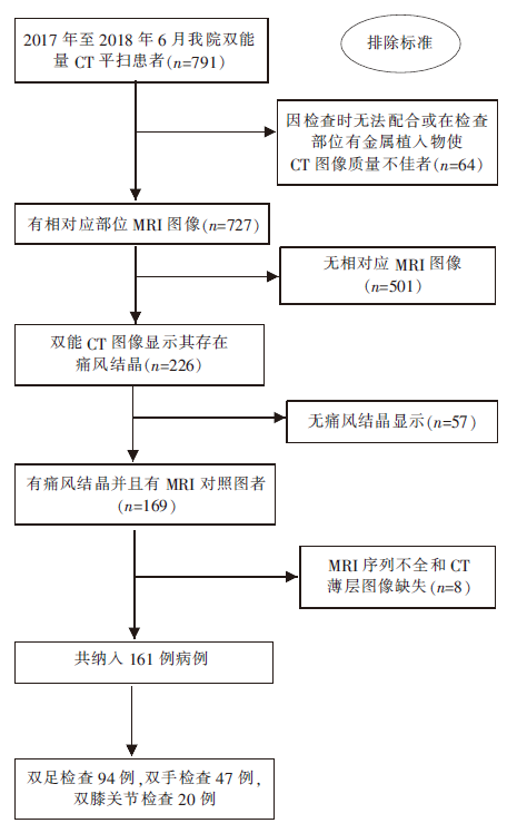
目的: 探讨采用双能量CT检查评价痛风性关节炎患者的骨髓水肿的可行性。方法: 回顾性分析2017 年 1月至2018 年 6月,在本院进行双能量CT平扫和MRI检查的161例痛风患者。所有患者进行双能量CT检查时均采用Sn140 kVp和80 kVp的管电压,并将图像传输至Syngo.via(Siemens,Germany)后处理工作站骨髓水肿模块进行处理,以评估是否有骨髓水肿及骨髓水肿区域的双能量虚拟去钙化(double energy-virtual non-calcium, DE-VNC)CT值,同时并分析同一例患者其相应部位的MRI图像, 以T1加权像(T1 weighted image, T1WI)低信号、T2加权像(T2 weighted image, T2WI)和短反转时间反转恢复序列(short T1 inversion recovery,STIR)高信号作为诊断骨髓水肿的金标准,来并明确其范围和程度。结果: 在Syngo.via后处理工作站骨髓水肿模块下,骨髓水肿部位CT值[(-5.32±8.19) HU]与邻近非骨髓水肿部位CT值[(-60.55±12.50) HU]及远处非骨髓水肿部位CT值[(-64.59±12.71) HU]间比较,差异均有统计学意义(P<0.05)。以其MRI诊断是否有骨髓水肿为金标准,双能量CT诊断痛风性关节炎患者骨髓水肿的灵敏度为82.2%,特异度为90.7%,阳性预测值为86%,阴性预测值为88.1%,其中12例骨质破坏广泛者被漏诊。结论: 双能量CT在痛风性关节炎患者的骨髓水肿诊断中也具有较高的价值,但当患者骨质破坏明显时可能出现漏诊。

关键词: 双能量CT, 痛风, 骨髓水肿

Abstract:

Objective: To investigate the feasibility of dual-energy CT in the evaluation of marrow edema of gouty arthritis. Methods: One hundred and sixty-one patients who had undergone plain dual-energy CT and diagnosed as gout between January 2017 and June 2018 were retrospectively analyzed. All examinations were performed on dual-source CT (Somaton Definition Flash, Siemens, Germany) with tube voltage of Sn140 kVp and 80 kVp. Then the images were transferred to Syngo. via (Siemens, Germany) postprocessing workstation for further analysis with 'marrow edema' module to diagnose marrow edema and acquire CT values on (double energy-virtual non-calcium, DE-VNC) images. MRI images of corresponding sites were also analyzed by the sequence of T1WI (T1 weighted imaging), T2WI (T2 weighted imaging) and STIR (short T1 inversion recovery). Results: After being analyzed by Syngo.via postprocessing workstation, it had been demonstrated that CT values between sites with marrow edema, sites adjacent to marrow edema and sites distant to marrow edema had significant differences (P<0.05). Based on the golden standard of MRI images, the diagnostic sensitivity, specificity, positive predictive value and negative predictive value of dual-energy CT was 82.2%, 90.7%, 86% and 88.1%, respectively. Twelve missed cases was due to widespread bone destruction. Conclusions: Not only could dual-energy CT detect gouts, it also has good diagnostic accuracy in the evaluation of marrow edema of gouty arthritis. Cases with widespread bone destruction may be missed.

Key words: Dual-energy CT, Gout, Marrow edema

中图分类号: