诊断学理论与实践 ›› 2020, Vol. 19 ›› Issue (05): 494-498.doi: 10.16150/j.1671-2870.2020.05.009

• 论著 • 上一篇    下一篇

非对比剂增强磁共振血管成像流入反转恢复序列在肝硬化门静脉高压患者门静脉系统成像的可行性研究

王丽娟, 潘自来(), 苏文婷, 徐敬慈, 饶敏, 刘宵   

  1. 上海交通大学医学院附属瑞金医院北院放射科,上海 201801
  • 收稿日期:2020-02-01 出版日期:2020-10-25 发布日期:2022-07-14
  • 通讯作者: 潘自来 E-mail:zilaipanlilly@163.com
  • 基金资助:
    上海市卫生和计划生育委员会重点课题(201640022)

Feasibility study on the portal vein imaging using non-contrast-enhanced magnetic resonance angiography with flow inversion recovery sequence in the cirrhotic portal hypertension

WANG Lijuan, PAN Zilai(), SU Wenting, XU Jingci, RAO Min, LIU Xiao   

  1. Department of Radiology, Ruijin Hospital North, Shanghai Jiao Tong University School of Medicine, Shanghai 201801, China
  • Received:2020-02-01 Online:2020-10-25 Published:2022-07-14
  • Contact: PAN Zilai E-mail:zilaipanlilly@163.com

摘要:

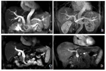
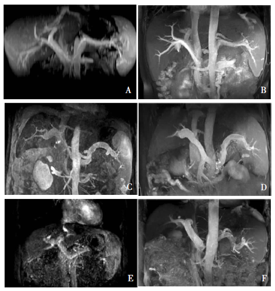
目的: 比较非对比剂增强磁共振血管成像(non-contrast-enhanced magnetic resonance angiography, NC-MRA)与对比剂增强磁共振血管成像(contrast enhancement magnetic resonance angiography,CE-MRA)在肝硬化门静脉高压患者门静脉系统检查中的应用价值。方法: 收集经临床或影像学诊断为肝硬化门静脉高压的患者20例,对其门静脉系统分别进行CE-MRA(采用快速梯度回波三维冠状位成像)检查及NC-MRA[采用流入反转恢复(flow inversion recovery,FIR)序列]检查,比较2种检查方法对门静脉、脾静脉及肠系膜上静脉显示情况,并计算图像信噪比。结果: 2种方法间显示的门静脉主干宽径、脾静脉主干宽径差异无统计学意义(P值分别为0.330、0.090);肠系膜上静脉近端及脾静脉的图像评分间差异无统计学意义(P=0.677,P=0.077)。NC-MRA图像显示的门静脉及1、2级分支的管壁清晰度和光滑度上优于CE-MRA,但门静脉图像评分在3分及以上例数少于CE-MRA组(分别为9例和17例),差异有统计学意义(P=0.008),提示CE-MRA对较远端分支的显示方面较NC-MRA有优势。NC-MRA图像的门静脉信号强度及信噪比高于CE-MRA组(P=0.040,P<0.01)。结论: NC-MRA采用FIR序列能清晰显示门静脉高压患者门静脉系统主要血管的效能与CE-MRA相似,虽然其显示较远端分支的能力比CE-MRA低,但其具有无创、安全的特点。FIR序列可作为临床门静脉系统NC-MRA检查序列的选择之一。

关键词: 肝硬化, 门静脉高压, 非对比剂增强磁共振血管成像

Abstract:

Objective: To compare the efficacy of the non-contrast-enhanced magnetic resonance angiography (NC-MRA) with flow inversion recovery (FIR) sequence and contrast enhancement MRA(CE-MRA) on portal vein imaging in the cirrhotic portal hypertension. Methods: Twenty patients diagnosed as the cirrhotic portal hypertension by the clinical criteria or radiology were enrolled, and CE-MRA with fast field echo 3D coronal imaging and NC-MRA with FIR were performed to display the portal vein system, respectively. The imaging quality of the portal vein, splenic vein and superior mesenteric vein in the portal vein system on NC-MRA was compared with CE-MRA, and the signal-to-noise ratios of images the were calculated. Results: The width of the main portal vein and splenic vein measured by NC-MRA was not different from CE-MRA(P value was 0.330 and 0.090, respectively), and the scores of the imaging quality in splenic vein and proximal superior mesenteric vein had no difference either(P=0.677, P=0.077). The imaging quality showed the clarity and the smooth degree in the portal vein and the branches of grade 1-2 on NC-MRA was superior to CE-MRA. However, the cases with image quality score ≥3 detected by NC-MRA were less than CE-MRA (9 vs 17, P=0.008), indicating CE-MRA had advantage in displaying distal veins over NC-MRA. The signal intensity and signal-to-noise ratios on NC-MRA were higher than CE-MRA(P=0.040, P<0.01). Conclusions: The NC-MRA with FIR sequence could clearly display the main blood vessels of the portal vein system in patients with the portal hypertension and shows similar efficacy as CE-MRA. NC-MRA with FIR sequence could be used as a safe and invasive method for displaying the portal vein system in spite of the lower efficacy on displaying distal veins.

Key words: Liver cirrhosis, Portal hypertension, Non-contrast-enhanced magnetic resonance

中图分类号: