诊断学理论与实践 ›› 2021, Vol. 20 ›› Issue (04): 362-367.doi: 10.16150/j.1671-2870.2021.04.006
黄少华, 梁宗辉, 童欢, 管雪妮, 郭瑛, 张雁, 曹宾, 孙育民( )
)
收稿日期:2021-03-20
出版日期:2021-08-25
发布日期:2022-06-28
通讯作者:
孙育民
E-mail:munaiyisun@163.com
基金资助:
HUANG Shaohua, LIANG Zonghui, TONG Huan, GUAN Xueni, GUO Ying, ZHANG Yan, CAO Bin, SUN Yumin()
Received:2021-03-20
Online:2021-08-25
Published:2022-06-28
Contact:
SUN Yumin
E-mail:munaiyisun@163.com
摘要:
目的:对强直性肌营养不良1型(myotonic dystrophy type 1,DM1)患者进行心脏磁共振(cardiac magnetic resonance,CMR)检查,观察其心肌纤维化的发生情况,分析CMR及心电图检查在DM1患者心脏风险评估中的价值。 方法:收集14例经基因明确诊断的DM1患者,予同期行CMR、心电图、动态心电图检查。根据其心电图或动态心电图是否发现异常,将患者分为心电图正常组和心电图异常组,以CMR检查中观察到钆剂延迟强化(late gadolinium enhancement,LGE)作为诊断标准,判断其是否存在心肌纤维化,并比较心电图正常组与异常组间的心肌纤维化发生情况。 结果:14例DM1患者经CMR检查,其中有5例被检出心肌纤维化,检出率为5/14。与无心肌纤维化的患者相比,存在心肌纤维化的DM1患者的左心室质量指数[(47.1±5.4) g/m2比(40.2±3.4) g/m2,P=0.012]、左室收缩末期容积指数[(31.L5±5.5) mL/m2比(25.8±2.8) mL/m2,P=0.024]、左房容积指数[(43.8±7.1)mL/m2比(34.3±7.4) mL/m2,P=0.037]均较高,而左室射血分数(52.2%±11.1%比63.9%±5.3%,P=0.019)较低,心功能明显下降。心电图正常组(5例)与异常组(9例)间比较, CMR检出异常率差异有统计学意义(7/9比1/5,P=0.036),但心肌纤维化发生率差异无统计学意义(4/9比1/5,P=0.36)。 结论:DM1可累及心肌导致心肌纤维化,且常规心电图筛查结果正常的患者并不能完全排除心肌纤维化,需要加行CMR检查以明确其是否存在心肌纤维化。
中图分类号:
黄少华, 梁宗辉, 童欢, 管雪妮, 郭瑛, 张雁, 曹宾, 孙育民. 心脏磁共振评估强直性肌营养不良1型患者心肌纤维化的临床价值[J]. 诊断学理论与实践, 2021, 20(04): 362-367.
HUANG Shaohua, LIANG Zonghui, TONG Huan, GUAN Xueni, GUO Ying, ZHANG Yan, CAO Bin, SUN Yumin. Evaluation of myocardial fibrosis by cardiac magnetic resonance imaging in patient with myotonic dystrophy type 1[J]. Journal of Diagnostics Concepts & Practice, 2021, 20(04): 362-367.
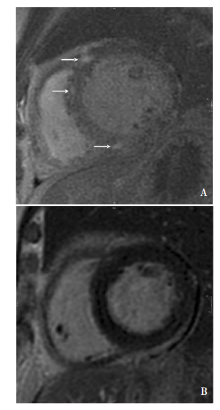
图1
心脏短轴切面LGE图像 A:室间隔LGE阳性(箭头),提示存在心肌纤维化;B:未见LGE阳性表现,提示无心肌纤维化。

表1
LGE阳性与LGE阴性患者的基线资料及CMR、心电图结果比较
| 参数 | LGE阳性 (n=5) | LGE阴性 (n=9) | P值 |
|---|---|---|---|
| 年龄 (岁) | 48.4±14.8 | 40.4±11.1 | 0.270 |
| 男/女 | 4/1 | 5/4 | 0.360 |
| CMR检查 | |||
| LVEF(%) | 52.2±11.1 | 63.9±5.3 | 0.019 |
| LGE成像质量(g) | 4.2±2.2 | 0 | 0.014 |
| 左室舒张末期容积指数(mL/m2) | 80.4±25.2 | 73.3±16.8 | 0.540 |
| 左室收缩末期容积指数(mL/m2) | 31.5±5.5 | 25.8±2.8 | 0.024 |
| 左室质量指数(g/m2) | 47.1±5.4 | 40.2±3.4 | 0.012 |
| 左房容积指数(mL/m2) | 43.8±7.1 | 34.3±7.4 | 0.037 |
| 心电图异常 | 4 | 5 | 0.360 |
表2
异常心电图组与正常心电图组CMR结果比较
| 检查 | 心电图异常组(n=9) | 心电图正常组(n=5) | P值 |
|---|---|---|---|
| CMR结果异常[n(%)] | 7 | 1 | 0.036 |
| LVEF (%) | 58.0±11.3 | 62.8±4.0 | 0.380 |
| 左室舒张末期容积指数(mL/m2) | 76.7±21.2 | 65.6±18.1 | 0.350 |
| 左室收缩末期容积指数(mL/m2) | 28.2±5.6 | 27.3±2.8 | 0.760 |
| 左室质量指数(g/m2) | 43.4±6.0 | 41.6±4.6 | 0.570 |
| 左房容积指数(mL/m2) | 37.1±9.3 | 38.6±7.7 | 0.760 |
| LGE阳性 | 4 | 1 | 0.360 |
| [1] | 王新德. 神经病学:肌肉疾病[M]. 北京: 人民军医出版社, 2007:16-637. |
| [2] |
Turner C, Hilton-Jones D. The myotonic dystrophies: dia-gnosis and management[J]. J Neurol Neurosurg Psychiatry, 2010,81(4):358-367.
doi: 10.1136/jnnp.2008.158261 pmid: 20176601 |
| [3] |
Brook JD, McCurrach ME, Harley HG, et al. Molecular basis of myotonic dystrophy: expansion of a trinucleotide (CTG) repeat at the 3′ end of a transcript encoding a protein kinase family member[J]. Cell, 1992,69(2):385.
doi: 10.1016/0092-8674(92)90418-c pmid: 1568252 |
| [4] | Hermans MC, Pinto YM, Merkies IS, et al. Hereditary muscular dystrophies and the heart[J]. Neuromuscul Di-sord, 2010,20(8):479-492. |
| [5] |
Chaudhry SP, Frishman WH. Myotonic dystrophies and the heart[J]. Cardiol Rev, 2012,20(1):1-3.
doi: 10.1097/CRD.0b013e31821950f9 URL |
| [6] |
Kawel-Boehm N, Maceira A, Valsangiacomo-Buechel ER, et al. Normal values for cardiovascular magnetic resonance in adults and children[J]. J Cardiovasc Magn Reson, 2015,17(1):29.
doi: 10.1186/s12968-015-0111-7 URL |
| [7] |
Verhaert D, Richards K, Rafael-Fortney JA, et al. Cardiac involvement in patients with muscular dystrophies: magnetic resonance imaging phenotype and genotypic considerations[J]. Circ Cardiovasc Imaging, 2011,4(1):67-76.
doi: 10.1161/CIRCIMAGING.110.960740 pmid: 21245364 |
| [8] |
Turkbey EB, Gai N, Lima JA, et al. Assessment of cardiac involvement in myotonic muscular dystrophy by T1 mapping on magnetic resonance imaging[J]. Heart Rhythm, 2012,9(10):1691-1697.
doi: 10.1016/j.hrthm.2012.06.032 pmid: 22710483 |
| [9] |
Choudhary P, Nandakumar R, Greig H, et al. Structural and electrical cardiac abnormalities are prevalent in asymptomatic adults with myotonic dystrophy[J]. Heart, 2016,102(18):1472-1478.
doi: 10.1136/heartjnl-2015-308517 pmid: 27164920 |
| [10] |
Luetkens JA, von Landenberg C, Isaak A, et al. Comprehensive cardiac magnetic resonance for assessment of cardiac involvement in myotonic muscular dystrophy type 1 and 2 without known cardiovascular disease[J]. Circ Cardiovasc Imaging, 2019,12(6):e009100.
doi: 10.1161/CIRCIMAGING.119.009100 URL |
| [11] |
Petri H, Ahtarovski KA, Vejlstrup N, et al. Myocardial fibrosis in patients with myotonic dystrophy type 1: a cardiovascular magnetic resonance study[J]. J Cardiovasc Magn Reson, 2014,16(1):59.
doi: 10.1186/s12968-014-0059-z URL |
| [12] |
Cardona A, Arnold WD, Kissel JT, et al. Myocardial fibrosis by late gadolinium enhancement cardiovascular magnetic resonance in myotonic muscular dystrophy type 1: highly prevalent but not associated with surface conduction abnormality[J]. J Cardiovasc Magn Reson, 2019,21(1):26.
doi: 10.1186/s12968-019-0535-6 URL |
| [13] |
Hermans MC, Faber CG, Bekkers SC, et al. Structural and functional cardiac changes in myotonic dystrophy type 1: a cardiovascular magnetic resonance study[J]. J Cardiovasc Magn Reson, 2012,14(1):48.
doi: 10.1186/1532-429X-14-48 URL |
| [14] |
de Ambroggi L, Raisaro A, Marchianó V, et al. Cardiac involvement in patients with myotonic dystrophy: characteristic features of magnetic resonance imaging[J]. Eur Heart J, 1995,16(7):1007-1010.
pmid: 7498193 |
| [15] |
Nazarian S, Bluemke DA, Wagner KR, et al. QRS prolongation in myotonic muscular dystrophy and diffuse fibrosis on cardiac magnetic resonance[J]. Magn Reson Med, 2010,64(1):107-114.
doi: 10.1002/mrm.22417 pmid: 20572151 |
| [16] |
Chmielewski L, Bietenbeck M, Patrascu A, et al. Non-invasive evaluation of the relationship between electrical and structural cardiac abnormalities in patients with myo-tonic dystrophy type 1[J]. Clin Res Cardiol, 2019,108(8):857-867.
doi: 10.1007/s00392-019-01414-0 pmid: 30767060 |
| [17] |
Lau JK, Sy RW, Corbett A, et al. Myotonic dystrophy and the heart: a systematic review of evaluation and management[J]. Int J Cardiol, 2015,184:600-608.
doi: 10.1016/j.ijcard.2015.03.069 pmid: 25769007 |
| [18] |
Groh WJ, Groh MR, Saha C, et al. Electrocardiographic abnormalities and sudden death in myotonic dystrophy type 1[J]. N Engl J Med, 2008,358(25):2688-2697.
doi: 10.1056/NEJMoa062800 URL |
| [19] |
Segawa I, Suzuki T, Kato M, et al. Relation between myo-cardial histological changes and ventricular tachycardia in cardiomyopathy: a study by 24-hour ECG-monitoring and endomyocardial biopsy[J]. Heart Vessels Suppl, 1990,5:37-40.
pmid: 2093710 |
| [20] |
Shiozaki AA, Senra T, Arteaga E, et al. Myocardial fibrosis detected by cardiac CT predicts ventricular fibrillation/ventricular tachycardia events in patients with hypertrophic cardiomyopathy[J]. J Cardiovasc Comput Tomogr, 2013,7(3):173-181.
doi: 10.1016/j.jcct.2013.04.002 pmid: 23849490 |
| [21] |
Nguyen HH, Wolfe JT 3rd, Holmes DR Jr, et al. Pathology of the cardiac conduction system in myotonic dystrophy: a study of 12 cases[J]. J Am Coll Cardiol, 1988,11(3):662-671.
pmid: 3278037 |
| [1] | 刘鹏, 严福华, 秦乐, 肖瑞杰. 肥厚型心肌病左室舒张功能的心脏磁共振心肌应变率参数与猝死风险关系的研究[J]. 诊断学理论与实践, 2022, 21(03): 317-325. |
| [2] | 陈瑞珍, 解玉泉,. 病毒性心肌炎向扩张型心肌病演变的免疫学机制[J]. 诊断学理论与实践, 2011, 10(05): 414-417. |
| 阅读次数 | ||||||
|
全文 |
|
|||||
|
摘要 |
|
|||||