诊断学理论与实践 ›› 2022, Vol. 21 ›› Issue (05): 575-580.doi: 10.16150/j.1671-2870.2022.05.005
刁雪红a, 申艳b, 陈林a( ), 詹嘉a, 方靓a, 蔡剑飞b, 陈悦a
收稿日期:2021-08-20
出版日期:2022-10-25
发布日期:2023-01-29
通讯作者:
陈林
E-mail:cl_point@126.com
基金资助:
DIAO Xuehonga, SHEN Yanb, CHEN Lina(), ZHAN Jiaa, FANG Lianga, CAI Jianfeib, CHEN Yuea
Received:2021-08-20
Online:2022-10-25
Published:2023-01-29
Contact:
CHEN Lin
E-mail:cl_point@126.com
摘要:
目的:观察超声微血流成像(superb mircovascular imaging,SMI)技术对类风湿性关节炎(rheumatoid arthritis,RA)临床缓解期患者滑膜细小血管的显示情况。方法:应用SMI观察42例临床缓解期RA患者滑膜血流的量和滑膜血流分级,以超声造影(contrast enhanced ultrasound, CEUS)血流模式为对照,比较这2种超声检查间的差异,并分析2种检查评估的血流分级与炎症指标红细胞沉降率(erythrocyte sedimentation rate,ESR)、C-反应蛋白(C reactive protein,CRP)间的相关性。结果:SMI及CEUS检查对临床缓解期RA患者滑膜血流的检出率(血流分级1级以上者)分别为73.8%、83.3%,两者对滑膜血流的检出率差异无统计学意义(P=0.160),两者间一致性较强(Kappa=0.723,P<0.001);两者对滑膜血管的血流分级结果差异也无统计学意义(P=0.083),呈中等一致性(Kappa=0.654,P<0.001)。患者接受进一步强化治疗后,SMI检查发现的滑膜血流数均较强化治疗前明显减少(P<0.001)。亚临床滑膜炎患者的SMI血流半定量评分与炎性指标CRP、ESR间也无相关性,这种结果均与CEUS表现一致。结论:SMI检查可灵敏地探测到滑膜的微小血管,发现炎症指标不升高的局部炎症,评价效能与CEUS一致,相对CEUS,其应用简便,可用于RA缓解期病情监测。
中图分类号:
刁雪红, 申艳, 陈林, 詹嘉, 方靓, 蔡剑飞, 陈悦. 超声微血流成像技术在临床缓解期类风湿性关节炎诊断中的应用[J]. 诊断学理论与实践, 2022, 21(05): 575-580.
DIAO Xuehong, SHEN Yan, CHEN Lin, ZHAN Jia, FANG Liang, CAI Jianfei, CHEN Yue. Application of superb microvascular imaging technology in diagnosing rheumatoid arthritis in the clinical remission stage[J]. Journal of Diagnostics Concepts & Practice, 2022, 21(05): 575-580.
表1
SMI和CEUS模式对RA临床缓解期患者滑膜血流分级的比较[例(n)]
| 超声血流模式 | 例数(n) | 滑膜血流分级 | t值 | P值 | |||
|---|---|---|---|---|---|---|---|
| 0级 | 1级 | 2级 | 3级 | ||||
| SMI | 42 | 8 | 20 | 10 | 4 | 1.776 | 0.083 |
| CEUS | 42 | 7 | 18 | 11 | 6 | ||
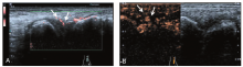
图1
SMI和CEUS对腕关节滑膜血流显示基本一致 A:SMI可以看到超过滑膜面积50%血流信号(1级),血流显示以分枝状为主;B:CEUS可以观察到少量滑膜有对比剂显影(1级),血流显示呈团块状增强。
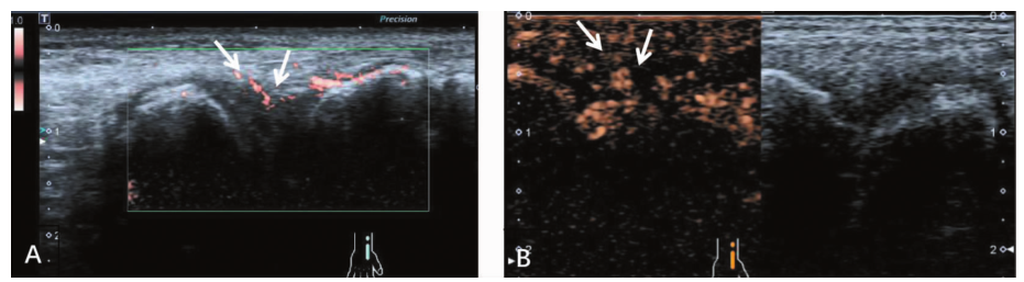
图2
SMI和CEUS对掌指关节滑膜血流显示一致 A:SMI显示短条状血流信号(1级);B:CEUS观察到短条状造影增强(1级)。

| [1] |
Sun C, Qi X, Yang Y, et al. Importance of baseline musculoskeletal ultrasound findings in the prognosis of rheumatoid arthritis[J]. Clin Rheumatol, 2022, 41(3):847-857.
doi: 10.1007/s10067-021-06017-7 URL |
| [2] |
Iagnocco A, Finucci A, Ceccarelli F, et al. Power Doppler ultrasound monitoring of response to anti-tumour necrosis factor alpha treatment in patients with rheumatoid arthritis[J]. Rheumatology (Oxford), 2015, 54(10):1890-1896.
doi: 10.1093/rheumatology/kev211 pmid: 26070937 |
| [3] |
Diao X, Zhan J, Chen L, et al. Role of superb microvascular imaging in differentiating between malignant and benign solid breast masses[J]. Clin Breast Cancer, 2020, 20(6):e786-e793.
doi: 10.1016/j.clbc.2020.06.009 pmid: 32863154 |
| [4] | 李伟伟, 吴迎, 周伟, 等. 超微血管三维立体成像技术对 BI-RADS 4 类乳腺肿块血流显示及鉴别良恶性的价值研究[J]. 诊断学理论与实践 2020, 6(19):583-587. |
| Li WW, Wu Y, Zhou W, et al. The diagnostic value of smart three-dimensional superb microvascular imaging in differentiating benign and malignant breast lesions of BI-RADS 4[J]. Journal of Diagnostics Concepts & Practice, 2020, 19(6):583-587. | |
| [5] |
Santos JA, Tee CA, Galsim RJG, et al. Musculoskeletal ultrasound using superb microvascular imaging documents treatment response to biosimilar infliximab in rheumatoid arthritis[J]. BMJ Case Rep, 2021, 14(2):e239112.
doi: 10.1136/bcr-2020-239112 URL |
| [6] |
Lee GY, Kim S, Choi ST, et al. The superb microvascular imaging is more sensitive than conventional power Doppler imaging in detection of active synovitis in patients with rheumatoid arthritis[J]. Clin Rheumatol, 2019, 38(9):2613-2620.
doi: 10.1007/s10067-019-04550-0 pmid: 31030360 |
| [7] |
Yu X, Li Z, Ren M, et al. Superb microvascular imaging (SMI) for evaluating hand joint lesions in patients with rheumatoid arthritis in clinical remission[J]. Rheumatol Int, 2018, 38(10):1885-1890.
doi: 10.1007/s00296-018-4112-3 pmid: 30062435 |
| [8] | 赵迎, 骈林萍, 杜明瑞, 等. 超声造影在类风湿性关节炎缓解期腕关节亚临床滑膜炎评估中的应用[J]. 河南医学研究, 2021, 30(14):2649-2651. |
| Zhao Y, Pian LP, Du RM, et al. Role of contrast-enhanced ultrasound in evaluation of subclinical synovitis of wrist joint in remission of rheumatoid arthritis[J]. Henan Med Res, 2021, 30(14):2649-2651. | |
| [9] |
Fakhfakh R, Elamri N, Baccouche K, et al. Ultrasound remission in patients with rheumatoid arthritis in clinical remission[J]. Reumatologia, 2021, 59(6):378-385.
doi: 10.5114/reum.2021.112237 URL |
| [10] |
Paulshus Sundlisæter N, Aga AB, Olsen IC, et al. Clinical and ultrasound remission after 6 months of treat-to-target therapy in early rheumatoid arthritis: associations to future good radiographic and physical outcomes[J]. Ann Rheum Dis, 2018, 77(10):1421-1425.
doi: 10.1136/annrheumdis-2017-212830 pmid: 29934373 |
| [11] |
Sconfienza LM, Silvestri E, Cimmino MA. High-resolution ultrasound evaluation of extrinsic wrist ligaments in patients affected by rheumatoid arthritis[J]. Eur Radiol, 2012, 22(7):1586-1591.
doi: 10.1007/s00330-012-2402-9 pmid: 22367473 |
| [12] |
Cohen G, Gossec L, Dougados M, et al. Radiological damage in patients with rheumatoid arthritis on sustained remission[J]. Ann Rheum Dis, 2007, 66(3):358-363.
doi: 10.1136/ard.2006.057497 pmid: 16935911 |
| [13] |
Bresnihan B, Kane D. Sonography and subclinical synovitis[J]. Ann Rheum Dis, 2004, 63(4):333-334.
pmid: 15020323 |
| [14] |
Saleem B, Brown AK, Keen H, et al. Should imaging be a component of rheumatoid arthritis remission criteria? A comparison between traditional and modified composite remission scores and imaging assessments[J]. Ann Rheum Dis, 2011, 70(5):792-798.
doi: 10.1136/ard.2010.134445 pmid: 21242236 |
| [15] |
Yu X, Li Z, Ren M, et al. Superb microvascular imaging (SMI) for evaluating hand joint lesions in patients with rheumatoid arthritis in clinical remission[J]. Rheumatol Int, 2018, 38(10):1885-1890.
doi: 10.1007/s00296-018-4112-3 pmid: 30062435 |
| [1] | 姚南, 杨丽春, 李支尧. 肾脏混合性上皮和间质肿瘤超声造影一例报告[J]. 诊断学理论与实践, 2020, 19(06): 610-612. |
| [2] | 何碧媛, 周毓青. 三维超声、超声造影及超声弹性成像在妇科疾病诊断中的应用进展及策略[J]. 诊断学理论与实践, 2020, 19(06): 626-629. |
| [3] | 周伟, 侯怡卿, 詹维伟. 超声造影及超声弹性成像在良恶性甲状腺结节鉴别诊断中的应用进展[J]. 诊断学理论与实践, 2020, 19(04): 344-349. |
| [4] | 卢叶君, 陈卉, 张剑, 徐斌, 王冲, 贺烨. 超微血管成像、超声弹性成像联合高频超声在微小乳腺癌中的诊断价值及相关高危超声特征的筛选[J]. 诊断学理论与实践, 2020, 19(04): 391-396. |
| [5] | 杨珉珉, 刘敏, 陈艳, 何甦晖, 郑丽雅. 经阴道四维子宫输卵管超声造影评价输卵管通畅性诊断效能的观察及误诊分析[J]. 诊断学理论与实践, 2018, 17(02): 202-206. |
| [6] | 吉日, 周春, 詹维伟, 杨志芳, 郭文佳. 超声造影评估老年男性2型糖尿病及糖耐量减低患者足部微循环的改变[J]. 诊断学理论与实践, 2017, 16(03): 287-291. |
| [7] | 王怡, 唐蕾, 詹维伟, 陈曼,. 超声造影在兔肝纤维化实质灌注的定量评估[J]. 诊断学理论与实践, 2013, 12(04): 470-473. |
| [8] | 周春, 周建桥, 詹维伟, 陈林, 戴军, 刘振华, 周伟, 柳俊, 董屹婕,. 彩色多普勒超声和超声造影在青春后期及成人睾丸扭转诊断中的价值[J]. 诊断学理论与实践, 2013, 12(03): 343-346. |
| [9] | 唐蕾, 王怡, 詹维伟, 陈曼,. 肝纤维化灰阶超声造影的临床应用评估[J]. 诊断学理论与实践, 2013, 12(02): 224-227. |
| [10] | 陈林, 陈悦, 詹维伟,. 多种超声技术在胰腺内分泌肿瘤中的应用进展[J]. 诊断学理论与实践, 2011, 10(04): 362-366. |
| [11] | 周萍, 周伟, 周建桥, 詹维伟,. 甲状腺乳头状癌的灰阶超声造影特征与颈部淋巴结转移的关系[J]. 诊断学理论与实践, 2011, 10(01): 45-49. |
| [12] | 缪怡, 徐雪亮, 黄秋愉, 胡朝英, 钱柳, 余奇文, 张继英,. 类风湿性关节炎患者抗胶原蛋白Ⅱ抗体及其自身反应性T淋巴细胞的检测[J]. 诊断学理论与实践, 2010, 9(04): 316-319. |
| [13] | 刘振华, 周建桥, 詹维伟,. 4例结核性附睾-睾丸炎的超声表现[J]. 诊断学理论与实践, 2010, 9(03): 267-270. |
| [14] | 金亚萍, 蒋莹, 刘燕萍, 周建桥, 詹维伟,. 囊性肾癌27例的超声诊断分析[J]. 诊断学理论与实践, 2009, 8(05): 515-519. |
| [15] | 吕琛, 詹维伟, 陈林, 周萍, 周密,. 超声造影对睾丸扭转的实验研究[J]. 诊断学理论与实践, 2008, 7(03): 278-282. |
| 阅读次数 | ||||||
|
全文 |
|
|||||
|
摘要 |
|
|||||