诊断学理论与实践 ›› 2022, Vol. 21 ›› Issue (06): 719-725.doi: 10.16150/j.1671-2870.2022.06.09
何燕燕, 李凤珠( )
)
收稿日期:2021-12-28
出版日期:2022-12-25
发布日期:2023-04-23
通讯作者:
李凤珠
E-mail:18800299580@163.com
HE Yanyan, LI Fengzhu()
Received:2021-12-28
Online:2022-12-25
Published:2023-04-23
Contact:
LI Fengzhu
E-mail:18800299580@163.com
摘要:
目的:探讨膀胱原发性上皮样血管肉瘤(epithelioid angiosarcoma, EA)患者的临床病理学特征、诊断与鉴别诊断、生物学行为、预后以及治疗方式的选择。方法:分析1例膀胱原发性EA患者的临床表现、组织学特征、免疫组织化学(免疫组化)检测结果及随访情况,并在PubMed上检索1998年至2021的相关文献,对完整的17例EA病例进行归纳总结。结果:膀胱原发性EA(以下简称EA)罕见,18例EA患者中,男性多于女性(男∶女为15∶3),中位发病年龄为65岁(32~85岁),8例患者(8/18)有明确的骨盆放疗史,多数患者短期内局部肿瘤复发,并出现肿瘤远处转移,随访中12例患者(12/18)死亡,中位生存时间仅6个月(5周~19个月),6例患者(6/18)带病或无病生存,随访时间从3个月到32个月不等。肿瘤细胞在光学显微镜下呈弥漫片状、血窦样排列,可见肿瘤细胞形成的空泡状单细胞性血管腔,背景广泛出血,细胞多边形,呈上皮样,胞质嗜双色性,细胞核大,核分裂象易见,可见病理性核分裂象。免疫组化检测结果显示,肿瘤细胞Vimentin、CD31、CD34、ERG、Fli-1和CK-P阳性,CK7部分阳性,Ki-67热点区约40%。结果:膀胱原发性EA罕见,部分患者有骨盆放疗病史,高度恶性,目前尚无有效的治疗方式,预后不良,根治性手术联合放化疗等多种治疗方式的选择有可能延长患者的生存期。因为细胞角蛋白的免疫组化染色阳性,膀胱原发性EA容易被误诊为高级别癌,需要与放疗后肉瘤、肉瘤样癌、假血管肉瘤样尿路上皮癌、上皮样血管内皮瘤等鉴别诊断。
中图分类号:
何燕燕, 李凤珠. 膀胱原发性上皮样血管肉瘤一例报道及文献复习[J]. 诊断学理论与实践, 2022, 21(06): 719-725.
HE Yanyan, LI Fengzhu. Primary epithelioid angiosarcoma of the bladder: clinicopathological analysis of a case and review of literature[J]. Journal of Diagnostics Concepts & Practice, 2022, 21(06): 719-725.
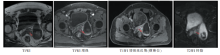
图1
术前膀胱MRI图片显示憩室内团块影
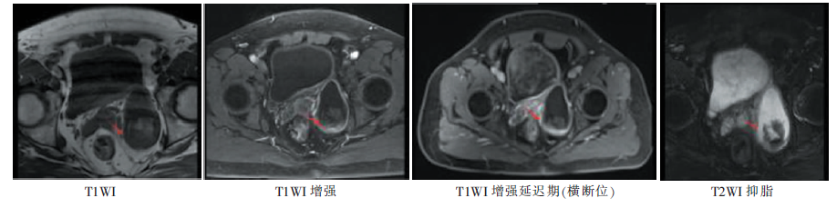
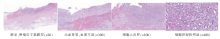
图2
膀胱EA病理图片(HE)


图3
膀胱EA部分免疫组化染色图片(×100)
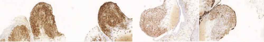
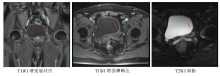
图4
术后膀胱MRI图片(2019年11月)显示膀胱左侧壁肿瘤复发
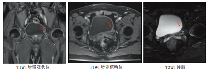

图5
PET-CT图片(2019年12月)提示膀胱左后壁肿瘤复发,伴左侧髂血管旁、腹膜后和腹腔淋巴结转移,骨多发转移
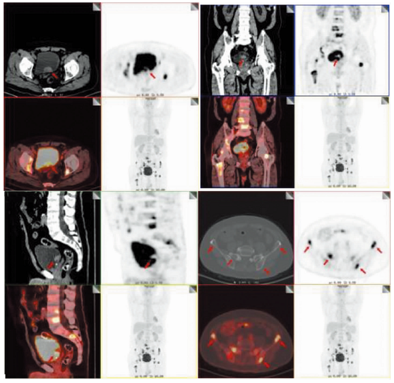
表1
18例膀胱EA临床病理特征
| 病例 号 | 作者, 年份 | 年龄 (岁) | 性别 | 首发症状 | 手术方式 | 部位 | 免疫组化 | 肿瘤史 | 放疗史 | 放疗至 EA时 间(y) | 联合治 疗方式 | 随访 (月) |
|---|---|---|---|---|---|---|---|---|---|---|---|---|
| 1 | Engel, 1998[ | 47 | 男 | 血尿,耻骨上 及左侧腹股 沟区疼痛 | 根治 | 膀胱三角区 | 不详 | 无 | 无 | 无 | 放化疗 | 32,带 病生存 |
| 2 | Seethala, 2006[ | 66 | 男 | 血尿 | 根治 | 膀胱左后壁、 顶壁 | CD31+CD34+ CK- | 不详 | 有 | 4 | 化疗 | 19,带 病生存 |
| 3 | Kulaga, 2007[ | 83 | 女 | 血尿 | 电切 | 膀胱后外侧壁 | Vimentin+CD31+ FⅧ-CD34- | 子宫内 膜癌 | 有 | 14 | 无 | 3,死亡 |
| 4 | Warne, 2011[ | 32 | 女 | 血尿,左侧 腹痛 | 电切 | 后壁 | CD31+FⅧ+ ULEX+CK+ | 无 | 无 | 无 | 放化疗 | 19, 死亡 |
| 5 | Abbasov, 2011[ | 51 | 男 | 血尿 | 电切+根治 | 右前壁、侧壁 | CK+FⅧ+CD31+ Vimentin+CD34- | 无 | 无 | 无 | 无 | 5周, 死亡 |
| 6 | Matoso, 2015[ | 73 | 女 | 血尿 | 电切+部分 切除 | 顶壁 | CD31+FⅧ+ | 宫颈鳞癌, 恶性间 皮瘤 | 有 | 10 | 无 | 6,死亡 |
| 7 | - | 77 | 男 | 血尿 | 电切 | 膀胱颈 | CD31+CD34+FⅧ- | 前列腺癌 | 有 | 9 | 无 | 14, 死亡 |
| 8 | - | 71 | 男 | 血尿 | 电切+根治 | 膀胱颈 | CD31+FⅧ+ | 前列腺癌 | 有 | 10 | 无 | 7,死亡 |
| 9 | - | 85 | 男 | 血尿 | 电切 | 右侧壁 | CD31+CD34+FⅧ+ | 前列腺癌 | 有 | 15 | 无 | 6,死亡 |
| 10 | - | 39 | 男 | 血尿 | 电切+根治 | 后壁 | CD31+ | 无 | 无 | 无 | 无 | 13, 死亡 |
| 11 | - | 64 | 男 | 血尿 | 电切+根 治+盆腔淋 巴结清扫 | 膀胱颈+ 左侧壁 | CD31+ERG+ | 前列腺癌 | 有 | 6 | 无 | 12,无 病生存 |
| 12 | - | 43 | 男 | 血尿 | 电切 | 左侧壁 | CD31+FⅧ+ERG+ CD34- | 无 | 无 | 无 | 无 | 6,带 病生存 |
| 13 | - | 73 | 男 | 血尿 | 电切 | 左侧尿道口 | CD31+ERG+ | 无 | 不详 | 不详 | 无 | 3,死亡 |
| 14 | - | 64 | 男 | 血尿 | 电切 | 膀胱颈 | CD34+ERG+ | 前列腺癌 | 有 | 15 | 3,带 病生存 | |
| 15 | Gerbaud, 2016[ | 72 | 男 | 血尿,急性 尿潴留 | 电切+根治 | 左侧壁 | Vimentin+CD31+ERG+ Fli-1+CD34-CK- | 无 | 无 | 无 | 放疗 | 5,死亡 |
| 16 | Wang, 2016[ | 79 | 男 | 血尿,刺激性 下尿路症状 | 电切+根治 | 三角区+ 前列腺 | CD31+CD34+ERG+ Fli-1+CK部分+ | 前列腺癌 | 有 | 6 | 无 | 20,无 病生存 |
| 17 | NizamA, 2018[ | 57 | 男 | 血尿 | 部分切除 | 肾盂、输尿管、 膀胱 | CD31+FⅧ+Fli-1+CK+ CD34灶阳性 ERG灶阳性 | 无 | 无 | 无 | 无 | 3,死亡 |
| 18 | 本例 | 56 | 男 | 血尿 | 电切 | 左后壁 | CD31+CD34+ERG+ Fli-1+Vimentin+CK+ | 无 | 无 | 无 | 安罗 替尼 | 9,死亡 |
| [1] | 王坚. 血管肉瘤. 软组织肿瘤病理学.2版. 北京: 人民卫生出版社, 2017:819-857. |
| Wang J. Angiosarcoma[M]//Wang J, Zhu X Z. Pathology of soft tissue tumors. 2nd Ed, Beijing: People′s Health Publishing House, 2017:819-857. | |
| [2] | Humphrey P A, Moch H, Cubilla A L, et al. Tumors of the urinary tract[M]// WHO Classification of Tumors of the Urinary System and Male Gential Organs. Lyon: IARC Press, 2016:122-127. |
| [3] |
Matoso A, Epstein J I. Epithelioid Angiosarcoma of the Bladder: A Series of 9 Cases[J]. Am J Surg Pathol, 2015, 39(10):1377-1382.
doi: 10.1097/PAS.0000000000000444 pmid: 25929352 |
| [4] |
Abbasov B, Munguia G, Mazal P R, et al. Epithelioid angiosarcoma of the bladder: report of a new case with immunohistochemical profile and review of the literature[J]. Pathology, 2011, 43(3):290-293.
doi: 10.1097/PAT.0b013e328344e2fb URL |
| [5] |
Engel J D, Kuzel T M, Moceanu M C, et al. Angiosarcoma of the bladder: a review[J]. Urology, 1998, 52(5):778-784.
pmid: 9801098 |
| [6] |
Seethala R R, Gomez J A, Vakar-Lopez F. Primary angiosarcoma of the bladder[J]. Arch Pathol Lab Med, 2006, 130(10):1543-1547.
doi: 10.5858/2006-130-1543-PAOTB pmid: 17090199 |
| [7] |
Kulaga A, Yilmaz A, Wilkin R P, et al. Epithelioid angiosarcoma of the bladder after irradiation for endometrioid adenocarcinoma[J]. Virchows Arch, 2007, 450(2):245-246.
pmid: 17149614 |
| [8] | Warne R R, Ong J S, Snowball B, et al. Primary angiosarcoma of the bladder in a young female[J]. BMJ Case Rep, 2011, 2011:bcr1120103484. |
| [9] |
Gerbaud F, Ingels A, Ferlicot S, et al. Angiosarcoma of the bladder: review of the literature and discussion about a clinical case[J]. Urol Case Rep, 2017, 13:97-100.
doi: 10.1016/j.eucr.2016.12.007 pmid: 28480169 |
| [10] | Wang G, Black P C, Skinnider B F, et al. Post-radiation epithelioid angiosarcoma of the urinary bladder and prostate[J]. Can Urol Assoc J, 2016, 10(5-6):E197-E200. |
| [11] |
Nizam A, Paquette E L, Wang B G, et al. Epithelioid angiosarcoma of the bladder: a case report and review of the Literature[J]. Clin Genitourin Cancer, 2018, 16(6):e1091-e1095.
doi: 10.1016/j.clgc.2018.07.010 URL |
| [12] | 聂秀. 尿路上皮癌的组织学变异[M]//聂秀, 黄邦杏. 膀胱活检病理解读. 2版. 北京: 人民卫生出版社, 2012:131-135. |
| Nie X. Biopsy interpretation of the bladder.2nd ed. Beijing: People′s Health Publishing House, 2012:131-135. | |
| [13] | Pazona J F, Gupta R, Wysock J, et al. Angiosarcoma of bladder: long-term survival after multimodal therapy[J]. Urology, 2007, 69(3):575,e9-e10. |
| [14] | 祝冰晶, 罗虎, 唐春兰. 安罗替尼治疗原发性胸膜及心包血管肉瘤1例[J]. 肿瘤预防与治疗, 2020, 33(9):813-816. |
| Zhu B J, Luo H, Tang C L. Treatment of primary pleural and pericardial angiosarcoma with arotinib: a case report[J]. J Cancer Control Treat, 2020, 33(9):813-816. |
| [1] | 徐苓, 王根发, 张良. UroVysion 荧光原位杂交技术检测在泌尿系统肿瘤诊断及膀胱癌预后监测中应用的初步探索[J]. 诊断学理论与实践, 2018, 17(02): 159-164. |
| [2] | 李莉, 卞炳贤, 张良, 沈立松. 尿液多种microRNA检测方法的建立及其在膀胱癌诊断中的应用研究[J]. 诊断学理论与实践, 2017, 16(01): 93-97. |
| [3] | 范瑜, 刘晓晟, 路青, 姚秋英,. 膀胱癌3.0T MRI多b值弥散加权成像研究[J]. 诊断学理论与实践, 2013, 12(03): 334-338. |
| [4] | 李莉, 丁奕星, 徐苓, 马妍慧, 张良,. 染色体3、7、17号和9p21位点在膀胱尿路上皮癌中的畸变情况及其临床意义[J]. 诊断学理论与实践, 2012, 11(01): 47-51. |
| [5] | 周健, 李海, 陈金珍, 黄振华,. Survivin、FAS在膀胱移行细胞癌中的表达及临床意义[J]. 诊断学理论与实践, 2009, 8(06): 627-630. |
| [6] | 张磊,. 膀胱侵袭性血管黏液瘤1例[J]. 诊断学理论与实践, 2008, 7(02): 213-. |
| [7] | 陈华, 张华, 项明洁, 张志伟, 周文龙, 沈周俊, 黄海峰,. 尿膀胱癌抗原、CYFRA21-1和透明质酸在膀胱癌诊断中的价值[J]. 诊断学理论与实践, 2008, 7(01): 73-76. |
| [8] | 陈华, 张华, 项明洁,. 膀胱癌尿肿瘤标志物的研究进展[J]. 诊断学理论与实践, 2006, 5(05): 446-448. |
| [9] | 徐锋,曹文俊,吴华成,王枕亚,林佳菲,樊绮诗. 生存素检测在膀胱癌实验诊断中的意义[J]. 诊断学理论与实践, 2005, 4(03): 206-208. |
| 阅读次数 | ||||||
|
全文 |
|
|||||
|
摘要 |
|
|||||