诊断学理论与实践 ›› 2022, Vol. 21 ›› Issue (06): 735-740.doi: 10.16150/j.1671-2870.2022.06.12
闫化茹, 刘坤, 韩志宏, 房振, 吴丽莉( )
)
收稿日期:2021-06-09
出版日期:2022-12-25
发布日期:2023-04-23
通讯作者:
吴丽莉
E-mail:lilywu9ceh@126.com
YAN Huaru, LIU Kun, HAN Zhihong, FANG Zhen, WU Lili()
Received:2021-06-09
Online:2022-12-25
Published:2023-04-23
Contact:
WU Lili
E-mail:lilywu9ceh@126.com
摘要:
目的:报道1例肾上腺旁吻合状血管瘤(anastomosing hemangioma, AH)患者,并复习相关文献,对AH的临床病理特征、诊断及鉴别诊断要点进行总结。方法:收集本例AH患者的临床资料,观察其组织学形态及免疫组化特点,并结合相关文献进行综合分析。结果:大体检查见肿瘤位于肾上腺旁脂肪组织内,边界清楚,切面呈灰白、灰红色,质地中等。显微镜下可见肿瘤由相互吻合的窦隙样管腔构成,管腔内衬靴钉样内皮细胞,少数上皮伴有轻度不典型性,可见纤维蛋白性血栓,个别细胞质内可见嗜酸性小体。免疫组化检测示,肿瘤细胞表达CD31和CD34,不表达D2-40、CK5/6、calretinin、CKpan、EMA、Melan-A和HMB45,Ki-67增殖指数约为5%。最终该患者经病理诊断为(肾上腺旁)AH。结果:本文报道1例AH患者,其为罕见的良性血管源性肿瘤,因其血管高度吻合、管腔衬覆的靴钉样内皮细胞常伴不典型的特点,需要与高分化血管肉瘤相区别;泌尿生殖系统的AH还需与血管平滑肌脂肪瘤、腺瘤样瘤、继发于肾细胞癌的血管瘤样增生等疾病相鉴别。
中图分类号:
闫化茹, 刘坤, 韩志宏, 房振, 吴丽莉. 肾上腺旁吻合状血管瘤1例并文献复习[J]. 诊断学理论与实践, 2022, 21(06): 735-740.
YAN Huaru, LIU Kun, HAN Zhihong, FANG Zhen, WU Lili. Paradrenal anastomosing hemangioma: a case report with literature review[J]. Journal of Diagnostics Concepts & Practice, 2022, 21(06): 735-740.

图1
肿瘤组织学形态(HE) A:肿瘤位于肾上腺下方,周边及内部均可见厚壁血管(×10);B:肿瘤内可见毛细血管大小的管腔或裂隙形成,衬覆靴钉样内皮细胞,个别细胞核较大,箭头示微小纤维蛋白血栓(×400);C:肿瘤细胞核大小相对一致,箭头示细胞质内嗜酸性小体(×400);D:肿瘤细胞似有围绕厚壁血管排列趋势(×100);E:红细胞渗出现象(×200);F:局灶退行性变(×40)。
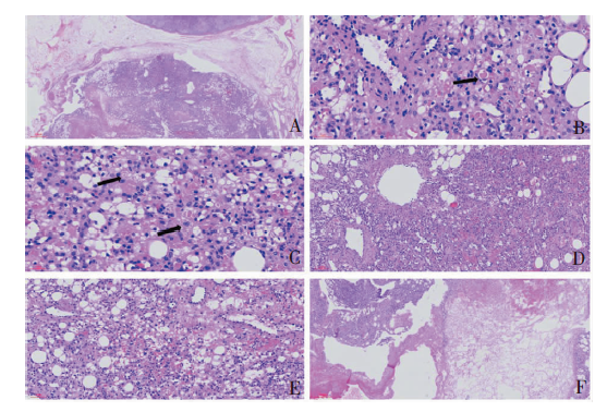

图2
肿瘤免疫组化结果 A:CD31可勾勒出大小不一的血管,部分血管有相互吻合趋势(×100);B:肌性血管壁平滑肌细胞和部分血管旁支持细胞表达SMA(×200);C:肿瘤细胞HMB45阴性(×40);D:Ki-67增殖指数约5%(×200)。
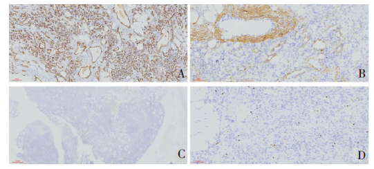
表1
94例AH临床特征
| 文献 | 例数(n) | 性别 | 年龄(岁) | 部位 | 最大径 (cm) | 术前诊断 | 处理 | 随访 |
|---|---|---|---|---|---|---|---|---|
| O′Neill AC et al[ | 32 | 18男 14女 | 17~81 | 腹膜后;肾脏;主动脉旁;肾旁;卵巢;肝脏;睾丸;肾上腺;肠系膜;精索 | 不详 | 肾上腺嗜铬细胞瘤;肠系膜脂肪肉瘤;肾癌;腹膜后脂肪肉瘤;淋巴结伴癌转移;其余未知 | 肿块切除术;粗针穿刺活检 | 4例2~26个月无复发或转移。 其余失访。 |
| John I,et al[ | 16 | 10男 6女 | 2~85 | 主动脉旁;椎体;腰大肌;胸椎旁;脊椎旁;腹膜后;纵隔 漏斗状骨盆韧带;子宫角;上臂;下腔静脉旁和右主动脉旁 | 1.5~6.7 4例不详 | 肉瘤;周围神经鞘瘤和淋巴瘤;其余未知 | 9例肿块切除术;7例粗针穿刺活检 | 10例1~46个月无复发转移或疾病进展。 其余失访。 |
| Bean GR, et al[ | 13 | 9男 4女 | 39~70 | 肾;肾上腺;脊柱; 椎旁;下腔静脉旁; 腹膜;卵巢;附件旁; 子宫肌层;肝脏 | 0.7~8.0 | 未知 | 肿块切除术 | 10例1~107个月无复发或转移。 其余失访。 |
| Kryvenko ON, et al[ | 7 | 1男 7女 | 39~77 | 肾(其中一例为双侧); 卵巢 | 0.6~5.0 | 卵巢子宫内膜样癌;卵巢浆液囊肿;卵巢浆液性囊腺瘤;其余因其它疾病行CT检查发现肾脏肿块 | 肿块切除术 | 10例随访3~122个月,无复发或转移。 其余失访。 |
| Montgomery E, et al[ | 6 | 4男 2女 | 49~75 | 肾;睾丸 | 1.3~2.0 | 未知 | 3例肾切除术,1例肿块切除术,2例睾丸切除术 | 5例8~36个月无复发或转移。1例失访。 |
| Lin J, et al[ | 6 | 2男 4女 | 46~71 | 肝;结肠小肠 | 0.2~6.0 | 3血管瘤;3血管肉瘤 | 肿块切除术 | 5例8~96个月无复发或转移。1例失访。 |
| 赵明等[ | 5 | 3男 2女 | 47~77 | 肾上腺;肾 | 1.6~2.5 | 肝癌肾转移;肾错构瘤;肾上腺嗜铬细胞瘤或皮质腺瘤 | 1例部分肾切除术,1例根治性肾切除术;3例肾上腺肿瘤切除术 | 5例6~52个月无复发或转移。 |
| Burton KR, et al[ | 1 | 男 | 68 | 多发:肾旁、下腔静脉旁、 髂内、双侧肾上腺 | 1.5~3.7 | 肾恶性肿瘤 | 左肾上腺 切除术、左 肾周和下 腔静脉旁 结节切除术 | 未知 |
| Huang ZY, et al[ | 1 | 男 | 37 | 鼻腔 | 2.3 | 鼻息肉、乳头状瘤 | 肿块切除术 | 22个月无复发转移。 |
| Ross M, et al[ | 1 | 男 | 49 | 肾上腺 | 2.0 | 肾上腺肿瘤 | 肾上腺切 除术 | 未知 |
| Rathore K, et al[ | 1 | 男 | 64 | 左心房 | 4.0 | 左心房壁肿瘤 | 肿块切除术 | 未知 |
| 姚建国等[ | 1 | 男 | 71 | 肾上腺 | 1.7 | 右肾上腺肿瘤 | 右肾上腺 切除术 | 10个月无复发转移。 |
| 虞义建等[ | 1 | 男 | 50 | 肾上腺 | 1.1 | 嗜铬细胞瘤 | 右肾上腺 切除术 | 未知 |
| 王小刚等[ | 1 | 男 | 55 | 肾 | 4.0 | 左肾占位 | 根治性左 肾切除术 | 12个月无复发转移。 |
| 党琳琳等[ | 1 | 女 | 44 | 肝脏 | 5.0 | 富于血供的肿瘤 | 肿块切除术 | 2个月无复发转移。 |
表2
57例AH组织病理特征
| 第一作者 | 例数(n) | 边界 | 吻合的 小血管 | 靴钉样内 皮细胞 | 细胞不 典型性 | 核分裂 | CD31/ CD34 | 微小血栓 | 嗜酸性小体 | 髓外造血 | 变性 |
|---|---|---|---|---|---|---|---|---|---|---|---|
| John I, et al[ | 16 | 1/16局灶不清 | 16/16+ | 16/16+ | 轻 | 少-无 | + | 16/16+ | 未知 | 7/16+ | 未知 |
| Bean GR, et al[ | 13 | 2/13局灶不清 | 13/13+ | 6/13+ | 轻-无 | 少-无 | 未知 | 13/13+ | 12/13+ | 7/13+ | 7/13+ |
| Kryvenko ON, et al[ | 7 | 清楚 | 7/7+ | 3/7+ | 无 | 无 | + | 未知 | 6/7+ | 3/17+ | 4/7+ |
| Montgomery E, et al[ | 6 | 3/6局灶浸润 | 6/6+ | 6/6+ | 轻-无 | 少-无 | + | 5/6+ | 2/6+ | 2/6+ | 5/6+ |
| Lin J, et al[ | 6 | 清楚 | 6/6+ | 6/6+ | 轻 | 无 | 3/3+ | 4/6+ | 1/6+ | 1/6+ | 未知 |
| 赵明等[ | 5 | 清楚 | 5/5+ | 5/5+ | 无 | 少-无 | + | + | 1/5+ | 2/5+ | 5/5+ |
| Huang ZY, et al[ | 1 | 清楚 | + | + | 轻 | 无 | + | 未知 | 未知 | 无 | 未知 |
| 姚建国等[ | 1 | 清楚 | + | + | 轻 | 未知 | + | + | 未知 | 未知+ | + |
| 虞义建等[ | 1 | 清楚 | + | + | 无 | 无 | + | + | 未知 | 未知 | 未知 |
| 党琳琳等[ | 1 | 欠清楚 | + | + | 轻 | 无 | + | + | 未知 | 未知 | + |
| [1] |
Montgomery E, Epstein J I. Anastomosing hemangioma of the genitourinary tract: a lesion mimicking angiosarcoma[J]. Am J Surg Pathol, 2009, 33(9):1364-1369.
doi: 10.1097/PAS.0b013e3181ad30a7 pmid: 19606014 |
| [2] |
Ross M, Polcari A, Picken M, et al. Anastomosing hemangioma arising from the adrenal gland[J]. Urology, 2012, 80(3):e27-e28.
doi: 10.1016/j.urology.2012.05.032 URL |
| [3] |
Kryvenko O N, Gupta N S, Meier F A, et al. Anastomosing hemangioma of the genitourinary system: eight cases in the kidney and ovary with immunohistochemical and ultrastructural analysis[J]. Am J Clin Pathol, 2011, 136(3):450-457.
doi: 10.1309/AJCPJPW34QCQYTMT pmid: 21846922 |
| [4] |
Lin J, Bigge J, Ulbright T M, et al. Anastomosing hemangioma of the liver and gastrointestinal tract: an unusual variant histologically mimicking angiosarcoma[J]. Am J Surg Pathol, 2013, 37(11):1761-1765.
doi: 10.1097/PAS.0b013e3182967e6c pmid: 23887160 |
| [5] | 党琳琳, 侯东省, 林存虎, 等. 肝吻合状血管瘤1例[J]. 临床与实验病理学杂志, 2020, 36(6):744-745. |
| Dang L L, Hou D S, Lin C H, et al. Anastomotic hemangioma of liver: a case report[J]. Chin J Clin Exp Pathol, 2020, 36(6):744-745. | |
| [6] | 姚建国, 潘红佳, 王春华, 等. 吻合状血管瘤一例[J]. 中华病理学杂志, 2015, 44(2):133-134. |
| Yao J G, Pan H J, Wang C H, et al. One case of anastomotic hemangioma[J]. Chin J Pathol, 2015, 44(2):133-134. | |
| [7] | 虞义建, 郑晶琼, 蒋武斌, 等. 原发肾上腺交织状血管瘤1例[J]. 临床与实验病理学杂志, 2020, 36(3):375-376. |
| Yu Y J, Zhen J Q, Jiang W B, et al. One case of primary adrenal interlaced hemangioma[J]. Chin J Clin Exp Pathol, 2020, 36(3):375-376. | |
| [8] | 王小刚, 徐仁芳, 薛钟, 等. 肾吻合性血管瘤一例报告[J]. 中华泌尿外科杂志, 2019, 40(6):468. |
| Wang X G, Xu R F, Xue Z, et al. Renal anastomotic hemangioma: a case report[J]. Chin J Urol, 2019, 40(6):468. | |
| [9] |
赵明, 孔梅, 余晶晶, 等. 肾脏及肾上腺交织状血管瘤临床病理分析[J]. 中华病理学杂志, 2016, 45(10):698-702.
pmid: 27760611 |
|
Zhao M, Kong M, Yu J J, et al. Clinicopathologic analysis of anastomosing hemangioma of the kidney and adrenal gland[J]. Chin J Pathol, 2016, 45(10):698-702.
doi: 10.3760/cma.j.issn.0529-5807.2016.10.006 pmid: 27760611 |
|
| [10] |
Huang Z Y, Chen C C, Thingujam B. Anastomo-sing Hemangioma of the Nasal Cavity[J]. Laryngoscope, 2020, 130(2):354-357.
doi: 10.1002/lary.v130.2 URL |
| [11] |
Rathore K, Yussouf R, Teh M, et al. Left atrial anastomosing hemangioma causing recurrent pericardial effusion[J]. Ann Thorac Surg, 2020, 109(3):e157-e159.
doi: 10.1016/j.athoracsur.2019.06.082 pmid: 31430463 |
| [12] |
Lappa E, Drakos E. Anastomosing Hemangioma: Short Review of a Benign Mimicker of Angiosarcoma[J]. Arch Pathol Lab Med, 2020, 144(2):240-244.
doi: 10.5858/arpa.2018-0264-RS pmid: 30958692 |
| [13] |
Büttner M, Kufer V, Brunner K, et al. Benign mesenchymal tumours and tumour-like lesions in end-stage renal disease[J]. Histopathology, 2013, 62(2):229-236.
doi: 10.1111/j.1365-2559.2012.04349.x pmid: 23020314 |
| [14] |
John I, Folpe A L. Anastomosing Hemangiomas Arising in Unusual Locations: A Clinicopathologic Study of 17 Soft Tissue Cases Showing a Predilection for the Paraspinal Region[J]. Am J Surg Pathol, 2016, 40(8):1084-1089.
doi: 10.1097/PAS.0000000000000627 pmid: 26945338 |
| [15] |
O′neill A C, Craig J W, Silverman S G, et al. Anastomosing hemangiomas: locations of occurrence, imaging features, and diagnosis with percutaneous biopsy[J]. Abdom Radiol (NY), 2016, 41(7):1325-1332.
doi: 10.1007/s00261-016-0690-2 pmid: 26960722 |
| [16] | Burton K R, Jakate K, Pace K T, et al. A case of recurrent, multifocal anastomosing haemangiomas[J]. BMJ Case Rep, 2017, 2017:bcr2017220076. |
| [17] |
Kryvenko O N, Roquero L, Gupta N S, et al. Low-grade clear cell renal cell carcinoma mimicking hemangioma of the kidney: a series of 4 cases[J]. Arch Pathol Lab Med, 2013, 137(2):251-254.
doi: 10.5858/arpa.2011-0615-OA pmid: 23368867 |
| [18] |
Liau J Y, Lee J C, Tsai J H, et al. Thrombotic Hemangioma With Organizing/Anastomosing Features: Expanding the Spectrum of GNA-mutated Hemangiomas With a Predilection for the Skin of the Lower Abdominal Regions[J]. Am J Surg Pathol, 2020, 44(2):255-262.
doi: 10.1097/PAS.0000000000001392 URL |
| [19] |
Bean G R, Joseph N M, Gill R M, et al. Recurrent GNAQ mutations in anastomosing hemangiomas[J]. Mod Pathol, 2017, 30(5):722-727.
doi: 10.1038/modpathol.2016.234 URL |
| [20] |
Liau J Y, Lee J C, Tsai J H, et al. High frequency of GNA14, GNAQ, and GNA11 mutations in cherry hemangioma: a histopathological and molecular study of 85 cases indicating GNA14 as the most commonly mutated gene in vascular neoplasms[J]. Mod Pathol, 2019, 32(11):1657-1665.
doi: 10.1038/s41379-019-0284-y URL |
| [21] |
Liau J Y, Tsai J H, Lan J, et al. GNA11 joins GNAQ and GNA14 as a recurrently mutated gene in anastomo-sing hemangioma[J]. Virchows Arch, 2020, 476(3):475-481.
doi: 10.1007/s00428-019-02673-y |
| [22] |
Sahni V A, LY A, Silverman S G. Usefulness of percutaneous biopsy in diagnosing benign renal masses that mimic malignancy[J]. Abdom Imaging, 2011, 36(1):91-101.
doi: 10.1007/s00261-009-9597-5 pmid: 20049430 |
| [1] | 王之倩, 李敏, 于一飞, 周建桥. 21-羟化酶缺陷先天性肾上腺皮质增生患者睾丸肾上腺残基瘤超声特征分析[J]. 诊断学理论与实践, 2022, 21(05): 588-591. |
| [2] | 中华医学会内分泌学分会. 新型冠状病毒肺炎疫情下肾上腺疾病管理专家建议[J]. 诊断学理论与实践, 2022, 21(02): 139-142. |
| [3] | 李芹芹, 金晓龙, 袁菲. 儿童系统性EB病毒阳性T细胞淋巴瘤临床病理分析一例及文献复习[J]. 诊断学理论与实践, 2020, 19(1): 63-68. |
| [4] | 张明华, 王荟, 翁香琴, 房振, 韩志宏, 吴丽莉. 腹水细胞学诊断髓系肉瘤1例报告附文献复习[J]. 诊断学理论与实践, 2020, 19(1): 88-91. |
| [5] | 胡哲, 陈歆, 罗芳秀, 初少莉, 王继光. 肾上腺醛固酮和皮质醇共分泌瘤一例报告[J]. 诊断学理论与实践, 2020, 19(05): 525-527. |
| [6] | 许海敏, 陈晓炎, 张静, 杨晓群, 王朝夫. 肺微囊性纤维黏液瘤一例临床病理分析及文献复习[J]. 诊断学理论与实践, 2020, 19(04): 381-385. |
| [7] | 金娇莺, 李倩玉, 蒋虹伟, 韩冬艳, 奚豪, 蔚青. 混合性嗜铬细胞瘤1例报道并文献复习[J]. 诊断学理论与实践, 2019, 18(2): 165-169. |
| [8] | 康健捷, 苏佩珣, 邓兵梅, 杨红军, 王卓才. 肾上腺腺瘤型原发性醛固酮增多症并发横纹肌溶解症一例[J]. 诊断学理论与实践, 2019, 18(05): 583-584. |
| [9] | 陈晓炎, 杨晓群, 袁菲, 张静, 王朝夫. 肺纤毛黏液结节性乳头状肿瘤2例临床病理分析及文献复习[J]. 诊断学理论与实践, 2018, 17(05): 575-580. |
| [10] | 顾斌, 王朝夫, 金晓龙, 袁菲, 张静, 许海敏, 任景丽, 陈晓炎. 肺硬化性肺细胞瘤23例临床病理分析[J]. 诊断学理论与实践, 2017, 16(02): 188-194. |
| [11] | Anoj Adhikari, 陈克敏,. 肾上腺偶发瘤的影像学诊断与治疗进展[J]. 诊断学理论与实践, 2015, 14(05): 473-478. |
| [12] | 木良善, 苏颋为, 姜蕾, 蒋怡然, 周薇薇, 王卫庆, 宁光,. 嗜铬细胞瘤患者血浆间甲肾上腺素类物质与血压及糖脂代谢的相关性研究[J]. 诊断学理论与实践, 2015, 14(04): 318-323. |
| [13] | 王晴柔, 陈克敏, 黄蔚, 徐学勤, 林晓珠, 柴维敏,. 肾上腺髓性脂肪瘤的CT诊断与鉴别诊断[J]. 诊断学理论与实践, 2014, 13(05): 491-494. |
| [14] | 方文强, 宋琦,. 肾上腺皮质增生的CT诊断与鉴别诊断[J]. 诊断学理论与实践, 2014, 13(05): 469-471. |
| [15] | 迟婧, 宋琦,. ACTH非依赖性库欣综合征的影像学表现[J]. 诊断学理论与实践, 2013, 12(06): 649-653. |
| 阅读次数 | ||||||
|
全文 |
|
|||||
|
摘要 |
|
|||||