诊断学理论与实践 ›› 2023, Vol. 22 ›› Issue (03): 283-291.doi: 10.16150/j.1671-2870.2023.03.12
尹永芳1, 唐永华2( ), 梁妍1, 陈志仁1, 费晓春3
收稿日期:2023-01-18
出版日期:2023-06-25
发布日期:2023-11-17
通讯作者:
唐永华 E-mail:
YIN Yongfang1, TANG Yonghua2(), LIANG Yan1, CHEN Zhiren1, FEI Xiaochun3
Received:2023-01-18
Online:2023-06-25
Published:2023-11-17
摘要:
目的:探讨炎性髓系肿瘤Erdheim-Chester病(Erdheim-Chester disease,ECD)患者的临床及影像学特征。方法:回顾性分析2019年3月至2022年2月上海交通大学附属瑞金医院经病理证实的6例ECD患者的临床及影像学资料。结果:6例ECD患者的年龄为11~64岁,其中4例患者的年龄为11~33岁,较年轻;男女比例为2∶1,临床症状以中枢性尿崩(2例)、共济失调(2例)、下肢疼痛(3例)以及眼眶周黄色斑块(3例)为主要特点。6例患者中5例累及骨骼系统;6例均累及骨骼外系统,包括累及皮肤和皮下软组织5例,累及中枢神经系统2例,累及心血管系统2例,累及胸部2例(其中1例伴有肺腺癌),腹膜后巨大肿块和肾脏纤维化改变各1例。6例患者在影像诊断为ECD 2例、眼部黄色肉芽肿1例、其他3例影像不能进行病因诊断。6例患者中2例临床误诊为恶性肿瘤,但未能明确疾病类型。1例初次行X线检查双下肢假阴性,经CT检查后发现骨质硬化性改变。ECD患者的骨骼系统特征性X线和CT表现为,双侧上下肢骨质硬化,其中1例为下肢对称性,1例为左上肢骨质硬化;非特征性X和CT表现溶骨性骨质破坏,其中1例为颞下颌关节和2例为上颌骨。骨骼外多系统病变(6例)表现为密度、信号、放射性浓聚异常。随访提示,2例预后良好,1例死亡,3例疾病进展。结论:ECD患者多以中枢性尿崩及共济失调为主要症状,伴皮肤黄色肉芽肿及其他皮肤病变等临床特征。影像学表现主要为双下肢骨质硬化,同时伴有全身其他系统多发病变。
中图分类号:
尹永芳, 唐永华, 梁妍, 陈志仁, 费晓春. Erdheim-Chester病6例临床及影像学特征分析[J]. 诊断学理论与实践, 2023, 22(03): 283-291.
YIN Yongfang, TANG Yonghua, LIANG Yan, CHEN Zhiren, FEI Xiaochun. Clinical and imaging manifestations of Erdheim-Chester disease (six cases)[J]. Journal of Diagnostics Concepts & Practice, 2023, 22(03): 283-291.
表1
患者临床资料、临床诊断及病理诊断
| Serial number | Gender | age(years) | Clinical manifestation | Systems involved | Clinical diagnosis | Pathologic diagnosis | Genetic testing |
|---|---|---|---|---|---|---|---|
| Case 1 | Male | 11 | Ataxia, diabetes insipidus | Skin, central nervous system, skeletal system | LCH | LCH merges ECD | BRAF V600E gene(+) |
| Case 2 | Male | 33 | Diabetes insipidus, ametropia, Cough from drinking water | Skin, central nervous system, skeletal system, urinary system | LCH | ECD | BRAF V600E gene(+) |
| Case 3 | Male | 17 | Wrist pain, aggravated after exercise | Skeletal system, cardiovascular system, respiratory system | LCH | ECD | BRAF V600E gene(-) |
| Case 4 | Male | 16 | Chest discomfort, right neck mass, fever, cough, dysphagia | Skin, skeletal system | LCH | ECD | BRAF V600E gene(+) |
| Case 5 | Female | 54 | Loss of appetite, edema of both lower limbs, general weakness, mass of left upper abdomen | Skin, retroperitoneal space | Retroperitoneal mesenchymal sarcoma | ECD | Not detected |
| Case 6 | Female | 64 | Periorbital yellow patch,chest space occupying | Skin, cardiovascular system, respiratory system | Xanthoma of orbit、 Lung cancer | ECD | Not detected |

图1
右侧上臂肿块病理检查结果 A~C:HE染色,放大倍数分别为×25、×100、×400,泡沫状或富含脂质的组织细胞,纤维化伴炎细胞浸润。D~G:免疫组化检测,CD68(+),S100(-),CD1α(个别+),Langerin(个别+)。
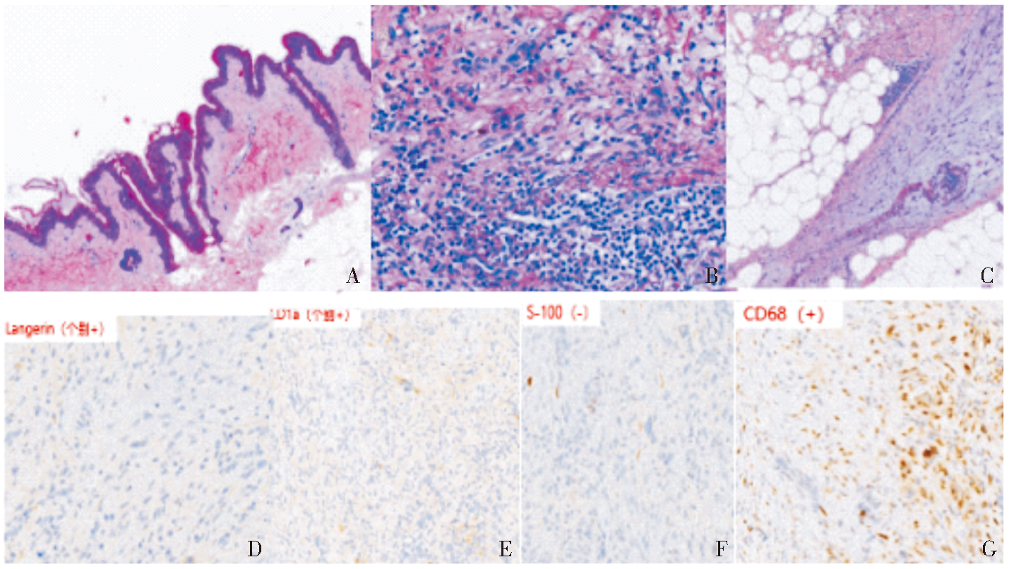
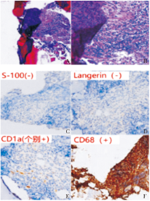
图2
病理结果 A、B:HE染色(×25、×100),见少量骨母细胞、脂肪细胞、血管内皮及造血成分;C~F:免疫组化染色, S-100(-),CD68(+),CD1α(个别+),Langerin(-)。

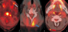
图3
皮肤及皮下软组织病变PET/CT图像 A:病例1左侧上颌窦前壁与外壁交界区近眼外眦皮下代谢增高灶(细箭头);B:病例2左后颈部皮下肌肉略增厚且代谢增高灶(蓝粗箭头),C:病例4右侧颈后皮下小结节状代谢增高灶(红粗箭头)。
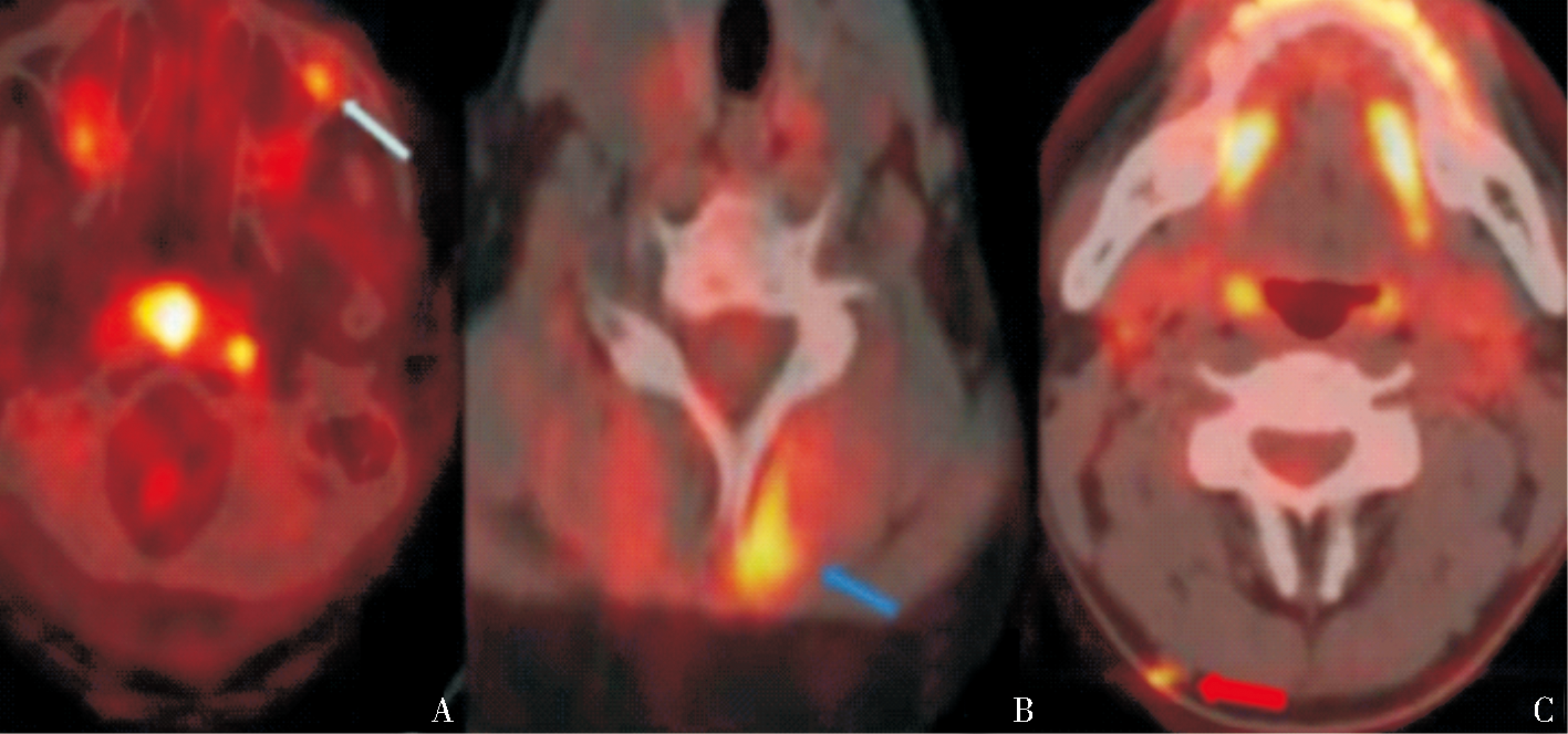
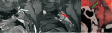
图4
垂体MRI和PET/CT图像 A:病例1,T1WI增强序列,垂体柄增粗且强化明显(蓝箭头),b:病例1,T1WI序列,正常神经垂体高信号消失(红箭头),C:病例2,PET/CT检查显示垂体代谢减低(绿箭头)。

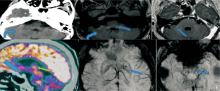
图5
小脑病变CT、MRI和PET/CT图像 A:病例1显示小脑萎缩(弯箭头); D:病例1 18F-FDG-PET/CT垂体(粗蓝箭头)、双侧小脑半球(红箭头)、背侧丘脑代谢不均匀减低,脑桥背盖部代谢增高(绿箭头); B、C、E、F:病例2 T2-FLAIR序列脑干(蓝箭头)、双侧小脑半球(弯箭头)多发斑片状高信号; C:病例2 T1WI增强序列右侧桥臂部分病灶小片状明显强化(蓝箭头),E、F:病例2示SWI序列双侧基底节区、脑干多发小斑点低信号(蓝箭头)。

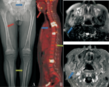
图6
骨骼系统X-ray、MRI和PET/CT图像 A:病例2,X线平片摄影,显示典型征象即骨盆、双下肢股骨及胫骨以对称性骨质硬化,骨松质硬化区边缘模糊不清,骨皮质光滑,无骨膜反应及穿凿样或虫蚀样骨质破坏,周围无软组织肿块(蓝红绿箭头);B:病例1,PET/CT检查,提示椎体、上下颌骨及棘突或棘间韧带、多发代谢增高灶(蓝红绿箭头),代谢极高SUVmax 15.2~17.1;C-D:病例1,MRI检查显示右侧颞下窝软组织T2WI呈高信号,T1WI增强不均匀强化,DWI扩散受限呈高信信号(蓝箭头)。
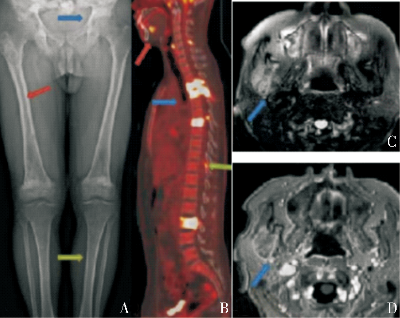

图7
骨骼系统X-ray、MRI和PET/CT图像 A :病例1,初次X线平片摄影肱骨呈假阴性; B:病例1,MRI阳性显示肱骨中下段FS-T2WI髓腔高信号(蓝箭头); C-D:病例5,溶骨性病变PET/CT示右侧颞下颌关节溶骨性骨质破坏,部分骨皮质模糊不连续伴代谢增高灶,SUV max约15.18,周围无软组织肿块(蓝箭头)。

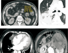
图8
其他部位病变CT图像 A:病例2,肾窦软组织密度,形态不规则,边缘毛糙,CT值约13 HU;B-C:病例3,右肺下叶实变、心包积液;D:病例5,左侧腹膜后肿物,左肾静脉受压。

| [1] |
CIVES M, SIMONE V, RIZZO F M, et al. Erdheim-Chester disease: a systematic review[J]. Crit Rev Oncol Hematol, 2015, 95(1):1-11.
doi: 10.1016/j.critrevonc.2015.02.004 pmid: 25744785 |
| [2] |
CHESTER W. Über Lipoidgranulomatose[J]. Virchows Arch. path Anat, 1930, 279:561-602.
doi: 10.1007/BF01942684 URL |
| [3] | WHO Classification of Tumours of Haematopoietic and Lymphoid Tissues. International Agency for Research on Cancer (IARC)[M].revised 4th edition. Lyon, 2017. |
| [4] | 付欣, 张丽英, 马静, 等. Erdheim-Chester病的临床病理分析[J]. 诊断病理学杂志, 2019, 26(12):822-826, 830. |
| FU X, ZHANG L Y, MA J, et al. Clinicopathological features of Erdheim-Chester disease[J]. J DIag Pathol, 2019, 26(12):822-826, 830. | |
| [5] | 刘玲春, 朴月善, 卢德宏. 几种常见的累及中枢神经系统的非朗格汉斯细胞组织细胞增生症[J]. 中华内科杂志, 2015, 54(12):1057-1059. |
| LIU L C, PIAO Y S, LU D H, et al. Several common Non-Langerhans cell histiocytosis involving the central nervous system[J]. Chin J Intern Med, 2015, 54(12):1057-1059. | |
| [6] |
HERVIER B, HAROCHE J, ARNAUD L, et al. Association of both Langerhans cell histiocytosis and Erdheim-Chester disease linked to the BRAFV600E mutation[J]. Blood, 2014, 124(7):1119-1126.
doi: 10.1182/blood-2013-12-543793 pmid: 24894769 |
| [7] |
MILNE P, BIGLEY V, BACON C M, et al. Hematopoie-tic origin of Langerhans cell histiocytosis and Erdheim-Chester disease in adults[J]. Blood, 2017, 130(2):167-175.
doi: 10.1182/blood-2016-12-757823 URL |
| [8] |
DURHAM B H, ROOS-WEIL D, BAILLOU C, et al. Functional evidence for derivation of systemic histiocytic neoplasms from hematopoietic stem/progenitor cells[J]. Blood, 2017, 130(2):176-180.
doi: 10.1182/blood-2016-12-757377 pmid: 28566492 |
| [9] |
CAVALLI G, GUGLIELMI B, BERTI A, et al. The multifaceted clinical presentations and manifestations of Erdheim-Chester disease: comprehensive review of the literature and of 10 new cases[J]. Ann Rheum Dis, 2013, 72(10):1691-1695.
doi: 10.1136/annrheumdis-2012-202542 pmid: 23396641 |
| [10] |
ARNAUD L, GOROCHOV G, CHARLOTTE F, et al. Systemic perturbation of cytokine and chemokine networks in Erdheim-Chester disease: a single-center series of 37 patients[J]. Blood, 2011, 117(10):2783-2790.
doi: 10.1182/blood-2010-10-313510 pmid: 21205927 |
| [11] |
HAROCHE J, COHEN-AUBART F, AMOURA Z. Erdheim-Chester disease[J]. Blood, 2020, 135(16):1311-1318.
doi: 10.1182/blood.2019002766 pmid: 32107533 |
| [12] |
DION E, GRAEF C, MIQUEL A, et al. Bone involvement in Erdheim-Chester disease: imaging findings including periostitis and partial epiphyseal involvement[J]. Radiology, 2006, 238(2):632-639.
pmid: 16371583 |
| [13] | 王志芳, 常春康, 章振林. 以双下肢疼痛为主要表现的Erdheim-Chester病一例[J]. 中华骨质疏松和骨矿盐疾病杂志, 2021, 14(2):169-174. |
| WANG Z F, CHANG C K, ZHANG Z L, et al. Erdhein-Chester disease with bilateral lower limb pain as the main manifestation: a case report[J]. Chin J Osteoporos Bone Miner Res, 2021, 14(2):169-174. | |
| [14] |
ARNAUD L, GOROCHOV G, CHARLOTTE F, et al. Systemic perturbation of cytokine and chemokine networks in Erdheim-Chester disease: a single-center series of 37 patients[J]. Blood, 2011, 117(10):2783-2790.
doi: 10.1182/blood-2010-10-313510 pmid: 21205927 |
| [15] |
HAROCHE J, COHEN-AUBART F, AMOURA Z. Erdheim-Chester disease[J]. Blood, 2020, 135(16):1311-1318.
doi: 10.1182/blood.2019002766 pmid: 32107533 |
| [16] | JULIEN H, FLEUR COHEN A, ZAHIRAMOU A, et al. Erdheim-Chester disease[J]. Blood Review Series, 2020, 135 (16):1311-1318. |
| [17] |
GOTTLIEB R, CHEN A. MR findings of Erdheim-Chester disease[J]. J Comput Assist Tomogr, 2002, 26(2):257-261.
pmid: 11884783 |
| [18] |
COHEN AUBART F, IDBAIH A, EMILE J F, et al. Histiocytosis and the nervous system: from diagnosis to targeted therapies[J]. Neuro Oncol, 2021, 23(9):1433-1446.
doi: 10.1093/neuonc/noab107 URL |
| [19] |
ALLEN T C, CHEVEZ-BARRIOS P, SHETLAR D J, et al. Pulmonary and ophthalmic involvement with Erdheim-Chester disease: a case report and review of the literature[J]. Arch Pathol Lab Med, 2004, 128(12):1428-1431.
doi: 10.5858/2004-128-1428-PAOIWE pmid: 15578889 |
| [20] |
ARNAUD L, PIERRE I, BEIGELMAN-AUBRY C, et al. Pulmonary involvement in Erdheim-Chester disease: a single-center study of thirty-four patients and a review of the literature[J]. Arthritis Rheum, 2010, 62(11):3504-3512.
doi: 10.1002/art.v62:11 URL |
| [21] |
PAPO M, DIAMOND E L, COHEN-AUBART F, et al. High prevalence of myeloid neoplasms in adults with non-Langerhans cell histiocytosis[J]. Blood, 2017, 130(8):1007-1013.
doi: 10.1182/blood-2017-01-761718 pmid: 28679734 |
| [22] |
DIAMOND E L, DAGNA L, HYMAN D M, et al. Consensus guidelines for the diagnosis and clinical management of Erdheim-Chester disease[J]. Blood, 2014, 124(4):483-492.
doi: 10.1182/blood-2014-03-561381 pmid: 24850756 |
| [23] |
VAGLIO A, SALVARANI C, BUZIO C. Retroperitoneal fibrosis[J]. Lancet, 2006, 367(9506):241-251.
doi: 10.1016/S0140-6736(06)68035-5 pmid: 16427494 |
| [24] | COHEN-AUBART F, EMILE J F, CARRAT F, et al. Phenotypes and survival in Erdheim-Chester disease: Results from a 165-patient cohort[J]. Am J Hematol, 2018, 93(5):E114-E117. |
| [25] |
FAN X, LIU T, ZHANG Z, et al. Comparison of neuroi-maging features of histiocytic neoplasms with central nervous system involvement: a retrospective study of 121 adult patients[J]. Eur Radiol, 2023, 33(11):8031-8042.
doi: 10.1007/s00330-023-09724-8 |
| [26] | AGGARWAL A, TAYCHERT M, HASANIN L, et al. Erdheim-Chester disease: a case report of BRAF V600E-negative, MAP2K1-positive ECD diagnosed by blood next-generation sequencing assay and a brief literature review[J]. Oncology (Williston Park), 2023, 37(7):298-302. |
| [1] | 唐文, 任刚, 蔡嵘,. 肾母细胞瘤CT征象与基因表达相关性的研究进展[J]. 诊断学理论与实践, 2016, 15(01): 69-71. |
| [2] | Anoj Adhikari, 陈克敏,. 肾上腺偶发瘤的影像学诊断与治疗进展[J]. 诊断学理论与实践, 2015, 14(05): 473-478. |
| [3] | 赵世华,. 心脏磁共振在冠心病诊断中的意义[J]. 诊断学理论与实践, 2011, 10(01): 22-25. |
| [4] | 方文强, 贺晓燕, 陈曦, 唐永华, 刘建民, 宁光, 陈克敏,. 原发性甲状旁腺功能亢进症的影像学诊断[J]. 诊断学理论与实践, 2006, 5(06): 487-491. |
| [5] | 严超,朱正纲,燕敏,陈军,尹浩然. 一例钙化性胃癌的影像学诊断[J]. 诊断学理论与实践, 2002, 1(03): 73-74. |
| 阅读次数 | ||||||
|
全文 |
|
|||||
|
摘要 |
|
|||||