诊断学理论与实践 ›› 2024, Vol. 23 ›› Issue (05): 494-499.doi: 10.16150/j.1671-2870.2024.05.005
李英1,2( ), 蒋晗1,2, 王晓雪1,2, 魏浩楠1,2
收稿日期:2024-03-20
接受日期:2024-08-08
出版日期:2024-10-25
发布日期:2025-02-25
通讯作者:
李英 E-mail:liying@ihcams.ac.cn
LI Ying1,2(), JIANG Han1,2, WANG Xiaoxue1,2, WEI Haonan1,2
Received:2024-03-20
Accepted:2024-08-08
Published:2024-10-25
Online:2025-02-25
摘要:
目的:分析血液病合并毛霉菌病临床诊治特点,供临床参考。方法:纳入2018年10月至2023年12月我院经临床综合诊断为血液病合并毛霉菌病的连续病例65例。所有患者均接受胸部CT检查,其中14例行头颅MRI检查。8例接受肺组织活检,57例行血、肺泡灌洗液或组织宏基因二代测序,分析其CT表现及总结诊断过程。结果:肺毛霉菌占我院同期确诊真菌感染的5.30%(65/1233例)。本组58例为肺毛霉菌病,7例为累及多器官的播散性毛霉菌病。肺毛霉菌病患者CT表现为单发肺内斑片状实变(23/65)、混合型(11/65)、多发结节(28/65)或弥漫渗出(3/65),伴有反晕征29例、晕征25例、胸腔积液20例、结节数目大于10个者11例,2例患者CT增强检查显示血管截断征。播散性毛霉菌病患者表现为肺内多发结节7例,同时伴脑内单发或多发结节6例、脑内弥漫性梗死灶1例、脾内和肾内结节2例。首次胸部CT到临床确诊平均时间(4.3±1.8)d,CT怀疑真菌感染后进行,血或组织、肺泡二代测序检查,经两性霉素B等抗真菌治疗后好转58例,手术切除肺组织2例,家属放弃治疗失访2例,死亡3例。结论:肺毛霉菌约占血液病住院患者合并真菌感染的5%;肺部CT影像主要表现为单发大片突出和多发结节。近45%患者CT影像出现反晕征,38.5%的患者晕征,可为诊断提供提示性参考,二代测序在临床诊断中发挥重要作用。
中图分类号:
李英, 蒋晗, 王晓雪, 魏浩楠. 65例血液病患者感染毛霉菌病的胸部CT表现及诊治分析[J]. 诊断学理论与实践, 2024, 23(05): 494-499.
LI Ying, JIANG Han, WANG Xiaoxue, WEI Haonan. Analysis of chest CT findings, diagnosis, and treatment of mucormycosis infection in 65 hematologic disease patients[J]. Journal of Diagnostics Concepts & Practice, 2024, 23(05): 494-499.
表1
胸部CT表现分型
| Thoracic CT manifestations | Cases | Reversed halo syndrome | Halo syndrome | Pleural effusion |
|---|---|---|---|---|
| Single shot large consolidation | 23(35%) | 18(78%) | 0 | 10(43%) |
| Multiple nodules | 28(43%) | 0 | 25(89%) | 20(71%) |
| Mixed | 11(17%) | 8(73%) | 0 | 2(18%) |
| Diffuse exudative type | 3(5%) | 3(100%) | 0 | 2(67%) |

图1
肺内毛霉菌灶 A、D分别显示病灶的反晕征、晕征;B、C显示增强检查局部血管截断;E、F显示10 d后病灶增大呈实变影伴反晕征
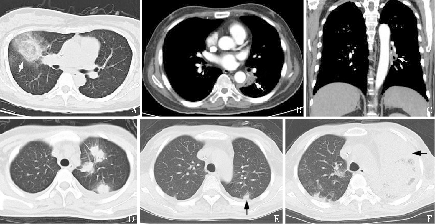

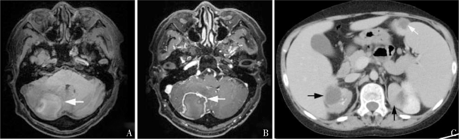
图2
颅脑及肾、脾内毛霉菌灶 A、B、C分别显示颅内、肾脏、脾脏病灶。
| [1] | VALENTINE J C, MORRISSEY C O, TACEY M A, et al. A population-based analysis of attributable hospitalisation costs of invasive fungal diseases in haematological malignancy patients using data linkage of state-wide regi-stry and costing databases: 2009-2015[J]. Mycoses, 2020, 63(2):162-171. |
| [2] | PETRIKKOS G, SKIADA A, LORTHOLARY O, et al. Epidemiology and clinical manifestations of mucormycosis[J]. Clin Infect Dis, 2012,54 Suppl 1:S23-34. |
| [3] | YANG N, ZHANG L, FENG S. Clinical features and treatment progress of invasive mucormycosis in patients with hematological malignancies[J]. J Fungi (Basel), 2023, 9(5):592. |
| [4] |
DANION F, AGUILAR C, CATHERINOT E, et al. Mucormycosis: new developments into a persistently devastating infection. Semin Respir Crit Care Med, 2015, 36(5):692-705.
doi: 10.1055/s-0035-1562896 pmid: 26398536 |
| [5] | 齐瑶, 赵明峰, 邓琦, 等. 血液病合并毛霉菌病七例报道并文献复习[J]. 中华血液学杂志, 2019, 40(11):943-947. |
| QI Y, ZHAO M F, DENG Q, et al. Report of seven cases of hematological diseases combined with trichoderma and literature review[J]. Chin J Hematol, 2019, 40(11):943-947. | |
| [6] | 中国医药教育协会真菌病专业委员会, 中国毛霉病专家共识工作组. 中国毛霉病临床诊疗专家共识(2022)[J]. 中华内科杂志, 2023, 62(6):597-605. |
| Fungal Disease Professional Committee of China Medical Education Association, Expert Consensus Working Group on Trichoderma in China. Consensus of clinical diagnosis and treatment experts on trichoderma in China (2022)[J]. Chin J Intern Med, 2023, 62(6):597-605. | |
| [7] | 吴挺, 周华, 顾海艇, 等. 肺毛霉菌病25例临床高危因素特征及预后分析[J]. 中华医学杂志, 2018, 98(32):2579-2582. |
| WU T, ZHOU H, GU H T, et al. Clinical high-risk factors and prognosis analysis of 25 cases of pulmonary mucormycosis[J]. Natl Med J China, 2018, 98(32):2579-2582. | |
| [8] |
AGRAWAL R, YELDANDI A, SAVAS H, et al. Pulmonary mucormycosis: risk factors, radiologic findings, and pathologic correlation[J]. Radiographics, 2020, 40(3):656-666.
doi: 10.1148/rg.2020190156 pmid: 32196429 |
| [9] | 许尚文, 陈自谦, 钟群, 等. 肺毛霉菌病的CT及正电子发射计算机体层成像-CT表现[J]. 中华放射学杂志, 2014, 48(7):551-554. |
| XU S W, CHEN Z Q, ZHONG Q, et al. CT and positron emission computed tomography CT manifestations of pulmonary mucormycosis[J]. Chin J Radiol, 2014, 48(7):551-554. | |
| [10] | HAMMER M M, MADAN R, HATABU H. Pulmonary mucormycosis: radiologic features at presentation and over time[J]. AJR Am J Roentgenol., 2018, 210(4):742-747. |
| [11] | JUNG J, KIM M Y, LEE H J, et al. Comparison of computed tomographic findings in pulmonary mucormycosis and invasive pulmonary aspergillosis[J]. Clin Microbiol Infect, 2015, 21(7):684.e11-684.e18. |
| [12] |
BOURCIER J, HEUDES P M, MORIO F, et al. Prevalence of the reversed halo sign in neutropenic patients compared with non-neutropenic patients: Data from a single-centre study involving 27 patients with pulmonary mucormycosis (2003-2016)[J]. Mycoses, 2017, 60(8):526-533.
doi: 10.1111/myc.12624 pmid: 28429890 |
| [13] | 石玉铸, 王鹏飞, 宋杰, 等. 白血病造血干细胞移植后肺部毛霉菌病CT特征表现[J]. 实用放射学杂志, 2017, 33(6):851-853. |
| SHI Y Z, WANG P F, SONG J, et al. CT features of pulmonary mucormycosis after leukemia hematopoietic stem cell transplantation[J]. J Pract Radiol, 2017, 33(6):851-853. | |
| [14] |
CHAMILOS G, MAROM E M, LEWIS R E, et al. Predictors of pulmonary zygomycosis versus invasive pulmonary aspergillosis in patients with cancer[J]. Clin Infect Dis, 2005, 41(1):60-66.
doi: 10.1086/430710 pmid: 15937764 |
| [15] |
NAM B D, KIM T J, LEE K S, et al. Pulmonary mucormycosis: serial morphologic changes on computed tomography correlate with clinical and pathologic findings[J]. Eur Radiol, 2018, 28(2):788-795.
doi: 10.1007/s00330-017-5007-5 pmid: 28812135 |
| [16] | 周红俐, 刘范林, 宋兰. 肺毛霉菌病的CT表现及动态随诊[J]. 实用放射学杂志, 2020, 36(8): 1216-1219. |
| ZHOU H L, LIU F L, SONG L. CT manifestations and dynamic follow-up of pulmonary mucormycosis[J]. J Pract Radiol, 2020, 36(8): 1216-1219. | |
| [17] | LERSY F, ROYER-LEBLOND J, LHERMITTE B, et al. Cerebral mucormycosis: neuroimaging findings and histopathological correlation[J]. J Neurol, 2022, 269(3):1386-1395. |
| [18] |
KHABA M C, NEVONDO L M, MOROATSHEHLA S M, et al. Disseminated mucormycosis presenting as a renal mass in an human immunodeficiency virus-infected patient: a case report[J]. S Afr J Infect Dis, 2021, 36(1):202.
doi: 10.4102/sajid.v36i1.202 pmid: 34485490 |
| [1] | 马文明, 李强, 卓然, 冒永鑫, 戴军, 罗艳, 孙福康. 嗜铬细胞瘤术前CT影像学参数与围手术期参数的相关性研究[J]. 诊断学理论与实践, 2020, 19(02): 172-176. |
| [2] | 韩毅, 黄洪晖, 应春妹, 汪雅萍, 韩晓凤, 朱坚轶, 肖菲, 陈芳源,. 血液病患者临床分离病原菌分布及耐药性特点[J]. 诊断学理论与实践, 2013, 12(02): 199-204. |
| [3] | 薛永权,. 谈谈怎样进行镜下染色体核型分析[J]. 诊断学理论与实践, 2012, 11(05): 539-540. |
| [4] | 薛永权,. 导致恶性血液病染色体R带核型分析失败的原因和对策[J]. 诊断学理论与实践, 2010, 9(04): 390-391. |
| [5] | 薛永权,. 恶性血液病细胞遗传学检测的标准方法及流程[J]. 诊断学理论与实践, 2009, 8(04): 390-392. |
| [6] | 张梅,姜叙诚. 免疫组织化学在骨髓活检中的应用[J]. 诊断学理论与实践, 2005, 4(03): 244-247. |
| [7] | 李莉,孔宪涛. 流式细胞术对血液病诊断的价值[J]. 诊断学理论与实践, 2004, 3(06): 80-84. |
| [8] | 刘隽,胡钧培,常春康,彭永军. 骨髓活检切片在恶性血液病诊断中的作用[J]. 诊断学理论与实践, 2002, 1(02): 0-. |
| 阅读次数 | ||||||
|
全文 |
|
|||||
|
摘要 |
|
|||||