诊断学理论与实践 ›› 2024, Vol. 23 ›› Issue (06): 602-611.doi: 10.16150/j.1671-2870.2024.06.007
周恒花a, 林斓a, 朱桂香a, 刘敏b, 黄文涛a( )
)
收稿日期:2024-01-17
出版日期:2024-12-25
发布日期:2024-12-25
通讯作者:
黄文涛 E-mail: wt.huang@hotmail.com
ZHOU Henghuaa, LIN Lana, ZHU Guixianga, LIU Minb, HUANG Wentaoa()
Received:2024-01-17
Published:2024-12-25
Online:2024-12-25
摘要:
目的: 探讨膀胱纯上皮性神经内分泌肿瘤(neuroendocrine neoplasms, NEN)的临床病理学特征和预后。方法: 回顾性分析2018年至2024年间收治的2例膀胱纯上皮性NEN患者的临床资料、组织病理学形态以及免疫组织化学,并进行鉴别诊断和详细地文献复习。结果: 本组2例均为女性,年龄分别为85岁(例1)和84岁(例2),分别因下肢水肿和血尿就诊。B超及MRI检查示,例1膀胱三角区见一息肉状低回声,MRI增强无强化,考虑良性病变可能;例2膀胱前壁内见一结节状低回声,MRI增强呈明显强化,考虑恶性肿瘤可能,副神经节瘤不除外。巨检,2例分别为息肉状组织及结节状组织各一枚,最大径0.6~3.0 cm,例1表面光滑,例2切面呈灰黄色、质中。镜下,2例肿瘤均呈典型NEN的组织病理学形态,表面均被覆膀胱黏膜。例1肿瘤完全位于黏膜固有层内,例2肿瘤位于固有肌层内呈浸润性生长。例1诊断为分化好的神经内分泌肿瘤(well-differentiated neuroendocrine tumors,WD-NET),例2为大细胞神经内分泌癌(large cell neuroendocrine carcinoma,LCNEC)。2例肿瘤细胞均弥漫阳性表达广谱细胞角蛋白及神经内分泌标志物(2/2),P53均呈野生型表达(2/2),例1肿瘤Ki-67增殖指数为3%~5%,局部表达PSAP,例2肿瘤Ki-67增殖指数达60%~70%,少量表达GATA-3。2例均未行特殊治疗,例1随访76个月,无瘤生存;例2术后3个月复发伴肺转移死亡。文献复习提示,膀胱纯上皮性NEN均具有典型NEN肿瘤的病理组织学形态及免疫表型,WD-NET多呈惰性经过,而LCNEC多具有高度侵袭性,易复发转移,与本文报道的2例病例一致。结论: 膀胱纯上皮性NEN非常少见,明确诊断主要依据组织病理学检查和免疫组织化学染色,WD-NET完整切除后预后良好,而LCNEC具有高度侵袭性,预后不良。
中图分类号:
周恒花, 林斓, 朱桂香, 刘敏, 黄文涛. 2例膀胱纯上皮性神经内分泌肿瘤临床病理特征差异及文献复习[J]. 诊断学理论与实践, 2024, 23(06): 602-611.
ZHOU Henghua, LIN Lan, ZHU Guixiang, LIU Min, HUANG Wentao. Pure epithelial neuroendocrine neoplasms of the bladder: clinicopathological characteristics of 2 cases and literature review[J]. Journal of Diagnostics Concepts & Practice, 2024, 23(06): 602-611.
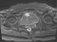
图1
MRI示例2膀胱前壁增厚,内见一结节影(箭头所示),最大径3.0 cm,边界尚清
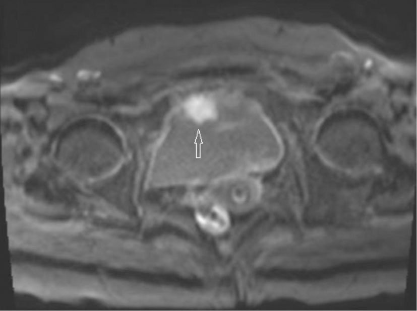
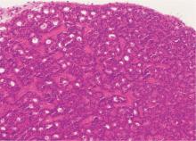
图2
例1肿瘤位于固有层内,呈经典型NENs组织学结构(梁状/假腺管样/筛状),细胞形态较一致,表面被覆正常黏膜(HE,×200)
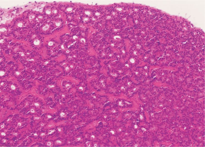
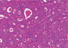
图3
例2肿瘤呈假腺管样/筛状/实性片状/巢团状排列,间质内富于纤细的薄壁血管,假腺腔内见浓染的嗜酸性分泌物聚积(HE,×200)
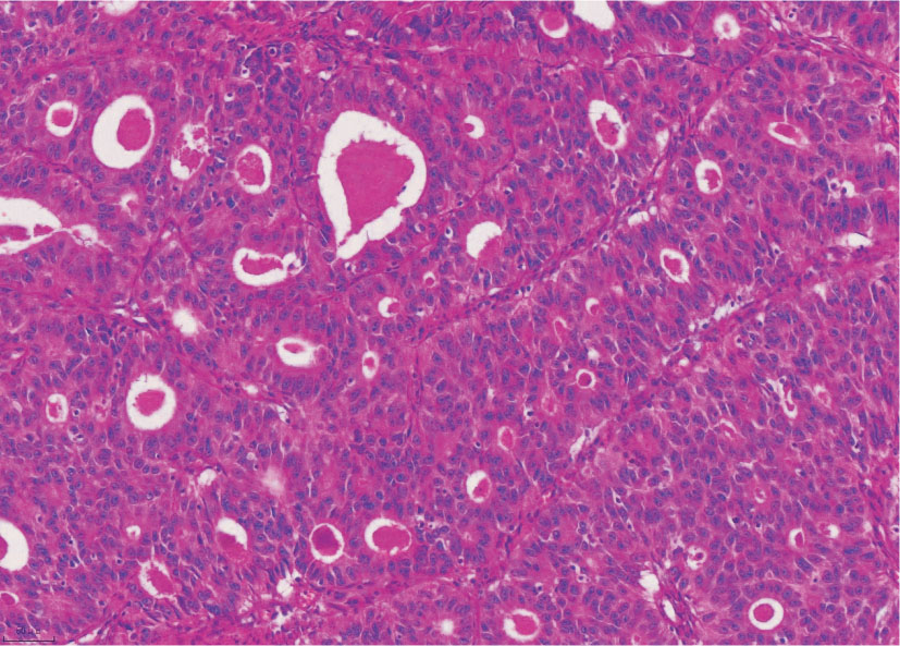
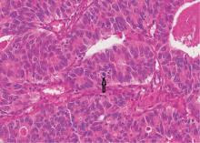
图4
例2肿瘤细胞较大,胞浆丰富、嗜酸性,空泡状核,核仁明显,可见核分裂象(箭头所示)(HE,×400)
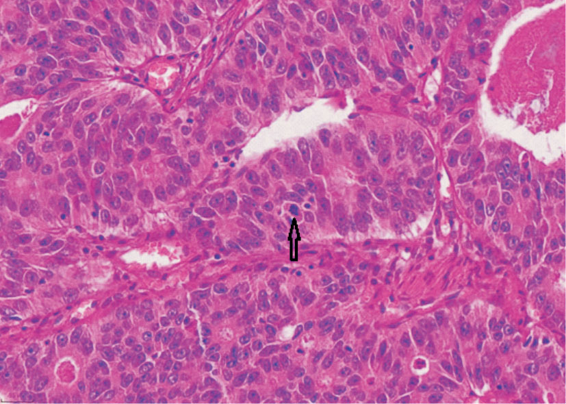
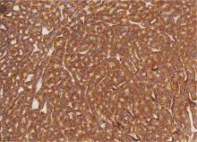
图5
例1肿瘤细胞胞浆Syn弥漫强阳性(EnVision两步法,×200)
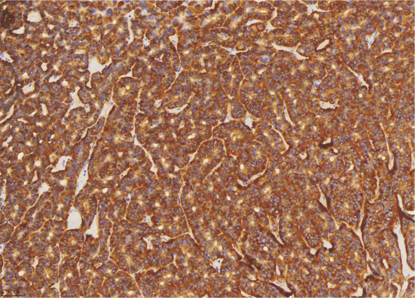
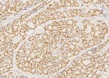
图6
例2肿瘤细胞胞核INSM1弥漫阳性(EnVision两步法,×200)

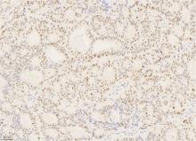
图7
例2肿瘤细胞胞核P53呈斑驳的野生型表达(EnVi-sion两步法,×200)
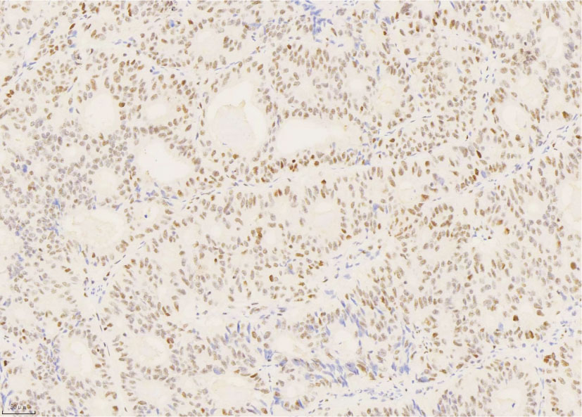
图8
例2肿瘤细胞胞核Ki-67增殖指数高达60%~70% (EnVision两步法,×200)
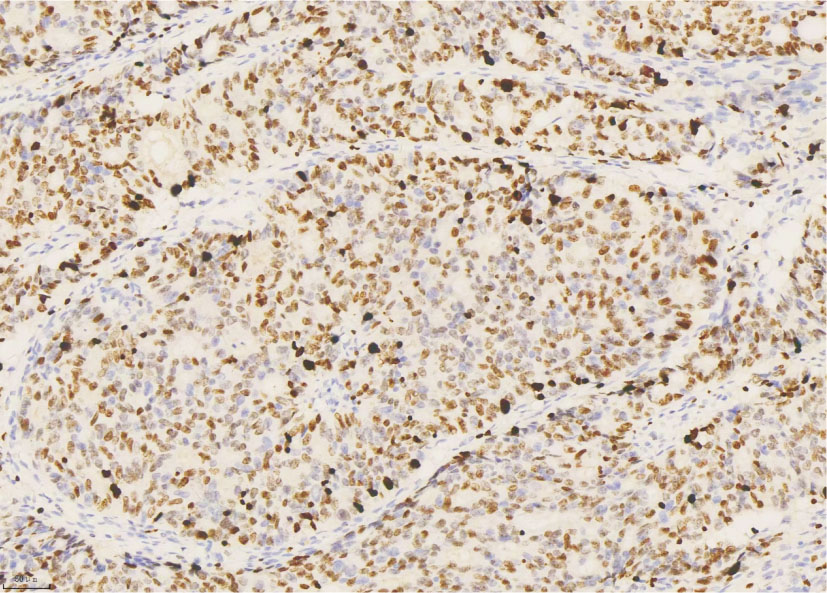
表1
30例膀胱WD-NET临床病理特征和预后
| Reference | Number | Age | Gender | Site and shape | Size (cm) | Clinical feature | Surgery | Staging | Associated urothelial alterations | Follow up and prognosis (mo) |
|---|---|---|---|---|---|---|---|---|---|---|
| [ | 1 | 30 | M | Neck, NA | 0.3 | Hematuria | Biopsy | NA | NA | 12,NED |
| [ | 1 | 61 | F | Trigone, pappilary | 0.3 | Hematuria | TURBT | pT1 | NA | NA |
| [ | 1 | 62 | F | Trigone, polypoid | 1.2 | Hematuria | TURBT | NA | CCG | NA |
| [ | 1 | 54 | F | Neck, polypoid | 0.9 | Hematuria and dysuria | TURBT | NA | Inverting papilloma | 6,NED |
| [ | 1 | 73 | M | Posterior wall, polypoid | 1.0 | Accidentally | TURBT | pT1 | NA | 22,NED |
| [ | 2 | 69/47 | M(2/2) | Neck(2/2),polypoid(2/2) | 0.3,0.7 | Hematuria(2/2) | TURBT(2/2) | pT1(2/2) | CCG/NA=1/1 | NA(2/2) |
| [ | 1 | 77 | M | Trigone, NA, | 1.5 | Accidentally | TURBT | pT1 | NA | NA |
| [ | 1 | 68 | M | Neck, polypoid | 0.4 | Hematuria | TURBT | pT1 | von Brunn’s nest | 14,NED |
| [ | 5 | Mean54.4 | M/F=4/1 | Neck/trigone=3/2, polypoid (5/5) | Mean0.34 | Hematuria/Accidentally=3/2 | TURBT(5/5) | pT1(5/5) | CCG(5/5) | Mean33.4,NED |
| [ | 1 | 49 | F | Trigone, NA | 3.0 | Hematuria | TURBT | pT2 | No | 6,NED |
| [ | 1 | 72 | M | Trigone, polypoid | 0.8 | Accidentally | TURBT | pT1 | CCG | 72,NED |
| [ | 1 | 71 | F | Trigone, polypoid | 2.8 | Vaginal pain and urinary incontinence | TURBT | pT1 | NA | NA |
| [ | 1 | 52 | M | Neck, polypoid | 0.7 | Urinary tract obstruction and hematuria | TURBT | pT1 | NA | 12,NED |
| [ | 1 | 44 | M | Left posterior lateral wall, nodule | 2.0 | Initial liver and peritoneal metastasis | Biopsy | pT2 | NA | 16,Died |
| [ | 1 | 83 | F | Neck, polypoid | 2.5 | Hematuria | TURBT | pT1 | CCG | NA |
| [ | 7 | Mean61.4 | M/F=5/2 | NA/neck=6/1,polypoid(7/7) | NA(7/7) | NA | Biopsy/TURBT=6/1 | pT1(7/7) | CCG/NA/papilloma=5/1/1 | Mean74.86,NED |
| [ | 1 | 51 | F | Neck, polypoid | 0.8 | Hematuria | TURBT | pT1 | CCG | 4,NED |
| [ | 1 | 90 | F | Bottom, polypoid | NA | Dysuria | TURBT | pT1 | CCG | 6,NED |
| Present case | 1 | 85 | F | Trigone, polypoid | 0.6 | Accidentally | TURBT | pT1 | CCG | 76,NED |
表2
32例膀胱LCNEC临床病理特征和预后
| Reference | Number | Age | Gender | Site | Size (cm) | Clinical feature | Staging | Treatment | Follow up and prognosis (mo) |
|---|---|---|---|---|---|---|---|---|---|
| [ | 1 | 73 | M | Posterior wall | 4.0 | Hematuria | pT3b | RC+LND | 2,died of RE/ME |
| [ | 1 | 32 | M | Anterior superior wall | 3.0 | Hematuria | pT3 | PC+CT | 12,alive with RE/ME |
| [ | 2 | 40/43 | M/F=1:1 | NA | NA | NA | pT2,pT4 | RC+CT,RC+RT | 13,NED;12,died of ME |
| [ | 1 | 37 | M | Posterior wall | 2.5 | Hematuria | pT3b | RC+LND+CT | 22,NED |
| [ | 1 | 19 | M | NA | NA | NA | NA | PC+CT | 14,died of ME |
| [ | 1 | 74 | M | Left wall | NA | Brain metastases | NA | PC+CT+RT | 5,died of pulmonary embolism |
| [ | 1 | 67 | M | Left posterior lateral wall | 5.0 | Hematuria | pT2 | TURBT | 0.5,died of heart failure |
| [ | 1 | 59 | M | Bottom | Huge | Dysuria and hematuria | pT4a | TURBT | NA |
| [ | 1 | 68 | M | Top wall | 2.8 | Hematuria | pT2b | RC+LND+CT | 30,died of RE/ME |
| [ | 1 | 84 | M | Posterior wall | NA | Hematuria | NA | TURBT+CT | NA |
| [ | 1 | 68 | M | Top wall | 2.8 | Hematuria | pT2b | RC+LND | NA |
| [ | 1 | 70 | M | Left wall | 3.5 | Hematuria | pT2b | TURBT | 7,died of ME |
| [ | 1 | 58 | M | Left and anterior walls | 6.5 | Hematuria | pT3b | RC | 5,died of ME |
| [ | 5 | Mean71.8 | M/F=4/1 | NA | NA | NA | 3,pT3a/2,pT4a | RC(5/5);1 NACT | 2.4-116.4(Mean14.4), 3 died of RE/2 died from other |
| [ | 1 | 72 | M | Bottom | NA | Back pain and acute renal failure | pT4a | RC+CT | 36,RE with regression |
| [ | 1 | 72 | M | Diffuse | NA | Hematuria | pT2 | RC+CT+IT | 11,died of ME |
| [ | 1 | 45 | M | Left wall | 4.0 | Acute renal failure | NA | TURBT+CT+RT | NA |
| [ | 1 | 39 | M | Right wall | 4.1 | Hematuria | pT2b | RC+LND+CT | 59,NED |
| [ | 1 | 30 | M | Anterior wall | 3.4 | Hematuria | pT3b | PC+CT | 24,NED |
| [ | 1 | 49 | M | Left wall | 7.0 | Vertebral metastasis | pT2 | TURBT+CT+RT | 12,Pain improvemen |
| [ | 1 | 66 | M | Left posterior wall | 4.3 | Hematuria | pT3b | NA | 10,NED |
| [ | 1 | 67 | M | Right posterior wall and right ureteral orifice | 4.4 | Hematuria | pT3a | RC+LND+CT | 39,NED |
| [ | 1 | 56 | F | Anterior wall, posterior wall, and trigone | 3.8 | Hematuria | pT2 | RC+CT | 6,Lost follow-up |
| [ | 1 | 79 | M | Left wall | 5.0 | Hematuria | pT3 | RC+IT | 24,RE |
| [ | 1 | 72 | M | Anterior wall | 2.0 | Hematuria | pT2b | PC | 10,NED |
| [ | 1 | 65 | M | Right anterior wall | 3.0 | Hematuria | pT2a | PC+CT | 7,NED |
| Present case | 1 | 84 | F | Anterior wall | 3.0 | Urinary frequency, urgency, and hematuria | pT3a | PC | 3, died of RE/ME |
| [1] |
KOUBA E, CHENG L. Neuroendocrine tumors of the urinary bladder according to the 2016 World Health Organization classification: molecular and clinical characteristics[J]. Endocr Pathol, 2016, 27(3):188-199.
doi: 10.1007/s12022-016-9444-5 pmid: 27334654 |
| [2] | MENON S, MOCH H. Neuroendocrine neoplasms[M]// WHO Classif. Tumours - Urin. Male Genit. Tumours, 5th ed. IARC,Lyon; 2022:386-391 |
| [3] |
WANG G, YUAN R, ZHOU C, et al. Urinary large cell neuroendocrine carcinoma: a clinicopathologic analysis of 22 cases[J]. Am J Surg Pathol, 2021, 45(10):1399-1408.
doi: 10.1097/PAS.0000000000001740 pmid: 34074810 |
| [4] |
COLBY T V. Carcinoid tumor of the bladder. A case report[J]. Arch Pathol Lab Med, 1980, 104(4):199-200.
pmid: 6892681 |
| [5] | HAILEMARIAM S, GASPERT A, KOMMINOTH P, et al. Primary, pure, large-cell neuroendocrine carcinoma of the urinary bladder[J]. Mod Pathol, 1998, 11(10):1016-1020. |
| [6] |
BURGESS N A, LEWIS D C, MATTHEWS P N. Primary carcinoid of the bladder[J]. Br J Urol, 1992, 69(2):213-214.
pmid: 1537038 |
| [7] |
WALKER B F, SOMEREN A, KENNEDY J C, et al. Primary carcinoid tumor of the urinary bladder[J]. Arch Pathol Lab Med, 1992, 116(11):1217-1220.
pmid: 1444752 |
| [8] |
STANFIELD B L, GRIMES M M, KAY S. Primary carcinoid tumor of the bladder arising beneath an inverted papilloma[J]. Arch Pathol Lab Med, 1994, 118(6):666-667.
pmid: 8204019 |
| [9] |
SUGIHARA A, KAJIO K, YOSHIMOTO T, et al. Primary carcinoid tumor of the urinary bladder[J]. Int Urol Nephrol, 2002, 33(1):53-57.
doi: 10.1023/a:1014400818905 pmid: 12090339 |
| [10] |
MARTIGNONI G, EBLE J N. Carcinoid tumors of the urinary bladder. Immunohistochemical study of 2 cases and review of the literature[J]. Arch Pathol Lab Med, 2003, 127(1):e22-24.
doi: 10.5858/2003-127-e22-CTOTU pmid: 12562289 |
| [11] |
MCCABE J E, DAS S, DOWLING P, et al. Oncocytic carcinoid tumour of the bladder[J]. J Clin Pathol, 2005, 58(4):446-447.
pmid: 15790719 |
| [12] |
MASCOLO M, ALTIERI V, MIGNOGNA C, et al. Calcitonin-producing well-differentiated neuroendocrine carcinoma (carcinoid tumor) of the urinary bladder: case report[J]. BMC Cancer, 2005, 5:88.
pmid: 16048646 |
| [13] | CHEN Y B, EPSTEIN J I. Primary carcinoid tumors of the urinary bladder and prostatic urethra: a clinicopathologic study of 6 cases[J]. Am J Surg Pathol, 2011, 35(3):442-446. |
| [14] | BAYDAR D E, TASAR C. Carcinoid tumor in the urinary bladder: unreported features[J]. Am J Surg Pathol, 2011, 35(11):1754-1757. |
| [15] |
ZOZUMI M, NAKAI M, MATSUDA I, et al. Primary carcinoid tumor of the urinary bladder with prominent subnuclear eosinophilic granules[J]. Pathol Res Pract, 2012, 208(2):109-112.
doi: 10.1016/j.prp.2011.10.008 pmid: 22115748 |
| [16] | KAPLAN A L, MARGOLIS D J, SAID J, et al. Primary carcinoid tumor of urinary bladder discovered on pelvic magnetic resonance imaging[J]. Urology, 2012, 80(5):e55-57. |
| [17] | MONDAL K, MANDAL R. A carcinoid tumor in the urinary bladder with uncommon clinicopathological presentation[J]. Iran J Pathol, 2017, 12(3):277-280. |
| [18] | DADHWAL R, JAIN S, SETH A, et al. Neuroendocrine tumour of urinary bladder: a rare case of aggressively behaving primary well-differentiated neuroendocrine tumour with review of literature[J]. BMJ Case Rep, 2019, 12(11):e231061. |
| [19] |
WARNCKE J, WHITE S, O'KEEFE M, et al. Primary carcinoid tumor of the bladder[J]. Can J Urol, 2018, 25(4):9421-9423.
pmid: 30125523 |
| [20] |
RODRIGUEZ PENA M D C, SALLES D C, EPSTEIN J I, et al. Well-differentiated neuroendocrine tumors of the lower urinary tract: biologic behavior of a rare entity[J]. Hum Pathol, 2021, 109:53-58.
doi: 10.1016/j.humpath.2020.11.014 pmid: 33301750 |
| [21] | MARLETTA S, MARTIGNONI G, GHIMENTON C, et al. Well-differentiated neuroendocrine tumor of the urinary bladder expressing GATA 3[J]. Virchows Arch, 2023, 482(4):783-788. |
| [22] | XU Q, WANG C, WANG Q, et al. Primary well-differentia-ted neuroendocrine tumor of the urinary bladder: report of a very rare case and literature review[J]. Asian J Surg, 2024, 47(12):5450-5451. |
| [23] |
LEE K H, RYU S B, LEE M C, et al. Primary large cell neuroendocrine carcinoma of the urinary bladder[J]. Pathol Int, 2006, 56(11):688-693.
doi: 10.1111/j.1440-1827.2006.02031.x pmid: 17040293 |
| [24] |
ALIJO SERRANO F, SÁNCHEZ-MORA N, ANGEL ARRANZ J, et al. Large cell and small cell neuroendocrine bladder carcinoma: immunohistochemical and outcome study in a single institution[J]. Am J Clin Pathol, 2007, 128(5):733-739.
doi: 10.1309/HTREM6QYQDYGNWYA pmid: 17951193 |
| [25] |
BERTACCINI A, MARCHIORI D, CRICCA A, et al. Neuroendocrine carcinoma of the urinary bladder: case report and review of the literature[J]. Anticancer Res, 2008, 28(2B):1369-1372.
pmid: 18505081 |
| [26] |
LEE W J, KIM C H, CHANG S E, et al. Cutaneous metastasis from large-cell neuroendocrine carcinoma of the urinary bladder expressing CK20 and TTF-1[J]. Am J Dermatopathol, 2009, 31(2):166-169.
doi: 10.1097/DAD.0b013e31818eba4c pmid: 19318803 |
| [27] |
TSUGU A, YOSHIYAMA M, MATSUMAE M. Brain metastasis from large cell neuroendocrine carcinoma of the urinary bladder[J]. Surg Neurol Int, 2011, 2:84.
doi: 10.4103/2152-7806.82250 pmid: 21748036 |
| [28] |
SARI A, ERMETE M, SADULLAHOĞLU C, et al. Large cell neuroendocrine carcinoma of urinary bladder; case presentation[J]. Turk Patoloji Derg, 2013, 29(2):138-142.
doi: 10.5146/tjpath.2013.01165 pmid: 23661352 |
| [29] | JAGGON J R, BROWN T A, MAYHEW R. Metastatic primary neuroendocrine carcinoma of the genitourinary tract: A case report of an uncommon entity[J]. Am J Case Rep, 2013, 14:147-149. |
| [30] | 杜元程, 孙美红, 杨栋嵛, 等. 长链非编码RNA作为竞争性内源RNA在膀胱癌中的研究进展[J]. 中国临床研究, 2022, 35(4):560-562,567. |
| DU YC, SUN MH, YANG DY, et al. Research progress of long chain non coding RNA as competitive endogenous RNA in bladder cancer[J]. Chin Clin Res, 2022, 35(4):560-562,567. | |
| [31] |
TREGLIA G, PAONE G, FLORES B, et al. A rare case of large cell neuroendocrine carcinoma of the urinary bladder evaluated by ¹⁸F-FDG-PET/CT[J]. Rev Esp Med Nucl Imagen Mol, 2014, 33(5):312-313.
doi: 10.1016/j.remn.2013.10.007 pmid: 24440201 |
| [32] |
PUSIOL T, MORICHETTI D, ZORZI M G. "Pure" primary large cell neuroendocrine carcinoma of the urinary bladder: case report, literature review and diagnostic criteria[J]. Pathologica, 2014, 106(2):82-85.
pmid: 25291874 |
| [33] | 蒋艳霞, 于文娟, 张伟, 等. 膀胱神经内分泌癌17例临床病理特征分析[J]. 中华病理学杂志, 2014(11):736-741. |
| JIANG Y X, YU W J, ZHANG W, et al. Clinical and pathological characteristics analysis of 17 cases of bladder neuroendocrine carcinoma[J]. Chin J Pathol, 2014(11):736-741. | |
| [34] | RADOVIĆ N, TURNER R, BACALJA J. Primary "pure" large cell neuroendocrine carcinoma of the urinary bladder: a case report and review of the literature[J]. Clin Genitourin Cancer, 2015, 13(5):e375-377. |
| [35] | GUPTA S, THOMPSON R H, BOORJIAN S A, et al. High grade neuroendocrine carcinoma of the urinary bladder treated by radical cystectomy: a series of small cell, mixed neuroendocrine and large cell neuroendocrine carcinoma[J]. Pathology, 2015, 47(6):533-542. |
| [36] | CHONG V, ZWI J, HANNING F, et al. A case of large cell neuroendocrine carcinoma of the bladder with prolonged spontaneous remission[J]. J Surg Case Rep, 2017, 2017(5):rjw179. |
| [37] | ZAKARIA A, AL SHARE B, KOLLEPARA S, et al. External beam radiation and brachytherapy for prostate cancer: is it a possible trigger of large cell neuroendocrine carcinoma of the urinary bladder?[J] Case Rep Oncol Med, 2017, 2017:1853985. |
| [38] |
AKDENIZ E, BAKIRTAS M, BOLAT M S, et al. Pure large cell neuroendocrine carcinoma of the bladder without urological symptoms[J]. Pan Afr Med J, 2018, 30:134.
doi: 10.11604/pamj.2018.30.134.13437 pmid: 30374380 |
| [39] |
XIA K, ZHONG W, CHEN J, et al. Clinical characteristics, treatment strategy, and outcomes of primary large cell neuroendocrine carcinoma of the bladder: a case report and systematic review of the literature[J]. Front Oncol, 2020, 10:1291.
doi: 10.3389/fonc.2020.01291 pmid: 32850401 |
| [40] |
LI W, SU Z Z, KANG J H, et al. Application of contrast-enhanced ultrasonography for large cell neuroendocrine carcinoma in the urinary bladder: a case report[J]. BMC Med Imaging, 2020, 20(1):46.
doi: 10.1186/s12880-020-00447-6 pmid: 32362278 |
| [41] | TLILI G, AMMAR H, MAJDOUB W, et al. Paraplegia due to medullary compression caused by a large cell neuroendocrine carcinoma of the urinary bladder: a case report[J]. Ann Med Surg (Lond), 2021, 67:102475. |
| [42] | XIAO P, LIU J, SUN W, et al. Large cell neuroendocrine carcinoma of the urinary bladder: A case report and literature review[J]. Asian J Surg, 2023, 6(12):6049-6050. |
| [43] | HE B, CHEN Y, HUI Z. Primary pure bladder large cell neuroendocrine carcinoma: a case report[J]. Asian J Surg, 202, 46(12):5454-5455. |
| [44] |
MOHANTY P, MOHAPATRA A S, SABAT D, et al. Unusual histomorphological spectrum of urinary bladder cancers and their treatment modalities revisited: Our experience with series of five cases[J]. J Cancer Res Ther, 2023, 19(3):617-623.
doi: 10.4103/jcrt.jcrt_134_21 pmid: 37470584 |
| [45] | SUN Z, LIANG X, ZHANG C, et al. Primary pure large cell neuroendocrine carcinoma of the urinary bladder: a case report and literature review[J]. Front Oncol, 2024, 14:1337997. |
| [46] | BAI L L, GUO Y X, SONG S Y, et al. Primary large cell neuroendocrine carcinoma of the bladder: a case report[J]. World J Clin Cases, 2024, 12(21):4783-4788. |
| [47] |
ZHOU Y, YANG L. Large-cell neuroendocrine carcinoma of the bladder: a case report[J]. World J Clin Oncol, 2024, 15(9):1239-1244.
doi: 10.5306/wjco.v15.i9.1239 pmid: 39351458 |
| [48] |
SANGUEDOLCE F, CALÒ B, CHIRICO M, et al. Urinary tract large cell neuroendocrine carcinoma: diagnostic, prognostic and therapeutic issues[J]. Anticancer Res, 2020, 40(5):2439-2447.
doi: 10.21873/anticanres.14213 pmid: 32366387 |
| [49] | REKHTMAN N. Lung neuroendocrine neoplasms: recent progress and persistent challenges[J]. Mod Pathol, 2022, 35(Suppl 1):36-50. |
| [50] | 中华医学会病理学分会消化疾病学组, 2020年中国胃肠胰神经内分泌肿瘤病理诊断共识专家组. 中国胃肠胰神经内分泌肿瘤病理诊断共识(2020版)[J]. 中华病理学杂志, 2021, 50(1):14-20. |
| Digestive Disease Group, Pathology Branch, Chinese Medical Association, 2020 Consensus Expert Group On Pathological Diagnosis of Gastrointestinal, Pancreatic, Neuroendocrine Tumors in China. Pancreatic, Neuroendocrine Tumors in China. Chinese consensus on pathological diagnosis of gastrointestinal pancreatic neuroendocrine tumors (2020 edition)[J]. Chin J Pathol, 2021, 50(1):14-20. |
| [1] | 周晓蝶, 陈巍魏, 余波, 王璇, 王建军, 石群立, 饶秋, 鲍炜. 尿路上皮癌的临床病理学特征[J]. 诊断学理论与实践, 2023, 22(03): 292-299. |
| [2] | 何燕燕, 李凤珠. 膀胱原发性上皮样血管肉瘤一例报道及文献复习[J]. 诊断学理论与实践, 2022, 21(06): 719-725. |
| [3] | 徐苓, 王根发, 张良. UroVysion 荧光原位杂交技术检测在泌尿系统肿瘤诊断及膀胱癌预后监测中应用的初步探索[J]. 诊断学理论与实践, 2018, 17(02): 159-164. |
| [4] | 李莉, 卞炳贤, 张良, 沈立松. 尿液多种microRNA检测方法的建立及其在膀胱癌诊断中的应用研究[J]. 诊断学理论与实践, 2017, 16(01): 93-97. |
| [5] | 范瑜, 刘晓晟, 路青, 姚秋英,. 膀胱癌3.0T MRI多b值弥散加权成像研究[J]. 诊断学理论与实践, 2013, 12(03): 334-338. |
| [6] | 李莉, 丁奕星, 徐苓, 马妍慧, 张良,. 染色体3、7、17号和9p21位点在膀胱尿路上皮癌中的畸变情况及其临床意义[J]. 诊断学理论与实践, 2012, 11(01): 47-51. |
| [7] | 周健, 李海, 陈金珍, 黄振华,. Survivin、FAS在膀胱移行细胞癌中的表达及临床意义[J]. 诊断学理论与实践, 2009, 8(06): 627-630. |
| [8] | 张磊,. 膀胱侵袭性血管黏液瘤1例[J]. 诊断学理论与实践, 2008, 7(02): 213-. |
| [9] | 陈华, 张华, 项明洁, 张志伟, 周文龙, 沈周俊, 黄海峰,. 尿膀胱癌抗原、CYFRA21-1和透明质酸在膀胱癌诊断中的价值[J]. 诊断学理论与实践, 2008, 7(01): 73-76. |
| [10] | 陈华, 张华, 项明洁,. 膀胱癌尿肿瘤标志物的研究进展[J]. 诊断学理论与实践, 2006, 5(05): 446-448. |
| [11] | 徐锋,曹文俊,吴华成,王枕亚,林佳菲,樊绮诗. 生存素检测在膀胱癌实验诊断中的意义[J]. 诊断学理论与实践, 2005, 4(03): 206-208. |
| 阅读次数 | ||||||
|
全文 |
|
|||||
|
摘要 |
|
|||||