诊断学理论与实践 ›› 2019, Vol. 18 ›› Issue (04): 436-441.doi: 10.16150/j.1671-2870.2019.04.011

• 论著 • 上一篇    下一篇

运用动态增强磁共振成像评估颈动脉粥样斑块稳定性的初步研究

曹烨, 刘晓晟, 葛晓乾, 周斌()   

  1. 上海交通大学医学院附属仁济医院放射科,上海 200127
  • 收稿日期:2019-02-28 出版日期:2019-08-25 发布日期:2019-08-25
  • 通讯作者: 周斌 E-mail:zbyeah2003@163.com

Preliminary study on dynamic contrast-enhanced MRI in identifying vulnerability of carotid atherosclerotic plaques

CAO Ye, LIU Xiaosheng, GE Xiaoqian, ZHOU Bin()   

  1. Depatrment of Radiology, Renji Hospital, Shanghai Jiao Tong University College of Medicine, 200127, Chian
  • Received:2019-02-28 Online:2019-08-25 Published:2019-08-25
  • Contact: ZHOU Bin E-mail:zbyeah2003@163.com

摘要:

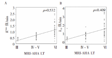
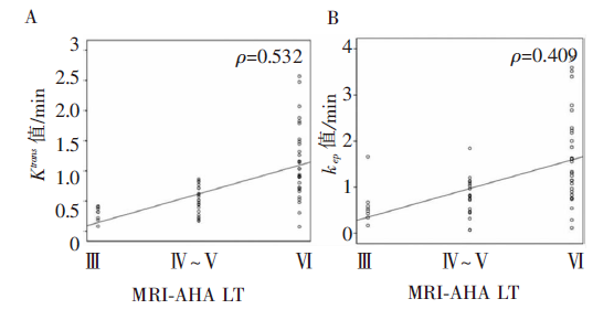
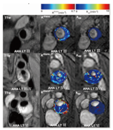
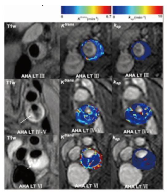
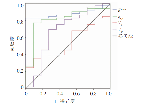
目的: 探索3.0T动态增强磁共振成像(dynamic contrast-enhanced magnetic resonance imaging,DCE-MRI)评价颈动脉粥样硬化斑块稳定性的效能。方法: 对66例超声检查显示颈动脉内膜增厚 ≥2 mm的患者,行颈动脉磁共振管壁成像(vessel wall magnetic resonance imaging,VW-MRI)及DCE-MRI扫描,并依据MRI修正的美国心脏协会(American Heart Association,AHA)斑块分型标准,对颈动脉斑块进行分型与定性(Ⅳ~Ⅵ型为不稳定斑块,余为稳定斑块),计算DCE-MRI药代动力学参数(Ktranskepvevp),评估各参数与斑块分型、斑块稳定性间的相关性,以及各参数区分稳定斑块与不稳定斑块的效能。结果: Ktranskep均与MRI 修正的AHA分型(Ⅲ~Ⅵ型)呈显著正相关(ρ=0.532, P<0.001; ρ=0.409, P<0.001)。Ktranskep可区分稳定斑块与易损斑块,其最佳临界值分别为0.043/min(此时诊断灵敏度为83%,特异度为100%)和0.741/min(诊断灵敏为77%,特异度为91%)。结论: DCE-MRI药代动力学参数Ktranskep可定量评估颈动脉斑块的稳定性,是斑块危险性分层的影像学标志物。

关键词: 动态增强磁共振成像, 颈动脉粥样硬化, 不稳定斑块

Abstract:

Objective: To explore the ability of 3.0T dynamic contrast-enhanced MR imaging (DCE-MRI) in identi-fying the vulnerability of carotid atherosclerotic plaques. Methods: From September 2016 to June 2017, 66 patients with carotid intima thickening ≥2 mm were enrolled in this study. All participants underwent vessel wall MR imaging (VW-MRI) and DCE-MRI. With the classification of plaques according to MRI-modified AHA Lesion-Type (LT), pharmacokine-tic parameters (PPs) of DCE-MRI were measured, including Ktrans, kep, ve and vp. Correlations between PPs and LTs and the ability of these parameters to distinguish stable and vulnerable plaques suspected on MRI were assessed. Results: Signi-ficant positive correlation between Ktrans and LT Ⅲ to Ⅵ was found (P=0.532, P<0.001), so as was the correlation between kep and LT Ⅲ to Ⅵ (P=0.409, P<0.001). Stable and vulnerable plaques suspected on MRI could potentially be distinguished by Ktrans (sensitivity 83%, specificity 100%) and kep (sensitivity 77%, specificity 91%). Conclusions: Ktrans and kep from DCE-MRI can provide quantitative information for the differentiation of vulnerable plaques suspected on MRI from stable ones. These two parameters could be adopted as imaging markers for plaque characterization and risk stratification.

Key words: Dynamic contrast-enhanced magnetic resonance imaging, Carotid atherosclerosis, Vulnerable plaque

中图分类号: