诊断学理论与实践 ›› 2020, Vol. 19 ›› Issue (06): 588-593.doi: 10.16150/j.1671-2870.2020.06.008

• 论著 • 上一篇    下一篇

结肠镜窄带成像技术对直径<2 cm结肠息肉样病变的诊断价值及误诊分析

吴友伟, 张健(), 赵素平, 吕飒美, 史丽萍   

  1. 陕西省人民医院消化内二科,陕西 西安 710068
  • 收稿日期:2019-07-24 出版日期:2020-12-25 发布日期:2022-07-14
  • 通讯作者: 张健 E-mail:jawgan@qq.com
  • 基金资助:
    西安市科技计划项目(2017113SF/YX007)

Diagnostic value of colonoscopic narrow-band imaging for the colon polypoid lesions with diameter<2 cm and analysis of misdiagnosis

WU Youwei, ZHANG Jian(), ZHAO Suping, LÜ Samei, SHI Liping   

  1. Second Department of Gastroenterology, Shaanxi Provincial People's Hospital, Shaanxi Xi'an 710068, China
  • Received:2019-07-24 Online:2020-12-25 Published:2022-07-14
  • Contact: ZHANG Jian E-mail:jawgan@qq.com

摘要:

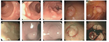
目的: 分析结肠镜窄带成像(narrow-band imaging,NBI)技术对直径<2 cm结肠息肉样病变的诊断价值。方法: 选取2018年2月至2019年2月期间在我院诊断的结肠息肉样病变患者86例,采用结肠镜常规模式和结肠镜NBI模式观察病灶的整体形态、腺管开口类型、毛细血管形态,并进行清晰度评分,同时在结肠镜下采集标本进行病理活检,以组织病理学诊断结果作为金标准,对NBI的诊断价值及误诊原因进行分析。结果: 86例患者共检出结肠息肉样病变104个,病灶直径为0.1~2.0 cm,平均直径为(0.72±0.34) cm,采用结肠镜NBI下国际结肠直肠病变内镜(NBI International Colorectal Endoscopic,NICE)分型,结果提示1型的结肠息肉样病变为39个(37.50%),2型为57个(54.81%),3型为8个(7.69%),不同NICE分型的结肠息肉样病变间的形态学特征差异无统计学意义(P>0.05)。病理学检查结果显示,非肿瘤性息肉为38个(36.54%),肿瘤性息肉为66个(63.46%)。结肠镜常规模式诊断结肠肿瘤性息肉的灵敏度为81.82%,特异度为81.57%,准确率为81.73%,NBI模式诊断结肠肿瘤性息肉的灵敏度为92.42%,特异度为89.47%,准确率为91.35%,2种方法间差异有统计学意义(P<0.05)。进一步分析显示,结肠镜NBI模式诊断直径<1 cm和1~2 cm肿瘤性息肉的灵敏度分别为92.59%和92.31%,特异度分别为86.67%和91.30%,准确率分别为90.48%和91.94%;不同直径的结肠息肉病变,采用结肠镜常规模式和NBI模式诊断结果与病理诊断间的一致性Kappa值分别为0.793和0.829。结肠镜NBI模式误诊的原因主要为肠道准备不足和病灶微结构显示不清。结论: 结肠镜NBI模式的结肠息肉检出率较结肠镜常规模式有所提升,在直径<2 cm的结肠息肉样病变鉴别诊断中具有较高价值,且对直径<1 cm的结肠息肉样病变性质鉴别诊断也具有较高准确率,可在临床推广应用。

关键词: 结肠息肉样病变, 结肠镜, 窄带成像

Abstract:

Objective: To study the diagnostic value of colonoscopic narrow-band imaging (NBI) for colon polypoid lesions with the diameter <2 cm. Methods: A total of 86 patients with the colon polypoid lesions diagnosed in our hospital during February 2018 to February 2019 were enrolled. Routine enteroscopy and colonoscopic NBI were performed in the patients, and the morphology of colonpolypoid lesions including the overall lesion, glandular opening type and capillary were observed, and the images were assessed with clarity score. Biopsy specimens obtained during performing colonoscopy were analyzed by pathological examination, and the diagnostic efficacy of NBI were assessed by comparing the results of pathological examination. Results: A total of 104 colon polypoid lesions were detected among 86 patients, and the diameters of lesions were 0.1-2.0 cm and the average value was(0.72±0.34) cm. The NBI International Colorectal Endoscopic(NICE) classification of lesions were summarized as follows: 39 (37.50%) colon polypoid lesions were type 1, 57 (54.81%) lesions were type 2 and 8 (7.69%) were type 3. There were no significant difference in the morphological characteristics of colon polypoid lesions between the different NICE types (P>0.05). The pathological examination showed that there were 38 non-neoplastic polyps (36.54%) and 66 neoplastic polyps (63.46%). The sensitivity, specificity and accuracy rate of colonoscopy for diagnosing the neoplastic polyps were 81.82%, 81.57% and 81.73%, respectively, while the values of NBI colonoscopy were 92.42%, 89.47% and 91.35%. The diagnostic efficacy between two methods were different (P<0.05). Further analysis showed that the diagnostic sensitivities of NBI colonoscopy for neoplastic polyps with the diameter<1cm and 1-2 cm were 92.59% and 92.31%, the specificities were 86.67% and 91.30%, and the accuracy rates were 90.48% and 91.94%. The consistency Kappa values of enteroscopy and colonoscopic NBI with results of pathology for the neoplastic polyps of different diameters were 0.793 and 0.829, respectively. The causes for misdiagnosing colon polypoid lesions by colonoscopic NBI were insufficient bowel preparations and blurred display of microstructure. Conclusions: Compared with the colonoscopy, the accuracy of NBI colonoscopy for diagnosing colon polyps is improved. The NBI colonoscopy is worth to be applied in clinical practice since it has high diagnostic value for colon polypoid lesions with the diameter <2 cm and also shows high accuracy for judging the characteristic of lesions with the diameter <1 cm.

Key words: Colon polypoid lesions, Colonoscopy, Narrow-band imaging

中图分类号: