诊断学理论与实践 ›› 2020, Vol. 19 ›› Issue (04): 407-413.doi: 10.16150/j.1671-2870.2020.04.016
高燕婷, 赵金艳, 王娟, 李佳, 许雯, 李莉, 蔺丽慧( )
)
收稿日期:2020-04-22
出版日期:2020-08-25
发布日期:2022-07-15
通讯作者:
蔺丽慧
E-mail:linlihui2014@126.com
基金资助:
GAO Yanting, ZHAO Jinyan, WANG Juan, LI Jia, XU Wen, LI Li, LIN Lihui()
Received:2020-04-22
Online:2020-08-25
Published:2022-07-15
Contact:
LIN Lihui
E-mail:linlihui2014@126.com
摘要:
目的: 分析急性髓细胞性白血病(acute myeloid leukemia,AML)患者的骨髓淋巴细胞亚群分布,比较不同疾病阶段、不同预后风险患者间的骨髓免疫功能差异,并探讨其临床意义。方法: 选取131例AML确诊患者治疗前的骨髓样本,采用多色流式细胞术分析样本中各淋巴细胞亚群(CD4+、CD8+、CD19+等)占总淋巴细胞的百分比及CD4+/CD8+比值,并将结果与公共数据平台获取的健康者骨髓样本淋巴细胞亚群结果进行比较。AML患者分别为初诊(94例)、继发(18例)及化疗缓解后复发患者(19例),共3组,比较各组间淋巴细胞亚群差异;进一步根据细胞遗传学异常将初诊AML分为低危、中危及高危组,比较3组间的淋巴细胞亚群差异。回溯复发组患者初发时的骨髓样本检测结果,配对比较初发与复发时样本中淋巴细胞亚群的变化。结果: 与健康者比较,AML患者骨髓样本中CD8+ T淋巴细胞百分比显著增高[(31.73%±12.38)%比(21.40%±7.33%),P<0.001],CD19+ B淋巴细胞百分比及CD4+/CD8+比值显著降低(9.62%比14.03%,P<0.01;1.04比1.48,P<0.05);复发患者骨髓样本中的CD8+ T淋巴细胞百分比较初诊患者显著增高[(41.56±11.64%)比(29.86±12.20%),P<0.001],CD19+ B淋巴细胞百分比及CD4+/CD8+比值显著降低(4.18%比11.82%,P<0.05;0.59比1.12,P<0.05)。配对比较显示,复发患者的CD19+ B细胞百分比与其初发时比较显著降低(2.40%比12.41%,P<0.05)。94例初诊AML不同预后风险度的3组间各淋巴细胞亚群比较,差异无统计学意义。各组间自然杀伤(natural killer, NK)细胞百分比差异无统计学意义。结论: AML患者骨髓呈明显的体液免疫及细胞免疫功能抑制状态,AML复发患者的骨髓免疫功能抑制较其初发时更为显著,提示检测AML患者骨髓淋巴亚群的分布情况可能是评估疾病预后的有效指标,对临床使用免疫调节药物治疗具有一定的指导意义。
中图分类号:
高燕婷, 赵金艳, 王娟, 李佳, 许雯, 李莉, 蔺丽慧. 急性髓细胞性白血病患者骨髓淋巴细胞亚群分析及其临床意义[J]. 诊断学理论与实践, 2020, 19(04): 407-413.
GAO Yanting, ZHAO Jinyan, WANG Juan, LI Jia, XU Wen, LI Li, LIN Lihui. Analysis of bone marrow lymphocyte subsets in patients with acute myeloid leukemia and its clinical significance[J]. Journal of Diagnostics Concepts & Practice, 2020, 19(04): 407-413.
表1
131例AML患者的细胞遗传学异常[n(%)]
| 分组 | 初诊AML | 继发AML(n=18) | 复发AML(n=19) | ||
|---|---|---|---|---|---|
| 低危组(n=36) | 中危组(n=27) | 高危组(n=31) | |||
| 性别(女/男) | 14/22 | 11/16 | 12/19 | 8/10 | 4/15 |
| 中位年龄(岁) | 53(18~77) | 38(26~68) | 54(25~72) | 53(33~78) | 48(17~70) |
| 融合基因 | |||||
| RUNX1-RUNX1T1 | 6(16.67%) | 1(5.26%) | |||
| KMT2A重排 | 2(6.45%) | 3(15.79%) | |||
| CBFB-MYH11 | 7(19.44%) | 3(15.79%) | |||
| 突变基因 | |||||
| NPM1 | 14(38.89%) | 1(3.70%) | 2(6.45%) | 1(5.26%) | |
| NPM1突变伴低水平FLT3-ITD | 2(5.56%) | 1(3.23%) | |||
| NPM突变伴高水平FLT3-ITD | 1(3.70%) | 1(3.23%) | |||
| 野生型NPM1伴低水平FLT3-ITD | 4(14.81%) | 4(12.90%) | 2(10.53%) | ||
| 野生型NPM1伴高水平FLT3-ITD | 1(3.23%) | ||||
| CEBPA双突变 | 9(25.00%) | ||||
| RUNX1 | 2(5.56%) | 7(22.58%) | 1(5.56%) | 2(10.53%) | |
| ASXL1 | 9(25.00%) | 21(67.74%) | 10(55.56%) | 2(10.53%) | |
| TP53 | 5(16.13%) | 5(27.78%) | 2(10.53%) | ||
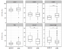
图1
健康者与AML患者骨髓样本中淋巴细胞亚群比较
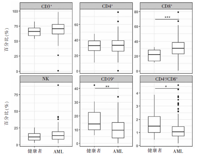
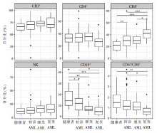
图2
健康者与不同疾病阶段AML患者骨髓样本中淋巴细胞亚群比较
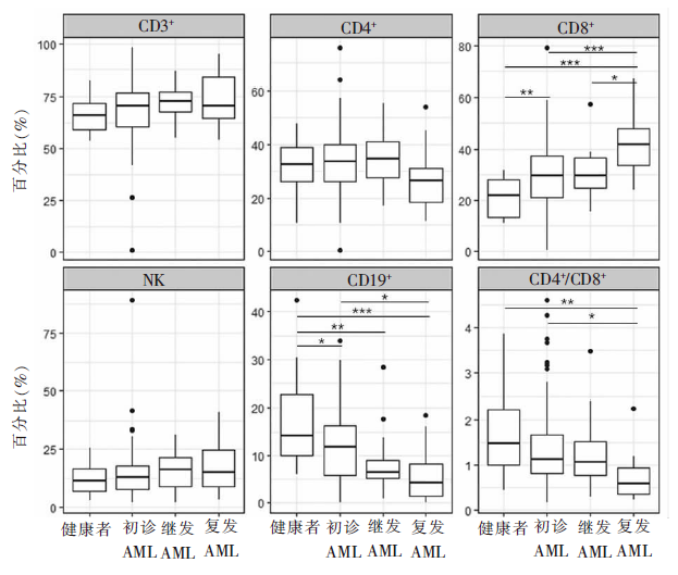
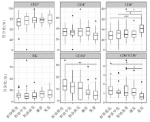
图3
不同危险度分层的初诊AML与继发、复发患者骨髓中淋巴细胞亚群比较


图4
15对AML患者初发与复发骨髓样本中淋巴细胞亚群配对比较

| [1] |
Döhner H, Weisdorf DJ, Bloomfield CD. Acute myeloid leukemia[J]. N Engl J Med, 2015, 373(12):1136-1152.
doi: 10.1056/NEJMra1406184 URL |
| [2] |
Global Burden of Disease Cancer Collaboration, Fitzmaurice C, Dicker D, et al. The global burden of cancer 2013[J]. JAMA Oncol, 2015, 1(4):505-527.
doi: 10.1001/jamaoncol.2015.0735 pmid: 26181261 |
| [3] |
Song X, Peng Y, Wang X, et al. Incidence, survival, and risk factors for adults with acute myeloid leukemia not otherwise specified and acute myeloid leukemia with recurrent genetic abnormalities: analysis of the surveillance, epidemiology, and end results (SEER) database, 2001-2013[J]. Acta Haematol, 2018, 139(2):115-127.
doi: 10.1159/000486228 URL |
| [4] |
Deschler B, Lübbert M. Acute myeloid leukemia: epidemiology and etiology[J]. Cancer, 2006, 107(9):2099-2107.
pmid: 17019734 |
| [5] | Institute NC. Cancer Stat Facts: Leukemia-Acute Myeloid Leukemia(AML)[DB/OL]. 2017 [2020-04-22]. https://seer.cancer.gov/statfacts/html/alyl.html. |
| [6] | Le Dieu R, Taussig DC, Ramsay AG, et al. Peripheral blood T cells in acute myeloid leukemia (AML) patients at diagnosis have abnormal phenotype and genotype and form defective immune synapses with AML blasts[J]. Blood, 2009, 114(18):3909-3916. |
| [7] |
Paczulla AM, Rothfelder K, Raffel S, et al. Absence of NKG2D ligands defines leukaemia stem cells and media-tes their immune evasion[J]. Nature, 2019, 572(7768):254-259.
doi: 10.1038/s41586-019-1410-1 URL |
| [8] |
Raulet DH, Gasser S, Gowen BG, et al. Regulation of li-gands for the NKG2D activating receptor[J]. Annu Rev Immunol, 2013, 31:413-441.
doi: 10.1146/annurev-immunol-032712-095951 pmid: 23298206 |
| [9] |
Goswami M, Prince G, Biancotto A, et al. Impaired B cell immunity in acute myeloid leukemia patients after chemotherapy[J]. J Transl Med, 2017, 15(1):155.
doi: 10.1186/s12967-017-1252-2 URL |
| [10] |
Gabert J, Beillard E, van der Velden VH, et al. Standardization and quality control studies of 'real-time' quantitative reverse transcriptase polymerase chain reaction of fusion gene transcripts for residual disease detection in leukemia-a Europe Against Cancer program[J]. Leukemia, 2003, 17(12):2318-2357.
doi: 10.1038/sj.leu.2403135 pmid: 14562125 |
| [11] |
Beillard E, Pallisgaard N, van der Velden VH, et al. Evaluation of candidate control genes for diagnosis and residual disease detection in leukemic patients using 'real-time' quantitative reverse-transcriptase polymerase chain reaction (RQ-PCR)-a Europe against cancer program[J]. Leukemia, 2003, 17(12):2474-2486.
pmid: 14562124 |
| [12] |
Duncavage EJ, Abel HJ, Szankasi P, et al. Targeted next generation sequencing of clinically significant gene mutations and translocations in leukemia[J]. Mod Pathol, 2012, 25(6):795-804.
doi: 10.1038/modpathol.2012.29 URL |
| [13] |
Luthra R, Patel KP, Reddy NG, et al. Next-generation sequencing-based multigene mutational screening for acute myeloid leukemia using MiSeq: applicability for diagnostics and disease monitoring[J]. Haematologica, 2014, 99(3):465-473.
doi: 10.3324/haematol.2013.093765 URL |
| [14] |
Arber DA, Orazi A, Hasserjian R, et al. The 2016 revision to the World Health Organization classification of myeloid neoplasms and acute leukemia[J]. Blood, 2016, 127(20):2391-2405.
doi: 10.1182/blood-2016-03-643544 URL |
| [15] |
Döhner H, Estey E, Grimwade D, et al. Diagnosis and management of AML in adults: 2017 ELN recommendations from an international expert panel[J]. Blood, 2017, 129(4):424-447.
doi: 10.1182/blood-2016-08-733196 URL |
| [16] |
Oetjen KA, Lindblad KE, Goswami M, et al. Human bone marrow assessment by single-cell RNA sequencing, mass cytometry, and flow cytometry[J]. JCI Insight, 2018, 3(23):e124928.
doi: 10.1172/jci.insight.124928 URL |
| [17] | 黄方, 郝思国. 急性髓系白血病患者外周血T淋巴细胞亚群的水平变化及临床意义[J]. 第二军医大学学报, 2020, 41(5):546-550. |
| [18] | 杨莉, 何浩明. 急性髓细胞白血病患者外周血淋巴细胞亚群的检验分析[J]. 国际检验医学杂志, 2016, 37(6):817-819. |
| [19] | 晁丹阳. 68例急性髓细胞白血病患者淋巴细胞亚群的变化分析[J]. 临床医学, 2017, 37(10):66-68. |
| [20] |
Austin R, Smyth MJ, Lane SW. Harnessing the immune system in acute myeloid leukaemia[J]. Crit Rev Oncol Hematol, 2016, 103:62-77.
doi: 10.1016/j.critrevonc.2016.04.020 URL |
| [21] |
Williams P, Basu S, Garcia-Manero G, et al. The distribution of T-cell subsets and the expression of immune checkpoint receptors and ligands in patients with newly diagnosed and relapsed acute myeloid leukemia[J]. Cancer, 2019, 125(9):1470-1481.
doi: 10.1002/cncr.31896 pmid: 30500073 |
| [22] |
Bozzano F, Perrone C, Moretta L, et al. NK cell precursors in human bone marrow in health and inflammation[J]. Front Immunol, 2019, 10:2045.
doi: 10.3389/fimmu.2019.02045 URL |
| [23] |
Ribeiro VS, Hasan M, Wilson A, et al. Cutting edge: Thymic NK cells develop independently from T cell precursors[J]. J Immunol, 2010, 185(9):4993-4997.
doi: 10.4049/jimmunol.1002273 pmid: 20889548 |
| [24] | Vargas CL, Poursine-Laurent J, Yang L, et al. Development of thymic NK cells from double negative 1 thymocyte precursors[J]. Blood, 2011, 118(13):3570-3578. |
| [25] |
Freud AG, Yu J, Caligiuri MA. Human natural killer cell development in secondary lymphoid tissues[J]. Semin Immunol, 2014, 26(2):132-137.
doi: 10.1016/j.smim.2014.02.008 URL |
| [26] |
Jia B, Wang L, Claxton DF, et al. Bone marrow CD8 T cells express high frequency of PD-1 and exhibit reduced anti-leukemia response in newly diagnosed AML patients[J]. Blood Cancer J, 2018, 8(3):34.
doi: 10.1038/s41408-018-0069-4 URL |
| [27] |
Knaus HA, Berglund S, Hackl H, et al. Signatures of CD8+ T cell dysfunction in AML patients and their reversibility with response to chemotherapy[J]. JCI Insight, 2018, 3(21):e120974.
doi: 10.1172/jci.insight.120974 URL |
| [1] | 石峰, 郭竹英, 郭海艳. 新型冠状病毒肺炎患者外周血淋巴细胞亚群变化的临床意义[J]. 诊断学理论与实践, 2022, 21(05): 619-624. |
| [2] | 王晨琛, 方跃华, 施仲伟, 屈雪蒸. 25例主动脉瓣成形术后一年的超声心动图评价[J]. 诊断学理论与实践, 2022, 21(03): 395-398. |
| [3] | 罗晓颖, 许燕, 张凤如, 吴立群, 戚文航. P波离散度和N端脑钠肽前体预测房颤冷冻球囊导管消融术后复发的价值[J]. 诊断学理论与实践, 2020, 19(1): 32-36. |
| [4] | 郭娟娟, 吴涛, 贾占武, 马利, 白海, 王存邦. WT1阳性急性髓细胞性白血病合并唐氏综合征一例报告[J]. 诊断学理论与实践, 2020, 19(05): 531-533. |
| [5] | 彭真萍, 项喜喜, 张苏江, 李佳明. 以类白血病反应为首发表现的慢性中性粒细胞白血病二例并文献复习[J]. 诊断学理论与实践, 2020, 19(02): 122-128. |
| [6] | 来小音, 孙家兰, 胡荣郭, 杨雪莲, 吴国炉, 李龙宣, 卜碧涛. T淋巴细胞亚群失衡与全身型重症肌无力临床症状加重及缓解的相关性研究[J]. 诊断学理论与实践, 2019, 18(2): 199-203. |
| [7] | 吕良敬, 倪若柠. 重视复发性流产患者中未分化结缔组织病的诊断[J]. 诊断学理论与实践, 2018, 17(03): 235-237. |
| [8] | 王晨琛, 詹维伟. 甲状腺癌术后复发转移灶的超声特征及超声引导下细针穿刺的应用价值[J]. 诊断学理论与实践, 2018, 17(01): 111-114. |
| [9] | 董育玮, 李郑红, 汪佩文, 陆伦根. 良性复发性肝内胆汁淤积伴先天性黄疸一例报道[J]. 诊断学理论与实践, 2017, 16(04): 434-436. |
| [10] | 陈颖, 李翠, 应春妹. 复发性自然流产患者T细胞中T细胞免疫球蛋白和免疫受体酪氨酸抑制基序的表达[J]. 诊断学理论与实践, 2017, 16(03): 273-276. |
| [11] | 戴然然, 王林林, 时国朝,. 调节性T细胞表达CD39和CD73在小鼠哮喘模型发病中的作用[J]. 诊断学理论与实践, 2016, 15(03): 226-230. |
| [12] | 吴瑛婷, 张军, 陈慧芬,. 原发性胆汁性肝硬化合并妊娠的研究进展[J]. 诊断学理论与实践, 2015, 14(06): 573-576. |
| [13] | 罗晓颖, 权薇薇, 许燕, 张风如, 吴立群, 戚文航,. C反应蛋白与房颤冷冻球囊导管消融术后房颤复发的相关性研究[J]. 诊断学理论与实践, 2015, 14(03): 219-222. |
| [14] | 刘海艳, 沈莉菁, 陈芳源,. Notch及Wnt信号通路与急性T淋巴细胞白血病[J]. 诊断学理论与实践, 2014, 13(06): 624-627. |
| [15] | 费奇力, 王维维, 袁向亮, 张良,. 标本前处理因素对流式细胞术检测淋巴细胞亚群的影响[J]. 诊断学理论与实践, 2014, 13(03): 325-328. |
| 阅读次数 | ||||||
|
全文 |
|
|||||
|
摘要 |
|
|||||