诊断学理论与实践 ›› 2024, Vol. 23 ›› Issue (06): 580-586.doi: 10.16150/j.1671-2870.2024.06.004
徐梦迪1,2, 高峰1, 朱剑1, 陈蕾1, 秦雨萌1, 黄越1, 唐银萍1, 沙杰1( )
)
收稿日期:2024-06-25
出版日期:2024-12-25
发布日期:2024-12-25
通讯作者:
沙杰 E-mail: shajie0414@126.com基金资助:
XU Mengdi1,2, GAO Feng1, ZHU Jian1, CHEN Lei1, QIN Yumeng1, HUANG Yue1, TANG Yinping1, SHA Jie1()
Received:2024-06-25
Published:2024-12-25
Online:2024-12-25
摘要:
目的: 探讨新型海绵胶囊联合人工智能细胞DNA检测在早期食管癌筛查中的价值。方法: 2021年6月至 2022年 6月期间,向社会招募年龄>40岁,愿意行食管癌筛查的受试者。首先让受试者行新型海绵细胞胶囊检查,收集细胞标本,采用人工智能 (artificial intelligence,AI) 评测细胞学 DNA 指数(DNA index,DI),后均行内镜检查,评价细胞学DI值与内镜结果之间的关系。结果: 本研究共纳入连续1 369名受试者。经内镜确诊食管病变组25例,其中食管低级别上皮内瘤变15例,食管高级别上皮内瘤变1例,食管癌9例。正常食管组1 344例,正常食管组DI值为2.154±0.339,食管病变组DI值为2.832±0.479,食管病变组DI值明显高于食管正常组DI值(P<0.05)。Logistic回归分析显示,食管病变组与食管正常组的 DI值比值比(odds ratio,OR)为 0.04(95%CI为 0.017~0.096)。海绵胶囊联合AI评测DI值诊断食管病变的最佳临界值为2.450,DI>2.450诊断食管病变的受试者工作特征(receiver operating characteristic,ROC)曲线下面积为 0.914,特异度为 83.71%,灵敏度为 88.00%,准确率为 83.78%。结论: 采用新型海绵胶囊收集细胞行AI评测细胞学DI检测可用于早期食管疾病(尤其是食管癌)的筛查。
中图分类号:
徐梦迪, 高峰, 朱剑, 陈蕾, 秦雨萌, 黄越, 唐银萍, 沙杰. 新型海绵胶囊联合人工智能细胞DNA检测在早期食管癌筛查中的价值[J]. 诊断学理论与实践, 2024, 23(06): 580-586.
XU Mengdi, GAO Feng, ZHU Jian, CHEN Lei, QIN Yumeng, HUANG Yue, TANG Yinping, SHA Jie. Value of novel sponge capsules combined with AI-based cell DNA detection in early esophageal cancer screening[J]. Journal of Diagnostics Concepts & Practice, 2024, 23(06): 580-586.
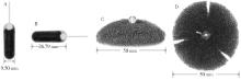
图1
食康1号海绵装置 如图A和B所示,装置由可消化的明胶胶囊、压缩的聚氨酯海绵网、60 cm长的绳子三部分构成,绳子通过小的玻璃珠固定在海绵细胞刷上。胶囊溶解后海绵网从中释放,释放后呈直径 50mm 椭圆形结构(图C和D)。

表1
受试者基线资料
| Item | Lesion group (n = 25) | Normal group (n = 75) | P |
|---|---|---|---|
| Sex | 0.640 | ||
| Female | 9 | 33 | |
| Male | 16 | 42 | |
| Age | 66.16±5.77 | 66.35±6.06 | 0.891 |
| Registered permanent residence | 1.000 | ||
| City | 22 | 65 | |
| Village | 3 | 10 | |
| Smoking | 1.000 | ||
| Yes | 2 | 5 | |
| No | 23 | 70 | |
| Tipple | 1.000 | ||
| Yes | 4 | 11 | |
| No | 21 | 64 | |
| Hot diet | 1.000 | ||
| Yes | 10 | 30 | |
| No | 15 | 45 | |
| Anodontia | 0.819 | ||
| Yes | 13 | 36 | |
| No | 12 | 39 | |
| Number of missing teeth | 0.954 | ||
| 0 | 12 | 39 | |
| 1-4 | 5 | 14 | |
| >5 | 8 | 22 | |
| Family history | 0.732 | ||
| Yes | 4 | 9 | |
| No | 21 | 66 |
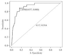
图2
细胞学DI诊断食管病变的ROC曲线下面积

| [1] |
WATERS J K, REZNIK S I. Update on management of squamous cell esophageal cancer[J]. Curr Oncol Rep, 2022, 24(3):375-385.
doi: 10.1007/s11912-021-01153-4 pmid: 35142974 |
| [2] | YU Z, BAI X, ZHOU R, et al. Differences in the incidence and mortality of digestive cancer between Global Cancer Observatory 2020 and Global Burden of Disease 2019[J]. Int J Cancer, 2024, 154(4):615-625. |
| [3] | 刘琳, 王钟怡, 黄楚殷, 等. 基于Web of Science数据库食管胃结合部腺癌研究的可视化及热点分析[J]. 中华消化外科杂志, 2023, 22(10):1243-1254. |
| LIU L, WANG Z Y, HUANG C Y, et al. Visualization and hotspots analysis of research on adenocarcinoma of esophagogastric junction based on Web of Science[J]. Chin J Dig Surg, 2023, 22(10):1243-1254. | |
| [4] | YANG H, HU B. Recent advances in early esophageal cancer: diagnosis and treatment based on endoscopy[J]. Postgrad Med, 2021, 133(6):665-673. |
| [5] |
ROTH M J, LIU S F, DAWSEY S M, et al. Cytologic detection of esophageal squamous cell carcinoma and precursor lesions using balloon and sponge samplers in asymptomatic adults in Linxian, China[J]. Cancer, 1997, 80(11):2047-2059.
doi: 10.1002/(sici)1097-0142(19971201)80:11<2047::aid-cncr3>3.0.co;2-u pmid: 9392326 |
| [6] |
LIU J, ZHAO H. Application of convolution neural network in medical image processing[J]. Technol Health Care, 2021, 29(2):407-417.
doi: 10.3233/THC-202657 pmid: 33386836 |
| [7] |
NGUYEN H N, SEVIN B U, AVERETTE H E, et al. The role of DNA index as a prognostic factor in early cervical carcinoma[J]. Gynecol Oncol, 1993, 50(1): 54-59.
pmid: 8349165 |
| [8] | 国家消化内镜专业质控中心, 国家消化系疾病临床医学研究中心(上海), 国家消化道早癌防治中心联盟, 等. 中国早期食管癌及癌前病变筛查专家共识意见(2019年,新乡)[J]. 中华消化内镜杂志, 2019, 36(11):793-801. |
| National Digestive Endoscopy Professional Quality Control Center, National Clinical Research Center for Digestive Diseases (Shanghai), National Alliance of Digestive Early Cancer Prevention and Control Centers, et al. Expert consensus on early esophageal cancer and precancerous lesions screening in China (2019, Xinxiang)[J]. Chin J Dig Endosc, 2019, 36(11):793-801. | |
| [9] | ZHOU M, WANG H, ZENG X, et al. Mortality, morbi-dity, and risk factors in China and its provinces, 1990-2017: a systematic analysis for the Global Burden of Disea-se Study 2017[J]. Lancet, 2019, 394(10204):1145-1158. |
| [10] |
MORGAN E, SOERJOMATARAM I, RUMGAY H, et al. The global landscape of esophageal squamous cell carcinoma and esophageal adenocarcinoma incidence and mortality in 2020 and projections to 2040: new estimates from GLOBOCAN 2020[J]. Gastroenterology, 2022, 163(3):649-658.e2.
doi: 10.1053/j.gastro.2022.05.054 pmid: 35671803 |
| [11] |
DIJKSTERHUIS W P M, KALFF M C, WAGNER A D, et al. Gender differences in treatment allocation and survival of advanced gastroesophageal cancer: a population-based study[J]. J Natl Cancer Inst, 2021, 113(11):1551-1560.
doi: 10.1093/jnci/djab075 pmid: 33837791 |
| [12] | LI R, SUN J, WANG T, et al. Comparison of secular trends in esophageal cancer mortality in China and Japan during 1990-2019: an age-period-cohort analysis[J]. Int J Environ Res Public Health, 2022, 19(16):10302. |
| [13] | CHAI T, SHEN Z, ZHANG P, et al. Comparison of high risk factors (hot food, hot beverage, alcohol, tobacco, and diet) of esophageal cancer: a protocol for a systematic review and meta-analysis[J]. Medicine (Baltimore), 2019, 98(17):e15176. |
| [14] | HUANG F L, YU S J. Esophageal cancer: risk factors, genetic association, and treatment[J]. Asian J Surg, 2018, 41(3):210-215. |
| [15] | LI B, LIU Y, PENG J, et al. Trends of esophageal cancer incidence and mortality and its influencing factors in China[J]. Risk Manag Healthc Policy, 2021, 14:4809-4821. |
| [16] | MMBAGA B T, MWASAMWAJA A, MUSHI G, et al. Missing and decayed teeth, oral hygiene and dental stai-ning in relation to esophageal cancer risk: ESCCAPE case-control study in Kilimanjaro, Tanzania[J]. Int J Cancer, 2021, 148(10):2416-2428. |
| [17] | 陈克能. 管饲要素营养在食管癌全程治疗中的特殊地位[J]. 中华消化外科杂志, 2023, 22(1):61-64. |
| CHEN K N. The special role of enteral nutrition in the comprehensive treatment of esophageal cancer[J]. Chin J Dig Surg, 2023, 22(1):61-64. | |
| [18] |
SU Z, ZOU G R, MAO Y P, et al. Prognostic impact of family history of cancer in Southern Chinese patients with esophageal squamous cell cancer[J]. J Cancer, 2019, 10(6):1349-1357.
doi: 10.7150/jca.26511 pmid: 31031844 |
| [19] | VISAGGI P, BARBERIO B, GHISA M, et al. Modern dia-gnosis of early esophageal cancer: from blood biomarkers to advanced endoscopy and artificial intelligence[J]. Cancers (Basel), 2021, 13(13):3162 |
| [20] |
DEISSOVÁ T, KALA Z, SLABÝ O, et al. Biomarkers for non-endoscopic examination of esophageal mucosa[J]. Vnitr Lek, 2020, 66(7):13-19.
pmid: 33380129 |
| [21] |
GAO Y, XIN L, FENG Y D, et al. Feasibility and accuracy of artificial intelligence-assisted sponge cytology for community-based esophageal squamous cell carcinoma screening in China[J]. Am J Gastroenterol, 2021, 116(11):2207-2215.
doi: 10.14309/ajg.0000000000001499 pmid: 34546186 |
| [22] | RAI V, ABDO J, AGRAWAL D K. Biomarkers for early detection, prognosis, and therapeutics of esophageal cancers[J]. Int J Mol Sci, 2023, 24(4):3316. |
| [23] | YUAN Z, WANG X, GENG X, et al. Liquid biopsy for esophageal cancer: Is detection of circulating cell-free DNA as a biomarker feasible?[J] Cancer Commun (Lond), 2021, 41(1):3-15. |
| [24] | SHAHEEN N J, KOMANDURI S, MUTHUSAMY V R, et al. Acceptability and adequacy of a non-endoscopic cell collection device for diagnosis of barrett's esophagus: lessons learned[J]. Dig Dis Sci, 2022, 67(1):177-186. |
| [25] |
FITZGERALD R C, DI PIETRO M, O'DONOVAN M, et al. Cytosponge-trefoil factor 3 versus usual care to identify Barrett's oesophagus in a primary care setting: a multicentre, pragmatic, randomised controlled trial[J]. Lancet, 2020, 396(10247):333-344.
doi: S0140-6736(20)31099-0 pmid: 32738955 |
| [26] | YAO B, FENG Y, ZHAO K, et al. Artificial intelligence assisted cytological detection for early esophageal squamous epithelial lesions by using low-grade squamous intraepithelial lesion as diagnostic threshold[J]. Cancer Med, 2023, 12(2):1228-1236. |
| [27] |
MIDDLETON D R S, MMBAGA B T, O'DONOVAN M, et al. Minimally invasive esophageal sponge cytology sampling is feasible in a Tanzanian community setting[J]. Int J Cancer, 2021, 148(5):1208-1218.
doi: 10.1002/ijc.33366 pmid: 33128785 |
| [1] | 洪烨娜, 张宇, SHI Kuangyu, 李彪, 郭睿. 放射性核素诊疗一体化的若干问题及对策[J]. 诊断学理论与实践, 2025, 24(03): 263-267. |
| [2] | 李卓含, 黄新韵, 郭睿, 李彪. 18F-FDG PET/CT在滤泡性淋巴瘤诊断和预后评估中的研究进展[J]. 诊断学理论与实践, 2024, 23(04): 439-444. |
| [3] | 吴娜明, 李军, 陶娟. 恶性黑色素瘤的诊断热点[J]. 诊断学理论与实践, 2023, 22(03): 215-220. |
| [4] | 唐静仪, 余群, 刘军. 结合人工智能的结构影像分析对阿尔茨海默病的早期预测及精准诊断研究进展[J]. 诊断学理论与实践, 2022, 21(01): 12-17. |
| [5] | 徐浩, 张治, 解学乾, 杨文艺, 刘少稳. 冠脉生理功能评估软件(DEEPVESSEL FFR)与有创FFR在评估冠脉缺血中的对比研究[J]. 诊断学理论与实践, 2021, 20(04): 384-390. |
| [6] | 许晶晶, 张敏鸣. 人工智能机器学习方法在阿尔茨海默病中的应用现状[J]. 诊断学理论与实践, 2018, 17(04): 466-470. |
| [7] | 周平红, 蔡明琰, 姚礼庆, 执笔,. 消化道黏膜病变内镜黏膜下剥离术的专家共识意见[J]. 诊断学理论与实践, 2012, 11(05): 531-535. |
| 阅读次数 | ||||||
|
全文 |
|
|||||
|
摘要 |
|
|||||