Journal of Diagnostics Concepts & Practice ›› 2024, Vol. 23 ›› Issue (02): 101-107.doi: 10.16150/j.1671-2870.2024.02.001
• Expert forum • Previous Articles Next Articles
ZHAO Xinxiang( ), ZHAO Xiaoying
Received:2024-01-29
Online:2024-04-25
Published:2024-07-04
Contact:
ZHAO Xinxiang
E-mail:zhaoxinxiang06@126.com
CLC Number:
ZHAO Xinxiang, ZHAO Xiaoying. Application norms and research progress of cardiac magnetic resonance imaging in the management of MINOCA patients[J]. Journal of Diagnostics Concepts & Practice, 2024, 23(02): 101-107.
Table 1
Diagnostic criteria of MINOCA
| 诊断条件 | 具体内容 |
|---|---|
| 符合AMI诊断 | Ⅰ.检测到心肌肌钙蛋白上升或下降,且至少有一个值超过第 99百分位数参考上限。 Ⅱ. MI确凿临床证据,至少表现为以下一项: a. 心肌缺血症状; b. 新的缺血性心电图变化; c. 出现病理性Q波; d. 影像学证据显示新的存活心肌丧失或新的区域室壁运动异常,其模式与缺血性病因一致; e. 通过血管造影或尸检发现冠状动脉血栓。 |
| 血管造影显示冠状动脉无明显阻塞 | 定义为血管造影显示任何主要心外膜血管无阻塞性疾病(即冠状动脉狭窄<50%),包括以下3项。 a. 冠状动脉正常(血管造影无狭窄); b. 轻度管腔不规则(血管造影狭窄率<30%); c. 中度冠状动脉粥样硬化病变(狭窄程度≥30%,但<50%)。 部分冠状动脉狭窄30%~50%的患者会出现有临床意义的功能性冠状动脉狭窄,对于这些患者,冠状动脉血流储备分数>0.8才能诊断为 MINOCA。 |
| 临床表现无明确的替代诊断 | 替代诊断包括但不限于败血症、肺栓塞和心肌炎等非缺血性病因。 |
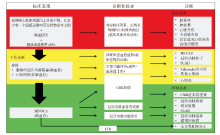
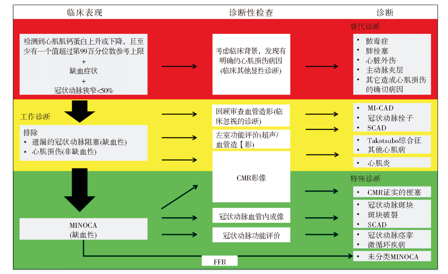
Figure 1
Clinical diagnostic process of MINOCA
| [1] | 中国心血管健康与疾病报告编写组. 《中国心血管健康与疾病报告2022》概要[J]. 中国介入心脏病学杂志, 2023, 31(7):485-508. |
| Committee of the Report on Cardiovascular Health and Diseases in China. Report on cardiovascular health and diseases in China 2022:an updated summary[J]. Chin J Intervent Cardiol, 2023, 31(7):485-508. | |
| [2] |
PASUPATHY S, AIR T, DREYER R P, et al. Systematic review of patients presenting with suspected myocardial infarction and nonobstructive coronary arteries[J]. Circulation, 2015, 131(10):861-870.
doi: 10.1161/CIRCULATIONAHA.114.011201 pmid: 25587100 |
| [3] |
LINDAHL B, BARON T, ALBERTUCCI M, et al. Myocardial infarction with non-obstructive coronary artery disease[J]. EuroIntervention, 2021, 17(11):e875-e887.
doi: 10.4244/EIJ-D-21-00426 pmid: 34870600 |
| [4] | GROSS H, STERNBERG W H. Myocardial infarction without significant lesions of coronary arteries[J]. Arch Intern Med (Chic), 1939, 64(1):249-267. |
| [5] |
ZHAO X, ZHANG Y, SUN Y, et al. Assessment of myocardial viability with delayed-enhancement MRI in coronary artery disease: A correlative study with coronary artery stenosis using digital subtraction angiography[J]. Exp Ther Med, 2016, 12(4):2285-2289.
pmid: 27698725 |
| [6] |
BELTRAME J F. Assessing patients with myocardial infarction and nonobstructed coronary arteries (MINOCA)[J]. J Intern Med, 2013, 273(2):182-185.
doi: 10.1111/j.1365-2796.2012.02591.x pmid: 22998397 |
| [7] |
AGEWALL S, BELTRAME J F, REYNOLDS H R, et al. ESC working group position paper on myocardial infarction with non-obstructive coronary arteries[J]. Eur Heart J, 2017, 38(3):143-153.
doi: 10.1093/eurheartj/ehw149 pmid: 28158518 |
| [8] |
KIM R J, FIENO D S, PARRISH T B, et al. Relationship of MRI delayed contrast enhancement to irreversible injury, infarct age, and contractile function[J]. Circulation, 1999, 100(19):1992-2002.
doi: 10.1161/01.cir.100.19.1992 pmid: 10556226 |
| [9] | PATHIK B, RAMAN B, MOHD AMIN N H, et al. Troponin-positive chest pain with unobstructed coronary arte-ries: incremental diagnostic value of cardiovascular magnetic resonance imaging[J]. Eur Heart J Cardiovasc Ima-ging, 2016, 17(10):1146-1152. |
| [10] | BULLUCK H, HAMMOND-HALEY M, FONTANA M, et al. Quantification of both the area-at-risk and acute myocardial infarct size in ST-segment elevation myocardial infarction using T1-mapping[J]. J Cardiovasc Magn Reson, 2017, 19(1):57. |
| [11] | JAHNKE C, SINN M, HOT A, et al. Differentiation of acute non-ST elevation myocardial infarction and acute infarct-like myocarditis by visual pattern analysis: a head-to-head comparison of different cardiac MR techniques[J]. Eur Radiol, 2023, 33(9):6258-6266. |
| [12] | DASTIDAR A G, BARITUSSIO A, DE GARATE E, et al. Prognostic role of CMR and conventional risk factors in myocardial infarction with nonobstructed coronary arteries[J]. JACC Cardiovasc Imaging, 2019, 12(10):1973-1982. |
| [13] | LINTINGRE P F, NIVET H, CLÉMENT-GUINAUDEAU S, et al. High-resolution late gadolinium enhancement magnetic resonance for the diagnosis of myocardial infarction with nonobstructed coronary arteries[J]. JACC Cardiovasc Imaging, 2020, 13(5):1135-1148. |
| [14] | JUNCÀ G, TEIS A, KASA G, et al. Timing of cardiac magnetic resonance and diagnostic yield in patients with myocardial infarction with nonobstructive coronary arte-ries[J]. Rev Esp Cardiol (Engl Ed), 2023:S1885-5857(23)00339-0. |
| [15] |
WILLIAMS M G L, LIANG K, DE GARATE E, et al. Peak troponin and CMR to guide management in suspected ACS and nonobstructive coronary arteries[J]. JACC Cardiovasc Imaging, 2022, 15(9):1578-1587.
doi: 10.1016/j.jcmg.2022.03.017 pmid: 36075617 |
| [16] |
REYNOLDS H R. Should every patient with MINOCA have cardiac magnetic resonance?[J]. JACC Cardiovasc Imaging, 2022, 15(9):1588-1590.
doi: 10.1016/j.jcmg.2022.07.009 pmid: 36075618 |
| [17] | PUSTJENS T F S, APPELMAN Y, DAMMAN P, et al. Guidelines for the management of myocardial infarction/injury with non-obstructive coronary arteries (MINOCA): a position paper from the Dutch ACS working group[J]. Neth Heart J, 2020, 28(3):116-130. |
| [18] | COLLET J P, THIELE H, BARBATO E, et al. 2020 ESC Guidelines for the management of acute coronary syndromes in patients presenting without persistent ST-segment elevation[J]. Eur Heart J, 2021, 42(14):1289-1367. |
| [19] | TAMIS-HOLLAND J E, JNEID H, REYNOLDS H R, et al. Contemporary diagnosis and management of patients with myocardial infarction in the absence of obstructive coronary artery disease: a scientific statement from the American Heart Association[J]. Circulation, 2019, 139(18):e891-e908. |
| [20] |
REYNOLDS H R, MAEHARA A, KWONG R Y, et al. Coronary optical coherence tomography and cardiac magnetic resonance imaging to determine underlying causes of myocardial infarction with nonobstructive coronary arteries in women[J]. Circulation, 2021, 143(7):624-640.
doi: 10.1161/CIRCULATIONAHA.120.052008 pmid: 33191769 |
| [21] | LIANG K, BISACCIA G, LEO I, et al. CMR reclassifies the majority of patients with suspected MINOCA and non MINOCA[J]. Eur Heart J Cardiovasc Imaging, 2023, 25(1):8-15. |
| [22] | KONST R E, PARKER M, BHATTI L, et al. Prognostic value of cardiac magnetic resonance imaging in patients with a working diagnosis of MINOCA-an outcome study with up to 10 years of follow-up[J]. Circ Cardiovasc Imagi-ng, 2023, 16(8):e014454. |
| [23] | MACHANAHALLI BALAKRISHNA A, ISMAYL M, THANDRA A, et al. Diagnostic value of cardiac magnetic resonance imaging and intracoronary optical cohe-rence tomography in patients with a working diagnosis of myocardial infarction with non-obstructive coronary arte-ries - a systematic review and meta-analysis[J]. Curr Probl Cardiol, 2023, 48(6):101126. |
| [24] | BYRNE R A, ROSSELLO X, COUGHLAN J J, et al. 2023 ESC Guidelines for the management of acute coronary syndromes[J]. Eur Heart J, 2023, 44(38):3720-3826. |
| [25] | WILLIAMS M G L, DASTIDAR A, LIANG K, et al. Sex differences in patients with acute coronary syndromes and non-obstructive coronary arteries: Presentation and outcome[J]. Int J Cardiol, 2023, 372:15-22. |
| [26] | QUESADA O, YILDIZ M, HENRY T D, et al. Mortality in ST-segment elevation myocardial infarction with nonobstructive coronary arteries and mimickers[J]. JAMA Netw Open, 2023, 6(11):e2343402. |
| [27] | PIZZI C, XHYHERI B, COSTA G M, et al. Nonobstructive versus obstructive coronary artery disease in acute coronary syndrome: a meta-analysis[J]. J Am Heart Assoc, 2016, 5(12):e004185. |
| [28] |
ANDERSSON H B, PEDERSEN F, ENGSTRØM T, et al. Long-term survival and causes of death in patients with ST-elevation acute coronary syndrome without obstructive coronary artery disease[J]. Eur Heart J, 2018, 39(2):102-110.
doi: 10.1093/eurheartj/ehx491 pmid: 29029035 |
| [29] |
DREYER R P, TAVELLA R, CURTIS J P, et al. Myocardial infarction with non-obstructive coronary arteries as compared with myocardial infarction and obstructive coronary disease: outcomes in a Medicare population[J]. Eur Heart J, 2020, 41(7):870-878.
doi: 10.1093/eurheartj/ehz403 pmid: 31222249 |
| [30] | BERGAMASCHI L, FOÀ A, PAOLISSO P, et al. Prognostic role of early cardiac magnetic resonance in myocardial infarction with nonobstructive coronary arteries[J]. JACC Cardiovasc Imaging, 2024, 17(2):149-161. |
| [31] | FEDELE D, CANTON L, BODEGA F, et al. Performance of Prognostic Scoring Systems in MINOCA: A comparison among GRACE, TIMI, HEART, and ACEF scores[J]. J Clin Med, 2023, 12(17):5687. |
| [32] | VICENTE-IBARRA N, FELIU E, BERTOMEU-MARTÍNEZ V, et al. Role of cardiovascular magnetic resonance in the prognosis of patients with myocardial infarction with non-obstructive coronary arteries[J]. J Cardiovasc Magn Reson, 2021, 23(1):83. |
| [33] | BUCCIARELLI V, BIANCO F, FRANCESCO A D, et al. Characteristics and prognosis of a contemporary cohort with myocardial infarction with non-obstructed coronary arteries (MINOCA) presenting different patterns of late gadolinium enhancements in cardiac magnetic resonance imaging[J]. J Clin Med, 2023, 12(6):2266. |
| [1] | CHANG Yuchen, LI Jingbo. Advances in biological markers of ferroptosis in myocardial infarction [J]. Journal of Diagnostics Concepts & Practice, 2023, 22(02): 197-202. |
| [2] | LIU Peng, YAN Fuhua, QIN Le, XIAO Ruijie. Study on correlation of cardiac magnetic resonance strain rate parameters of left ventricular diastolic function with risk of sudden death in hypertrophic cardiomyopathy [J]. Journal of Diagnostics Concepts & Practice, 2022, 21(03): 317-325. |
| [3] | LUO Xiaoying, XU Yan, ZHANG Jiansheng, WU Liqun, QI Wenhang. Predictive value of NT-proBNP for new-onset atrial fibrillation following acute myocardial infarction [J]. Journal of Diagnostics Concepts & Practice, 2020, 19(03): 303-307. |
| [4] | CHEN Yafen, CHEN Yuanyuan, WU Liping, XUE Qiqi, YANG Ke, LU Lin, CAO Jiumei. Effect of silencing information regulator factor 1 on mice with acute myocardial infarction [J]. Journal of Diagnostics Concepts & Practice, 2018, 17(06): 670-675. |
| [5] | WAN Yinglei, NI Yimin, GU Zhidong. Clinical value of heart-type fatty acid-binding protein in early diagnosis of acute myocardial infarction [J]. Journal of Diagnostics Concepts & Practice, 2016, 15(06): 582-585. |
| [6] | . [J]. Journal of Diagnostics Concepts & Practice, 2014, 13(06): 610-612. |
| [7] | . [J]. Journal of Diagnostics Concepts & Practice, 2011, 10(06): 517-522. |
| [8] | . [J]. Journal of Diagnostics Concepts & Practice, 2011, 10(01): 26-29. |
| [9] | . [J]. Journal of Diagnostics Concepts & Practice, 2009, 8(05): 506-509. |
| [10] | . [J]. Journal of Diagnostics Concepts & Practice, 2008, 7(05): 517-520. |
| [11] | . [J]. Journal of Diagnostics Concepts & Practice, 2008, 7(04): 390-393. |
| [12] | . [J]. Journal of Diagnostics Concepts & Practice, 2006, 5(05): 412-414. |
| [13] | . [J]. Journal of Diagnostics Concepts & Practice, 2006, 5(02): 142-146. |
| [14] | . [J]. Journal of Diagnostics Concepts & Practice, 2002, 1(04): 30-32. |
| [15] | . [J]. Journal of Diagnostics Concepts & Practice, 2002, 1(04): 25-26+43. |
| Viewed | ||||||
|
Full text |
|
|||||
|
Abstract |
|
|||||