Journal of Diagnostics Concepts & Practice ›› 2024, Vol. 23 ›› Issue (05): 484-493.doi: 10.16150/j.1671-2870.2024.05.004
• Original articles • Previous Articles Next Articles
YU Jin, WANG Jie, WANG Hujun, WANG Congxiao, LI Yingqi, FANG Boyan, WANG Yingpeng( )
)
Received:2023-12-04
Accepted:2024-06-07
Online:2024-10-25
Published:2025-02-25
Contact:
WANG Yingpeng
E-mail:ypwang@ccmu.edu.cn
CLC Number:
YU Jin, WANG Jie, WANG Hujun, WANG Congxiao, LI Yingqi, FANG Boyan, WANG Yingpeng. Study on the recognition of early-stage Parkinson’s disease patients using functional near-infrared spectroscopy signals based on machine learning[J]. Journal of Diagnostics Concepts & Practice, 2024, 23(05): 484-493.
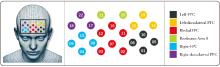
Figure 1
Light levels and probe distribution locations

Table 1
Hyperparameters of each algorithm model
| Algorithm | Hyperparameters |
|---|---|
| Support vector machine | - Kernel type: Linear kernel |
| - Regularization parameter C: 0.1 | |
| - Gamma (parameter for RBF kernel): 0.01 | |
| BP neural network | - Number of hidden layers: 2 layers |
| - Number of neurons in each hidden layer: 100, 50 | |
| - Learning rate: 0.001 | |
| - Activation function: ReLU | |
| Random forest | - Number of trees: 100 |
| - Maximum depth: 20 | |
| - Minimum number of samples required at a leaf node: 5 | |
| - Maximum number of features: sqrt | |
| Logistic regression | - Regularization type: L2 |
| - Regularization parameter C: 0.01 |
Table 2
Basic characteristics of subjects
| Item | PD Group (n=60) | Control Group (n=60) | P-value |
|---|---|---|---|
| Age (x±s, years) | 55.70±4.18 | 57.80±5.41 | 0.815 |
| Gender (Male/Female, n) | 37/23 | 31/29 | 0.725 |
| Height (x±s, cm) | 167.6±2.3 | 168.4±5.4 | 0.341 |
| Weight (x±s, kg) | 66.1±9.5 | 64.1±8.3 | 0.251 |
| Duration of Illness (x±s, years) | 0.4±0.3 | / | / |
| Hoehn Yahr Stage | 1.5±0.5 | / | / |
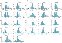
Figure 2
Histogram of dataset distribution
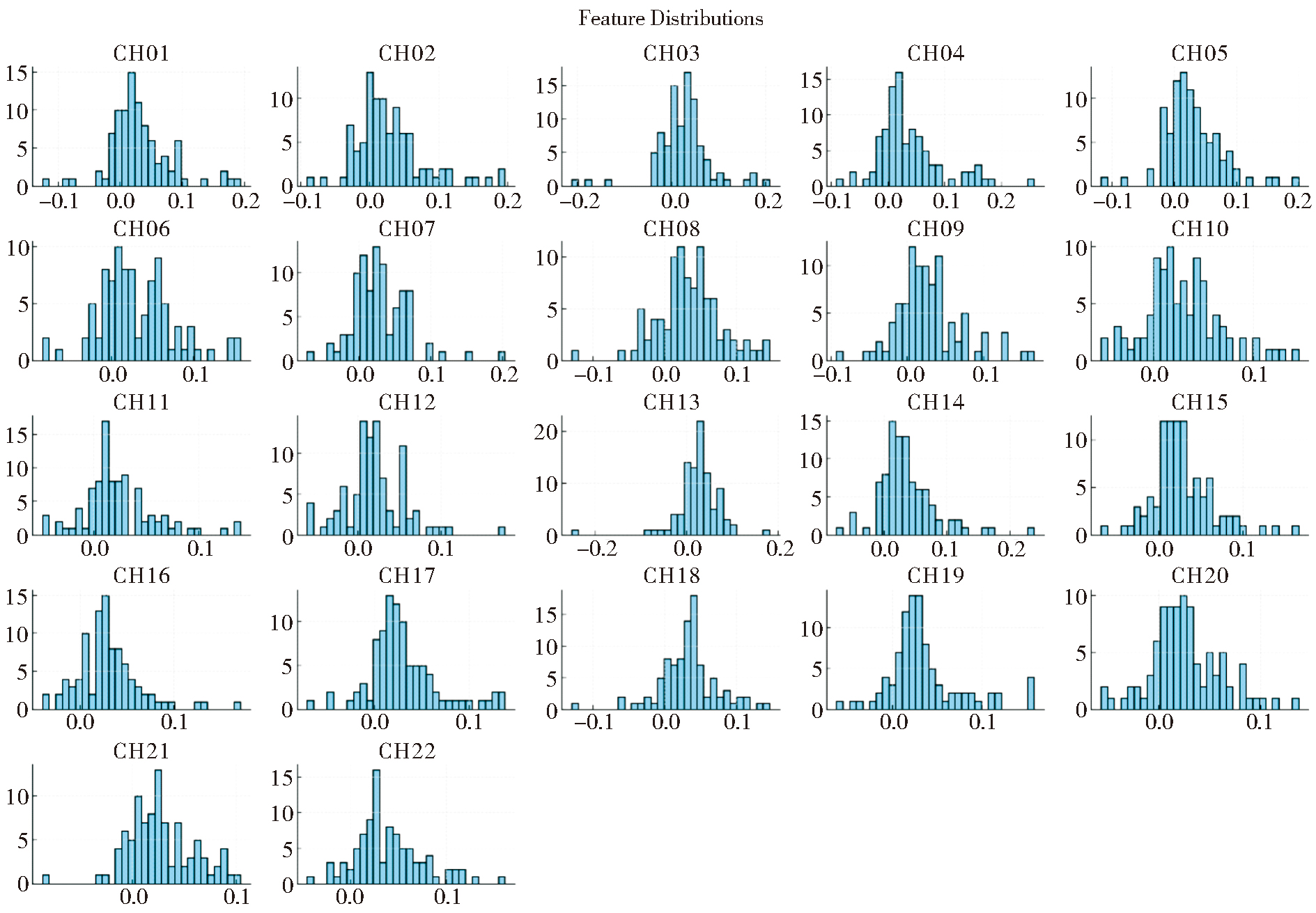
Table 3
Performance results of different classifiers
| Model | Accuracy | Sensitivity | Specificity | AUC |
|---|---|---|---|---|
| Support Vector Machine | 0.90 | 0.89 | 0.93 | 0.96 |
| BP Neural Network | 0.82 | 0.69 | 1.00 | 0.91 |
| Random Forest | 0.84 | 0.71 | 1.00 | 0.98 |
| Logistic Regression | 0.81 | 0.71 | 0.93 | 0.90 |
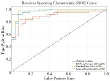
Figure 3
ROC curves for different classifiers
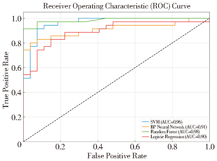
Figure 4
Confusion matrix results for each classifier
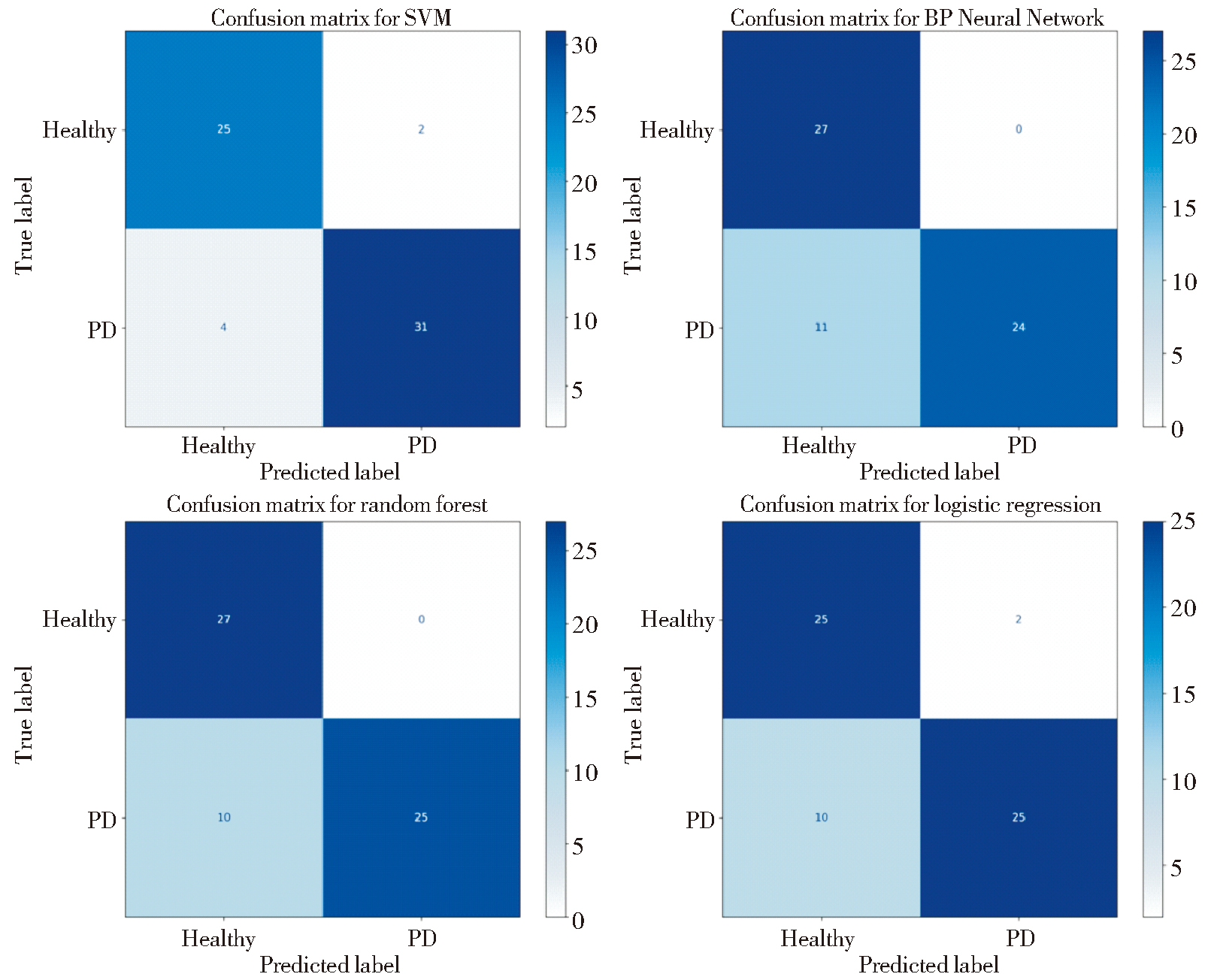
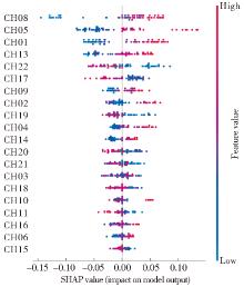
Figure 5
SHAP values scatter plot for model interpretability Note: Red dots indicate that the feature value has a positive impact on the prediction, whereas blue dots indicate a negative impact. The horizontal position of the dots indicates the specific contribution of the feature to the prediction, ranging from negative (left side) to positive (right side).
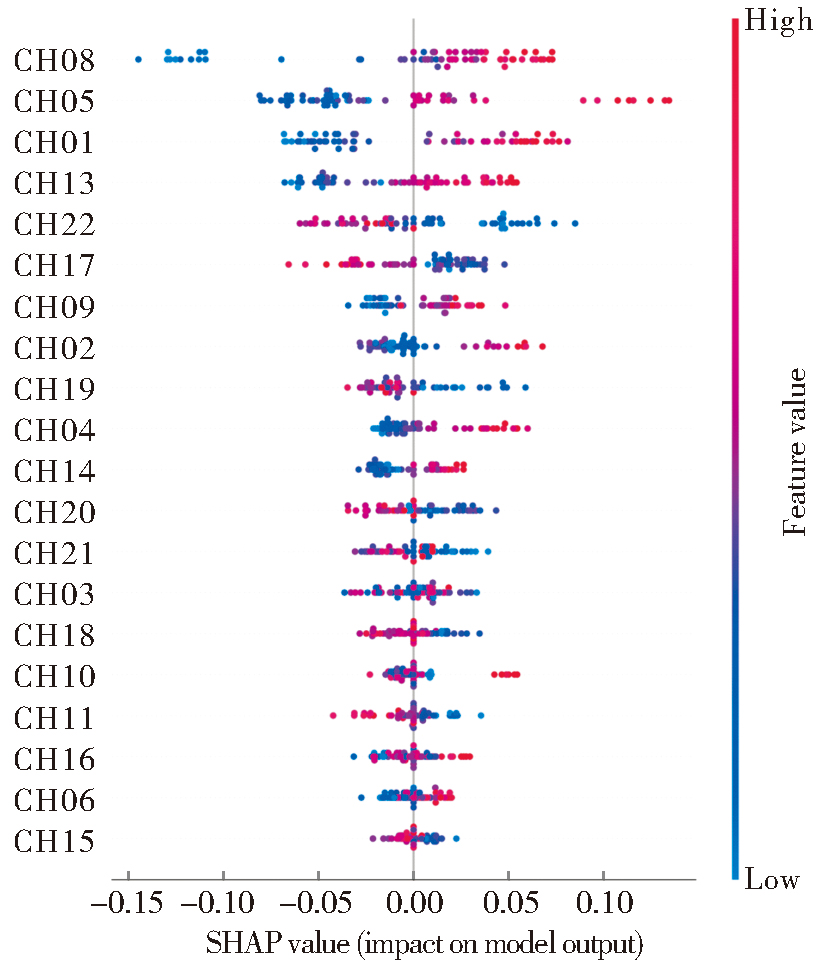
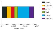
Figure 6
SHAP weighted average results

| [1] |
MCDONALD C, GORDON G, HAND A, et al. 200 Years of Parkinson's disease: what have we learnt from James Parkinson?[J]. Age Ageing, 2018, 47(2):209-214.
doi: 10.1093/ageing/afx196 pmid: 29315364 |
| [2] | TITOVA N, MARTINEZ-MARTIN P, KATUNINA E, et al. Advanced Parkinson's or "complex phase" Parkinson's disease? Re-evaluation is needed[J]. J Neural Transm (Vienna), 2017, 124(12):1529-1537. |
| [3] |
MARINO B L B, DE SOUZA L R, SOUSA K P A, et al. Parkinson's disease: a review from pathophysiology to treatment[J]. Mini Rev Med Chem, 2020, 20(9):754-767.
doi: 10.2174/1389557519666191104110908 pmid: 31686637 |
| [4] |
BARKHUIZEN M, ANDERSON D G, GROBLER A F. Advances in GBA-associated Parkinson's disease--Pathology, presentation and therapies[J]. Neurochem Int, 2016, 93:6-25.
doi: 10.1016/j.neuint.2015.12.004 pmid: 26743617 |
| [5] | 刘浩宇, 朋文佳, 芈静, 等. 1990-2019年全球帕金森病疾病负担的APC分析[J]. 中华全科医学, 2024, 22(1):154-157. |
| LIU H Y, PENG W J, MI J, et al. APC analysis of the global disease burden of Parkinson's disease from 1990 to 2019[J]. Chin J Gen Pract, 2024, 22(1):154-157. | |
| [6] | CHHOR V, KARACHI C, BONNET A M, et al. Anaesthesia and Parkinson's disease[J]. Ann Fr Anesth Reanim, 2011, 30(7-8): 559-68. |
| [7] | KHAN H, NASEER N, YAZIDI A, et al. Analysis of human gait using hybrid EEG-fNIRS-based BCI system: A review[J]. Front Hum Neurosci, 2021, 14:613254. |
| [8] | 李进, 艾芳, 刘媛, 等. 震颤分析在原发性震颤与帕金森病鉴别诊断中的准确性及价值[J]. 中国临床研究, 2021, 34(10):1354-1357. |
| LI J, AI F, LIU Y, et al. Tremor analysis in differential diagnosis of essential tremor and Parkinson's disease[J]. Chin J Clin Res, 2021, 34(10):1354-1357. | |
| [9] |
DOVONOU A, BOLDUC C, SOTO LINAN V, et al. Animal models of Parkinson's disease: bridging the gap between disease hallmarks and research questions[J]. Transl Neurodegener, 2023, 12(1):36.
doi: 10.1186/s40035-023-00368-8 pmid: 37468944 |
| [10] | WILCOX T, BIONDI M. fNIRS in the developmental scie-nces[J]. Wiley Interdiscip Rev Cogn Sci, 2015, 6(3):263-283. |
| [11] | COCKX H, OOSTENVELD R, TABOR M, et al. fNIRS is sensitive to leg activity in the primary motor cortex after systemic artifact correction[J]. Neuroimage, 2023, 269:119880. |
| [12] | WEIBLEY H, DI FILIPPO M, LIU X, et al. fNIRS monitoring of infant prefrontal cortex during crawling and an executive functioning task[J]. Front Behav Neurosci, 2021, 15:675366. |
| [13] | GUNASEKARA N, GAETA G, LEVY A, et al. fNIRS neuroimaging in olfactory research: A systematic literature review[J]. Front Behav Neurosci, 2022, 16:1040719. |
| [14] |
ZIMEO MORAIS G A, BALARDIN J B, SATO J R. fNIRS Optodes' Location Decider (fOLD): a toolbox for probe arrangement guided by brain regions-of-interest[J]. Sci Rep, 2018, 8(1):3341.
doi: 10.1038/s41598-018-21716-z pmid: 29463928 |
| [15] |
VITORIO R, STUART S, ROCHESTER L, et al. fNIRS response during walking - Artefact or cortical activity? A systematic review[J]. Neurosci Biobehav Rev, 2017, 83:160-172.
doi: S0149-7634(17)30347-0 pmid: 29017917 |
| [16] |
NASEER N, HONG K S. fNIRS-based brain-computer interfaces: a review[J]. Front Hum Neurosci, 2015, 9:3.
doi: 10.3389/fnhum.2015.00003 pmid: 25674060 |
| [17] | LU J, WANG Y, SHU Z, et al. fNIRS-based brain state transition features to signify functional degeneration after Parkinson's disease[J]. J Neural Eng, 2022, 19(4):10.1088/1741-2552/ac861e. |
| [18] | NIEUWHOF F, REELICK M F, MAIDAN I, et al. Measuring prefrontal cortical activity during dual task walki-ng in patients with Parkinson's disease: feasibility of using a new portable fNIRS device[J]. Pilot Feasibility Stud, 2016, 2:59. |
| [19] | KAUSHIK C, MCRAE A D, DAVENPORT M, et al. New equivalences between interpolation and SVMs: Kernels and Structured Features[J]. ArXiv, 2023, abs/2305.02304. |
| [20] | JANG R. Learning representations by forward-propagating errors[J]. ArXiv, 2023, abs/2308.09728. |
| [21] | CHEN C, HUANG T S, HUANG J C, et al. Design of music style classification teaching system based on BP neural network[C]. International Conference on Information System, 2022. |
| [22] | LIMAM H, ZOUHAIR A, OUESLATI W. A new hybrid multiclass approach based on KNN and SVM[J]. J Inf Knowl Manag, 2022(21): 2250061:1-2250061:16. |
| [23] | DEDJA K, NAKANO F K, PLIAKOS K, et al. Explaining random forest prediction through diverse rulesets[J]. ArXiv, 2022, abs/2203.15511. |
| [24] | ZAIDI A, LUHAYB ASM A L. Two statistical approaches to justify the use of the logistic function in binary logistic regression[J]. Math probl Eng, 2023, 5525675, 11pages. |
| [25] | NAGASAWA T, SATO T, NAMBU I, et al. fNIRS-GANs: data augmentation using generative adversarial networks for classifying motor tasks from functional near-infrared spectroscopy[J]. J Neural Eng, 2020, 17(1):016068. |
| [26] | HAN Y, HUANG J, YIN Y, et al. From brain to worksite: the role of fNIRS in cognitive studies and worker safety[J]. Front Public Health, 2023, 11:1256895. |
| [27] |
KUMAR V, SHIVAKUMAR V, CHHABRA H, et al. Functional near infra-red spectroscopy (fNIRS) in schizophrenia: A review[J]. Asian J Psychiatr, 2017, 27:18-31.
doi: S1876-2018(16)30434-8 pmid: 28558892 |
| [28] | BEHBOODI B, LIM S-H, LUNA M, et al. Artificial and convolutional neural networks for assessing functional connectivity in resting-state functional near infrared spectroscopy[J]. J Near Infrared Spectrosc, 2019, 27(3):191-205. |
| [29] |
FERRARI M, QUARESIMA V. A brief review on the history of human functional near-infrared spectroscopy (fNIRS) development and fields of application[J]. Neuroimage, 2012, 63(2):921-935.
doi: 10.1016/j.neuroimage.2012.03.049 pmid: 22510258 |
| [30] | WANG K, TIAN J, ZHENG C, et al. Interpretable prediction of 3-year all-cause mortality in patients with heart failure caused by coronary heart disease based on machine learning and SHAP[J]. Comput Biol Med, 2021, 137:104813. |
| [31] | ARREDONDO M M. Shining a light on cultural neuros-cience: Recommendations on the use of fNIRS to study how sociocultural contexts shape the brain[J]. Cultur Dive-rs Ethnic Minor Psychol, 2023, 29(1):106-117. |
| [32] | LI Y, ZHANG X, MING D. Early-stage fusion of EEG and fNIRS improves classification of motor imagery[J]. Front Neurosci, 2023, 16:1062889. |
| [33] | BLASI A, LLOYD-FOX S, KATUS L, et al. fNIRS for tracking brain development in the context of global health projects[J]. Photonics, 2019, 6(3):89. |
| [34] |
ALEXANDER R E, GAGE T W. Parkinson's disease: an update for dentists[J]. Gen Dent, 2000, 48(5):572-582.
pmid: 11199638 |
| [35] | 杜静, 吴铁妤, 严孙宏, 等. 脑白质病变与帕金森病患者临床症状的相关性研究[J]. 重庆医科大学学报, 2024, 49(5):558-562. |
| DU J, WU T Y, YAN S H, et al. Association between white matter lesion and clinical symptoms in patients with Parkinson's disease[J]. J Chongqing Med Univ, 2024, 49(5):558-562. | |
| [36] | VIRAMETEEKUL S, REVESZ T, JAUNMUKTANE Z, et al. Clinical diagnostic accuracy of Parkinson's disease: where do we stand?[J]. Mov Disord, 2023, 38(4):558-566. |
| [37] |
FILIPPI M, ELISABETTA S, PIRAMIDE N, et al. Functional MRI in idiopathic Parkinson's disease[J]. Int Rev Neurobiol, 2018, 141:439-467.
doi: S0074-7742(18)30072-2 pmid: 30314606 |
| [38] |
CORDES D, ZHUANG X, KALEEM M, et al. Advances in functional magnetic resonance imaging data analysis methods using Empirical Mode Decomposition to investigate temporal changes in early Parkinson's disease[J]. Alzheimers Dement (N Y), 2018, 4:372-386.
doi: 10.1016/j.trci.2018.04.009 pmid: 30175232 |
| [39] |
SYED NASSER N, IBRAHIM B, SHARIFAT H, et al. Incremental benefits of EEG informed fMRI in the study of disorders related to meso-corticolimbic dopamine pathway dysfunction: A systematic review of recent literature[J]. J Clin Neurosci, 2019, 65:87-99.
doi: S0967-5868(19)30367-4 pmid: 30955950 |
| [40] | LU J, WANG Y, SHU Z, et al. fNIRS-based brain state transition features to signify functional degeneration after Parkinson's disease[J]. J Neural Eng, 2022, 19(4):10.1088/1741-2552/ac861e. |
| [41] | ABTAHI M, BAHRAM BORGHEAI S, JAFARI R, et al. Merging fNIRS-EEG brain monitoring and body motion capture to distinguish parkinson’s disease[J]. IEEE Trans Neural Syst Rehabil Eng, 2020, 28(6):1246-1253. |
| [42] |
HOLPER L, TEN BRINCKE R H, WOLF M, et al. fNIRS derived hemodynamic signals and electrodermal responses in a sequential risk-taking task[J]. Brain Res, 2014, 1557:141-154.
doi: 10.1016/j.brainres.2014.02.013 pmid: 24530267 |
| [43] | CHEN Z, LI G, LIU J. Autonomic dysfunction in Parkinson's disease: Implications for pathophysiology, diagnosis, and treatment[J]. Neurobiol Dis, 2020, 134:104700. |
| [44] | GHOUSE A, NARDELLI M, VALENZA G. fNIRS Complexity analysis for the assessment of motor imagery and Mental Arithmetic Tasks[J]. Entropy (Basel), 2020, 22(7):761. |
| [45] | LIU W Y, TUNG T H, ZHANG C, et al. Systematic review for the prevention and management of falls and fear of falling in patients with Parkinson’s disease[J]. Brain Behav, 2022, 12(8):e2690. |
| [46] | 张玉玲, 陈安安, 张海涵, 等. 中西医结合治疗帕金森病伴发睡眠障碍的临床研究进展[J]. 神经病学与神经康复学杂志, 2023, 19(4):127-134. |
| ZHANG Y L, CHEN A A, ZHANG H H, et al. Progress of clinical research on the treatment of Parkinson’s di-sease accompanied by sleep disorder with integrative medicine[J]. J Neurol Neurorehabil, 2023, 19(4):127-134. | |
| [47] | 李杨夏, 张克忠. 帕金森病睡眠障碍研究进展[J]. 神经病学与神经康复学杂志, 2022, 18(1):22-28. |
| LI Y X, ZHANG K Z. Advances in Parkinson's disease sleep disorder[J]. J Neurol Neurorehabil, 2022, 18(1):22-28. |
| [1] | WU Dongdong, LI Shuhua, SU Wen, LIU Yinghong, CHEN Haibo, CHEN Di. Serotonin syndrome induced by anti-parkinsonism drugs:a case report [J]. Journal of Diagnostics Concepts & Practice, 2023, 22(03): 303-305. |
| [2] | WEI Jian, SUN Jie, CUI Shishuang. Development of a Nomogram model for early diagnosis of Parkinson disease [J]. Journal of Diagnostics Concepts & Practice, 2023, 22(03): 277-282. |
| [3] | HE Xin, CHEN Hui, FENG Weiwei. Research progress on the application of machine learning in assisted ultrasound diagnosis of adnexal masses [J]. Journal of Diagnostics Concepts & Practice, 2022, 21(04): 541-546. |
| Viewed | ||||||
|
Full text |
|
|||||
|
Abstract |
|
|||||