Journal of Diagnostics Concepts & Practice ›› 2022, Vol. 21 ›› Issue (04): 541-546.doi: 10.16150/j.1671-2870.2022.04.022
• Review articles • Previous Articles Next Articles
HE Xin, CHEN Hui, FENG Weiwei( )
)
Received:2022-02-20
Online:2022-08-25
Published:2022-11-07
Contact:
FENG Weiwei
E-mail:fww12066@rjh.comc.n
CLC Number:
HE Xin, CHEN Hui, FENG Weiwei. Research progress on the application of machine learning in assisted ultrasound diagnosis of adnexal masses[J]. Journal of Diagnostics Concepts & Practice, 2022, 21(04): 541-546.
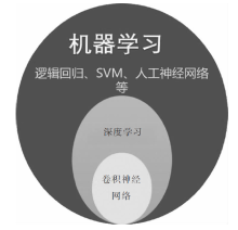
| 研究 | 年份 | AUC | 灵敏度 | 特异度 | 图像数量(恶性/良性) | 患者例数(恶性/良性) | 模型 | |
|---|---|---|---|---|---|---|---|---|
| LR | Timmerman[ | 2005 | 0.936 | 93% | 77.0% | 1 066(266/800) | LR1 | |
| 0.916 | 92% | 75.0% | LR2 | |||||
| ANN | Biagiotti[ | 1999 | 96% | 97.7% | 226(51/175) | |||
| Timmermann[ | 1999 | 0.979 | 95.9% | 93.5% | 173(49/124) | |||
| Moszynski[ | 2006 | 0.968 | 85.7% | 93.1% | 686(255/431) | |||
| SVM | Acharya[ | 2012 | 100.0% | 99.8% | 2 000(1 000/1 000) | 20(10/10) | SVM, RBF | |
| 99.6% | 100% | SVM, linear | ||||||
| Khazendar[ | 2019 | 80% | 77.0% | 187(75/112) | 177 | |||
| CNN | Zhang[ | 2019 | 0.997 | 99.73% | 95.85% | 428(357/71) | ||
| Christiansen[ | 2021 | 0.950 | 96.0% | 86.7% | 3 077 | 758(309/449) | Ovry-Dx1 | |
| 0.958 | 97.1% | 93.7% | Ovry-Dx2 | |||||
| Gao[ | 2022 | 0.911 | 83.1% | 86.8% | 592 275(39 258 /553 017) | 107 624(4 254/103 370) | ||
| Chen[ | 2022 | 0.930 | 92% | 85.0% | 422(118/304) | DLfeature | ||
| 0.900 | 92% | 80.0% | DLdecision |
| 算法 | 优点 | 缺点 |
|---|---|---|
| 逻辑回归算法 | (1)便于理解和实现,可以观测样本的概率分数。 (2)训练速度快 | (1)容易欠拟合。 (2)在一些非线性的数据上表现欠佳 |
| 人工神经网络 | (1)分类的准确度高,学习能力强。 (2)有较强的鲁棒性和容错能力。 | (1)需要大量的训练样本及参数。 (2)浅层神经网络对于特征学习的表达能力有限,深层神经网络的参数繁多,易导致过拟合,甚至可能因为梯度消失而导致不可学习。 |
| 支持向量机 | (1)SVM 是一种小样本学习方法。 (2)算法简单,而且具有较好的鲁棒性。 | (1) 常规SVM的分类结果是二分类的,用SVM解决多分类问题存在困难,且不能直接提供概率估计。 (2)SVM算法只有当核函数与数据的分布较为吻合时才能得到好的效果 |
| 深度卷积神经网络 | (1)具有自学习功能,可以直接从训练数据中提取特征。 (2)可以通过神经网络的深层结构来表示特征之间的关系。 (3)能够获取大量数据中包含的信息,同时实现特征提取、特征选择和分类3个核心步骤,并构建模型。 | (1)具有黑盒性,目前研究无法确定DCNN网络内部是如何运行的。 (2)网络模型复杂程度越高,其对计算设备的硬件要求就越高,对于数据质量的要求也更高 |

| [1] |
Webb PM, Jordan SJ. Epidemiology of epithelial ovarian cancer[J]. Best Pract Res Clin Obstet Gynaecol, 2017, 41:3-14.
doi: S1521-6934(16)30091-8 pmid: 27743768 |
| [2] | Lheureux S, Braunstein M, Oza AM. Epithelial ovarian cancer: Evolution of management in the era of precision medicine[J]. CA Cancer J Clin, 2019, 69(4):280-304. |
| [3] |
Fischerova D. Ultrasound scanning of the pelvis and abdomen for staging of gynecological tumors: a review[J]. Ultrasound Obstet Gynecol, 2011, 38(3):246-266.
doi: 10.1002/uog.10054 pmid: 21898632 |
| [4] | Tavoraitè I, Kronlachner L, Opolskienè G, et al. Ultrasound Assessment of Adnexal Pathology: Standardized Methods and Different Levels of Experience[J]. Medicina (Kaunas), 2021, 57(7):708. |
| [5] |
Timmerman D, Planchamp F, Bourne T, et al. ESGO/ISUOG/IOTA/ESGE Consensus Statement on pre-operative diagnosis of ovarian tumors[J]. Int J Gynecol Cancer, 2021, 31(7):961-982.
doi: 10.1136/ijgc-2021-002565 pmid: 34112736 |
| [6] |
Timmerman D, Valentin L, Bourne TH, et al. Terms, definitions and measurements to describe the sonographic features of adnexal tumors: a consensus opinion from the International Ovarian Tumor Analysis (IOTA) Group[J]. Ultrasound Obstet Gynecol, 2000, 16(5):500-505.
doi: 10.1046/j.1469-0705.2000.00287.x URL |
| [7] |
Timmerman D, Testa AC, Bourne T, et al. Logistic regression model to distinguish between the benign and malignant adnexal mass before surgery: a multicenter study by the International Ovarian Tumor Analysis Group[J]. J Clin Oncol, 2005, 23(34):8794-8801.
pmid: 16314639 |
| [8] |
Timmerman D, Testa AC, Bourne T, et al. Simple ultrasound-based rules for the diagnosis of ovarian cancer[J]. Ultrasound Obstet Gynecol, 2008, 31(6):681-690.
doi: 10.1002/uog.5365 pmid: 18504770 |
| [9] |
van Calster B, van Hoorde K, Valentin L, et al. Evalua-ting the risk of ovarian cancer before surgery using the ADNEX model to differentiate between benign, borderline, early and advanced stage invasive, and secondary metastatic tumours: prospective multicentre diagnostic study[J]. BMJ, 2014, 349:g5920.
doi: 10.1136/bmj.g5920 URL |
| [10] |
Amor F, Vaccaro H, Alcázar JL, et al. Gynecologic ima-ging reporting and data system: a new proposal for classifying adnexal masses on the basis of sonographic findings[J]. J Ultrasound Med, 2009, 28(3):285-291.
doi: 10.7863/jum.2009.28.3.285 URL |
| [11] |
Andreotti RF, Timmerman D, Strachowski LM, et al. O-RADS US Risk Stratification and Management System: A Consensus Guideline from the ACR Ovarian-Adnexal Reporting and Data System Committee[J]. Radiology, 2020, 294(1):168-185.
doi: 10.1148/radiol.2019191150 pmid: 31687921 |
| [12] | Awad M, Khanna R. Machine Learning[M]// Efficient Learning Machines. CA:Apress, Berkeley, 2015:1-18. |
| [13] |
Rajkomar A, Dean J, Kohane I. Machine Learning in Medicine[J]. N Engl J Med, 2019, 380(14):1347-1358.
doi: 10.1056/NEJMra1814259 URL |
| [14] | Brattain LJ, Telfer BA, Dhyani M, et al. Machine learning for medical ultrasound: status, methods, and future opportunities[J]. Abdom Radiol(NY), 2018, 43(4):786-799. |
| [15] | Moustafa AF, Cary TW, Sultan LR, et al. Color Doppler Ultrasound Improves Machine Learning Diagnosis of Breast Cancer[J]. Diagnostics (Basel), 2020, 10(9):631. |
| [16] |
Zhao CK, Ren TT, Yin YF, et al. A Comparative Analysis of Two Machine Learning-Based Diagnostic Patterns with Thyroid Imaging Reporting and Data System for Thyroid Nodules: Diagnostic Performance and Unnecessary Biopsy Rate[J]. Thyroid, 2021, 31(3):470-481.
doi: 10.1089/thy.2020.0305 URL |
| [17] |
Sone K, Toyohara Y, Taguchi A, et al. Application of artificial intelligence in gynecologic malignancies: A review[J]. J Obstet Gynaecol Res, 2021, 47(8):2577-2585.
doi: 10.1111/jog.14818 URL |
| [18] |
Xie HN, Wang N, He M, et al. Using deep-learning algorithms to classify fetal brain ultrasound images as normal or abnormal[J]. Ultrasound Obstet Gynecol, 2020, 56(4):579-587.
doi: 10.1002/uog.21967 pmid: 31909548 |
| [19] |
Al′Aref SJ, Anchouche K, Singh G, et al. Clinical applications of machine learning in cardiovascular disease and its relevance to cardiac imaging[J]. Eur Heart J, 2019, 40(24):1975-1986.
doi: 10.1093/eurheartj/ehy404 pmid: 30060039 |
| [20] | Verhulst PF. Recherches mathématiques sur la loi d′acroissement de la population[J]. Nouveaux mémoires de l′académie royale des sciences et belles-lettres de Bruxel-les, 1845, 18:1-38. |
| [21] | Namburi S. Logistic regression with conjugate gradient descent for document classification[D]. Kansas State University, 2016. |
| [22] |
Meys EM, Kaijser J, Kruitwagen RF, et al. Subjective assessment versus ultrasound models to diagnose ovarian cancer: A systematic review and meta-analysis[J]. Eur J Cancer, 2016, 58:17-29.
doi: 10.1016/j.ejca.2016.01.007 pmid: 26922169 |
| [23] |
Biagiotti R, Desii C, Vanzi E, et al. Predicting ovarian malignancy: application of artificial neural networks to transvaginal and color Doppler flow US[J]. Radiology, 1999, 210(2):399-403.
pmid: 10207421 |
| [24] |
Timmerman D, Verrelst H, Bourne TH, et al. Artificial neural network models for the preoperative discrimination between malignant and benign adnexal masses[J]. Ultrasound Obstet Gynecol, 1999, 13(1):17-25.
doi: 10.1046/j.1469-0705.1999.13010017.x URL |
| [25] |
Moszynski R, Szpurek D, Smolen A, et al. Comparison of diagnostic usefulness of predictive models in preliminary differentiation of adnexal masses[J]. Int J Gynecol Cancer, 2006, 16(1):45-51.
pmid: 16445609 |
| [26] |
McCulloch WS, Pitts W. A logical calculus of the ideas immanent in nervous activity. 1943[J]. Bull Math Biol, 1990, 52(1-2):99-115.
doi: 10.1016/S0092-8240(05)80006-0 URL |
| [27] | Moguerza JM, Muoz Alberto. Support Vector Machines with Applications 1[J]. Statistical Science, 2006, 21(3):358-362. |
| [28] |
Khazendar S, Sayasneh A, Al-Assam H, et al. Automated characterisation of ultrasound images of ovarian tumours: the diagnostic accuracy of a support vector machine and image processing with a local binary pattern operator[J]. Facts Views Vis Obgyn, 2015, 7(1):7-15.
pmid: 25897367 |
| [29] |
Acharya UR, Sree SV, Krishnan MM, et al. Ovarian tumor characterization using 3D ultrasound[J]. Technol Cancer Res Treat, 2012, 11(6):543-552.
doi: 10.7785/tcrt.2012.500272 URL |
| [30] | Khazendar S, Al-assam H, Du H, et al. Automated classification of static ultrasound images of ovarian tumors based on decision level fusion[C]. proceedings of the 2014 6th Computer Science and Electronic Engineering Conference (CEEC), 2014:25-26. |
| [31] |
Khan A, Sohail A, Zahoora U, et al. A survey of the recent architectures of deep convolutional neural networks[J]. Artificial intelligence review, 2020, 53(8):5455-5516.
doi: 10.1007/s10462-020-09825-6 URL |
| [32] |
Cai L, Gao J, Zhao D. A review of the application of deep learning in medical image classification and segmentation[J]. Ann Transl Med, 2020, 8(11):713.
doi: 10.21037/atm.2020.02.44 pmid: 32617333 |
| [33] |
Zhang L, Huang J, Liu L. Improved Deep Learning Network Based in combination with Cost-sensitive Learning for Early Detection of Ovarian Cancer in Color Ultrasound Detecting System[J]. J Med Syst, 2019, 43(8):251.
doi: 10.1007/s10916-019-1356-8 pmid: 31254110 |
| [34] |
Christiansen F, Epstein EL, Smedberg E, et al. Ultrasound image analysis using deep neural networks for discriminating between benign and malignant ovarian tumors: comparison with expert subjective assessment[J]. Ultrasound Obstet Gynecol, 2021, 57(1):155-163.
doi: 10.1002/uog.23530 pmid: 33142359 |
| [35] | Simonyan K, Zisserman A. Very deep convolutional networks for large-scale image recognition[C]. in International Conference on Learning Representations, 2015. |
| [36] | He K, Zhang X, Ren S, et al. Deep Residual Learning for Image Recognition[C]. IEEE Conference on Computer Vision and Pattern Recognition (CVPR), 2016. |
| [37] | Howard AG, Zhu M, Chen B, et al. Mobilenets: Efficient convolutional neural networks for mobile vision applications[J]. arXiv:1704.04861,2017. |
| [38] | Gao Y, Zeng S, Xu X, et al. Deep learning-enabled pelvic ultrasound images for accurate diagnosis of ovarian cancer in China: a retrospective, multicentre, diagnostic study[J]. Lancet Digit Health, 2022, 4(3):e179-e187. |
| [39] |
Chen H, Yang BW, Qian L, et al. Deep Learning Prediction of Ovarian Malignancy at US Compared with O-RADS and Expert Assessment[J]. Radiology, 2022, 304(1):106-113.
doi: 10.1148/radiol.211367 URL |
| [1] | WANG Wenhan, XIA Shujun, ZHAN Weiwei. Application of long non-coding RNA ENST00000489676 detection in ultrasonographic evaluation of cervical lymph node metastasis in papillary thyroid carcinoma [J]. Journal of Diagnostics Concepts & Practice, 2022, 21(04): 514-519. |
| [2] | GU Xuan, LIU Jun. Ultrasound screening to identify solid pseudopapillary tumours of the pancreas from pancreatic ductal adenocarcinoma [J]. Journal of Diagnostics Concepts & Practice, 2022, 21(04): 504-508. |
| [3] | MA Xuefei, WANG Xuefeng, WANG Kankan. Pan-cancer analysis of plasmacytoma variant translocation 1 and MYC gene expression pattern and survival prediction [J]. Journal of Diagnostics Concepts & Practice, 2022, 21(04): 490-496. |
| [4] | QU Qian, HAI Wangxi, HU Shengyan, ZHANG Min, CHEN Xiaoyue, ZHOU Yilei, WANG Jin, HU Xiaoping, LI Biao, HU Jiajia. Automated synthesis of dopamine transporters imaging probe 18F-FP-CIT based on allin one for Micro PET/CT imaging of rat basal ganglia [J]. Journal of Diagnostics Concepts & Practice, 2022, 21(04): 482-489. |
| [5] | CHE Wen, LIU Jiangshu, CHEN Xiaoyan, WANG Chaofu, YUAN Fei, WANG Xuan. Pulmonary mixed squamous cell and glandular papilloma clinicopathological characteristics of 2 cases and misdiagnosis analysis of frozen section [J]. Journal of Diagnostics Concepts & Practice, 2022, 21(04): 476-481. |
| [6] | XU Cheng, XU Xinxin, TIAN Ye, FAN Jiaying, SONG Zhen, YANG Ling. Effect of Haemophilus influenzae colonizing in lower respiratory tract on immune imbalance through TLR4 signaling pathway in asthmatic mice [J]. Journal of Diagnostics Concepts & Practice, 2022, 21(04): 470-475. |
| [7] | BAO Pingping, WU Chunxiao, GU Kai, PANG Yi, WANG Chunfang, SHI Liang, XIANG Yongmei, GONG Yangming, DOU Jianming, WU Mengyin, FU Chen, SHI Yan. Analysis on incidence of stomach cancer in 2016 and trend of incidence during 2002-2016 in Shanghai [J]. Journal of Diagnostics Concepts & Practice, 2022, 21(04): 462-469. |
| [8] | YANG Hui, LI Yunlu, YANG Kang, LI Shiju, HE Jin. Misdiagnosis by whole exome sequencing in progressive myoclonic ataxia consanguineous families: causes and strategies [J]. Journal of Diagnostics Concepts & Practice, 2022, 21(04): 456-461. |
| [9] | HU Jingjing, LÜ Haiwei, XUN Jingna, SHEN Yinzhong, LIU Li, LU Hongzhou. Characteristcs of mycobacterial species distribution in acquired immunodeficiency syndrome patients with mycobacterial infection in Shanghai [J]. Journal of Diagnostics Concepts & Practice, 2022, 21(04): 450-455. |
| [10] | CHEN Zhimin, LIU Bo, He Haolan, HE Yaozu, FENG Lizhi, LIU Xinhua, ZHANG Jiansheng, CAI Weiping, LI Linghua. Analysis of 133 dead cases of AIDS patients co-infected with Talaromycosis [J]. Journal of Diagnostics Concepts & Practice, 2022, 21(04): 444-449. |
| [11] | DU Yanran, JIAO Jing, REN Yunyun, ZHOU Jianqiao. Application of ultrasound-based radiomics technology in the evaluation of fetal lung maturity [J]. Journal of Diagnostics Concepts & Practice, 2022, 21(03): 326-330. |
| [12] | ZHANG Juanjuan, HE Qinyu, YANG Yuanyan, DONG Zhiya, XIAO Yuan, CHEN Lifen, ZHANG Caiping. Lamb-Shaffer syndrome presenting as short stature with delays in motor and language acquisition: a case report and literature review [J]. Journal of Diagnostics Concepts & Practice, 2022, 21(03): 336-342. |
| [13] | LIU Xin, QI Caihui, WANG Zhenjing, LÜ Na, WANG Shaoting, WANG Shuping. Transcriptome study of glucagon like peptide-1 agonist Exendin-4 on mouse embryonic osteoblast precursor MC3T3-E1 in vitro [J]. Journal of Diagnostics Concepts & Practice, 2022, 21(03): 367-373. |
| [14] | RUAN Yufeng, HU Liping, CHEN Shirong, YIN Jun, SUN Jing. Investigation on mastery status of standardized diagnosis and treatment of Helicobacter pylori(Hp) infection in general practitioners in Pudong New Aera, Shanghai [J]. Journal of Diagnostics Concepts & Practice, 2022, 21(03): 399-404. |
| [15] | MA Shaochen, GUO Xin, WANG Mingwei, WANG Huijun, YU Qijun, SU Wenyue, WANG Hualong, MA Qinying. Effect of game-based EEG neurofeedback training on improvement of cognitive function [J]. Journal of Diagnostics Concepts & Practice, 2022, 21(01): 41-45. |
| Viewed | ||||||
|
Full text |
|
|||||
|
Abstract |
|
|||||