Journal of Diagnostics Concepts & Practice ›› 2025, Vol. 24 ›› Issue (02): 194-203.doi: 10.16150/j.1671-2870.2025.02.011
Previous Articles Next Articles
QIN Yu1, LI Cheng2, HUA Qing1, ZHANG Huiting1, JIA Wanru1, DONG Yijie1, ZHOU Jianqiao1, XIA Shujun1( )
)
Received:2025-01-05
Accepted:2025-03-10
Online:2025-04-25
Published:2025-07-11
Contact:
XIA Shujun
E-mail:xiashu_jun@126.com
CLC Number:
QIN Yu, LI Cheng, HUA Qing, ZHANG Huiting, JIA Wanru, DONG Yijie, ZHOU Jianqiao, XIA Shujun. Ultrasound viscoelastic imaging in differentiation of benign and malignant breast tumors[J]. Journal of Diagnostics Concepts & Practice, 2025, 24(02): 194-203.
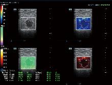
Figure 1
Measurement of shear wave modulus of elasticity and viscosity coefficient of breast lesions and their marginal tissues
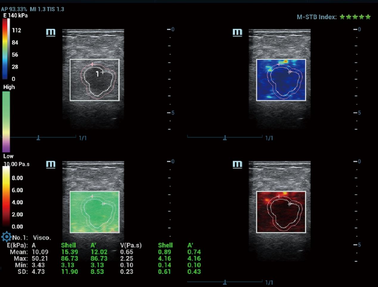
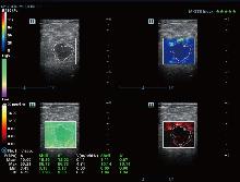
Figure 2
Shear wave elastic modulus and dispersion coefficient measurements of breast lesions and their marginal tissues

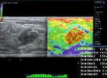
Figure 3
Measurement of ultrasound strain elasticity parameters of breast lesions and their marginal tissues
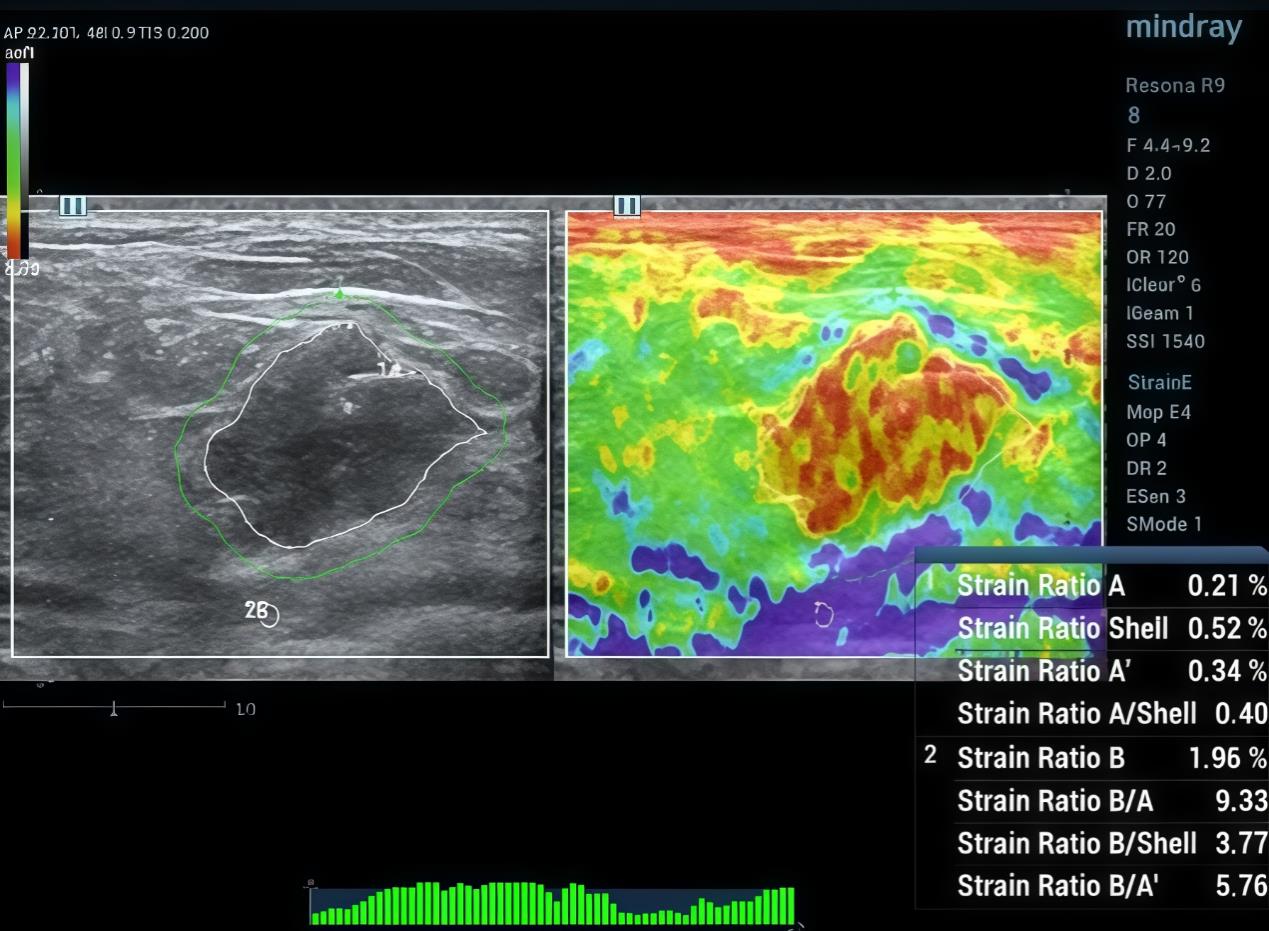
Table 1
Pathological results of the 717 cases
| Item | Number (%) | |
|---|---|---|
| Benign/Malignant | ||
| Malignant | 471 | (65.69) |
| Benign | 246 | (34.31) |
| Pathological type | ||
| Adenosis | 64 | (8.93) |
| Fibroadenoma | 93 | (12.97) |
| Intraductal papilloma | 52 | (7.25) |
| Other benign lesions | 33 | (4.60) |
| Borderline tumor | 6 | (0.84) |
| Ductal carcinoma in situ | 55 | (7.67) |
| Lobular carcinoma in situ | 8 | (1.12) |
| Invasive papillary carcinoma | 17 | (2.37) |
| Invasive ductal carcinoma | 334 | (46.58) |
| Invasive lobular carcinoma | 15 | (2.09) |
| Neuroendocrine tumor | 2 | (0.28) |
| Mucinous carcinoma | 13 | (1.81) |
| Mucinous carcinoma | 25 | (3.49) |
Table 2
Univariate analysis of ultrasound viscosity and elasticity variables on the benignity and malignancy of breast tumors
| Item | Total (N = 717) | Benign(n= 246) | Malignant(n= 471) | P-value |
|---|---|---|---|---|
| Age | 52.49±14.05 | 45.00 ± 13.17 | 56.40 ± 12.86 | <0.001 |
| BI-RADS | <0.001 | |||
| 4B and above | 516(71.97%) | 73(29.67%) | 443(94.06%) | |
| Below 4B | 201(28.03%) | 173(70.33%) | 28(5.94%) | |
| T-Emean | 25.93±14.65 | 23.12±13.30 | 27.40±15.12 | <0.001 |
| T-Emax | 126.63±84.63 | 90.34± 64.77 | 145.58±87.61 | <0.001 |
| T-Esd | 16.83 ± 11.33 | 12.83 ± 8.56 | 18.92 ±12.02 | <0.001 |
| Shell-Emean | 30.25 ±16.93 | 23.36 ±13.24 | 33.85 ± 17.53 | <0.001 |
| Shell-Emax | 139.84±85.38 | 98.11± 66.43 | 161.63±86.12 | <0.001 |
| Shell-Esd | 21.26 ± 13.57 | 15.18± 10.11 | 24.44 ± 14.06 | <0.001 |
| A-Emean | 27.74 ± 14.78 | 23.41± 12.85 | 30.00 ± 15.22 | <0.001 |
| A-Emax | 151.54±90.95 | 107.56±72.52 | 174.50±91.20 | <0.001 |
| A-Esd | 19.58 ± 12.02 | 14.56 ± 9.28 | 22.20 ± 12.46 | <0.001 |
| Shell/T-Emean | 1.21 ± 0.33 | 1.05 ± 0.27 | 1.29 ± 0.33 | <0.001 |
| Shell/T-Emax | 1.24 ± 0.59 | 1.22 ± 0.64 | 1.24 ± 0.56 | 0.400 |
| Shell/T-Esd | 1.37 ± 0.53 | 1.27 ± 0.55 | 1.42 ± 0.52 | <0.001 |
| T-Vmean | 1.74 ± 0.89 | 1.77 ± 0.87 | 1.72 ± 0.90 | 0.400 |
| T-Vmax | 8.15 ± 4.78 | 6.60 ± 3.92 | 8.95 ± 4.99 | <0.001 |
| T-Vsd | 1.15±0.71 | 1.02 ± 0.61 | 1.22 ± 0.75 | <0.001 |
| Shell-Vmean | 2.07±1.00 | 1.80 ± 0.85 | 2.21 ± 1.04 | <0.001 |
| Shell-Vmax | 8.97±4.77 | 7.10 ± 4.00 | 9.95 ± 4.85 | <0.001 |
| Shell-Vsd | 1.46±0.85 | 1.18 ± 0.69 | 1.60 ± 0.90 | <0.001 |
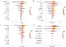
Figure 4
SHAP plot of viscoelastic parametersNotes: a. SHAP plot of viscosity coefficient parameter; b. SHAP plot of dispersion coefficient parameter; c. SHAP plot of elastic modulus parameter; d. SHAP plot of strain elasticity parameter.
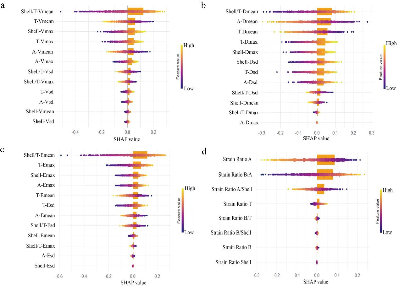
Table 3
Comparison of the predictive power of the three groups of models
| Model | AIC | AUC | 95%CI |
|---|---|---|---|
| Shell/T-Vmean+ Shell/T-Dmean Combined model | 0.755 | 0.718-0.789 | |
| Shell/T-Vmean Univariate model | 804.32 | 0.742 | 0.704-0.775 |
| Shell/T-Dmean Univariate model | 800.82 | 0.745 | 0.707-0.780 |
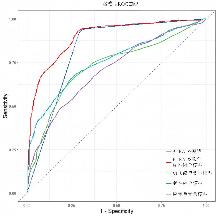
Figure 5
ROC curve of each prediction model for benign-malignant differentiation
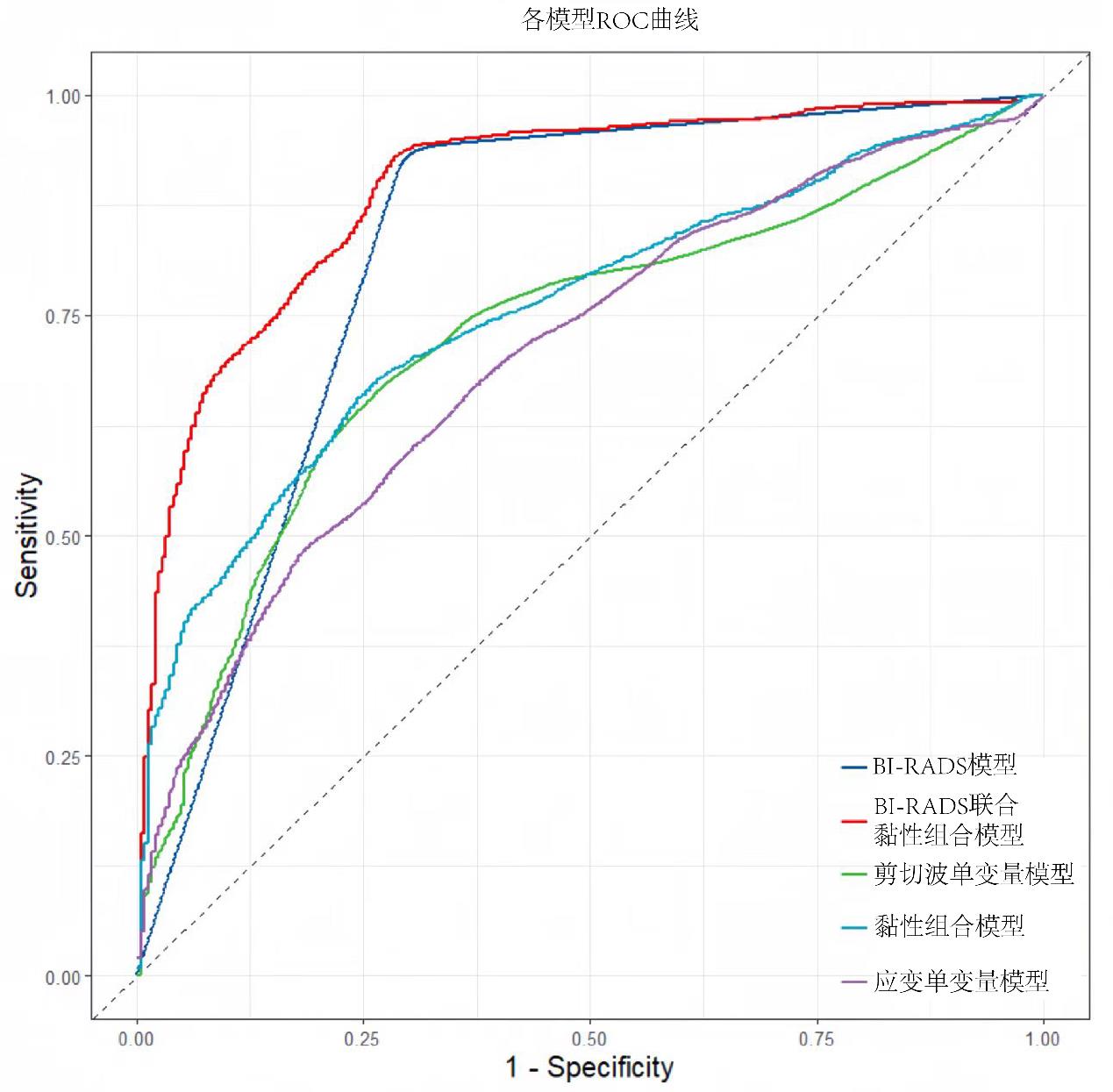
Table 4
Predictive value of each model for benign-malignant differentiation
| Model | AIC | AUC | 95%CI |
|---|---|---|---|
BI-RADS combined with viscoelastic model | 546.862 | 0.895 | 0.868-0.917 |
| BI-RADS model | 586.959 | 0.822 | 0.790-0.853 |
| Viscoelastic combined model | 788.584 | 0.755 | 0.718-0.789 |
| Shear wave univariate model | 830.377 | 0.726 | 0.685-0.764 |
| Strain univariate model | 848.274 | 0.705 | 0.663-0.744 |
| [1] | SUNG H, FERLAY J, SIEGEL R L, et al. Global Cancer Statistics 2020: GLOBOCAN estimates of incidence and mortality worldwide for 36 cancers in 185 countries[J]. CA Cancer J Clin, 2021, 71(3): 209-249. |
| [2] | CHEN W, ZHENG R, BAADE P D, et al. Cancer statistics in China, 2015[J]. CA Cancer J Clin, 2016, 66(2): 115-132. |
| [3] |
FELDMANN A, LANGLOIS C, DEWAILLY M, et al. Shear wave elastography (SWE): an analysis of breast lesion characterization in 83 breast lesions[J]. Ultrasound Med Biol, 2015, 41(10): 2594-2604.
doi: 10.1016/j.ultrasmedbio.2015.05.019 pmid: 26159068 |
| [4] |
RICCI P, MAGGINI E, MANCUSO E, et al. Clinical application of breast elastography: state of the art[J]. Eur J Radiol,2014, 83(3): 429-37.
doi: 10.1016/j.ejrad.2013.05.007 pmid: 23787274 |
| [5] |
SIGRIST R M S, LIAU J, KAFFAS A E, et al. Ultrasound elastography: review of techniques and clinical applications[J]. Theranostics, 2017, 7(5): 1303-1329.
doi: 10.7150/thno.18650 pmid: 28435467 |
| [6] |
SHIINA T, NIGHTINGALE K R, PALMERI M L, et al. WFUMB guidelines and recommendations for clinical use of ultrasound elastography: Part 1: basic principles and terminology[J]. Ultrasound Med Biol,2015,41(5):1126-1147.
doi: 10.1016/j.ultrasmedbio.2015.03.009 pmid: 25805059 |
| [7] | KUMAR V, DENIS M, GREGORY A, et al. Viscoelastic parameters as discriminators of breast masses: Initial human study results[J]. PLoS One, 2018,13(10): e0205717. |
| [8] |
LI W, JIANG J, CAO J, et al. The value of ultrasound viscosity imaging in preoperative differential diagnosis between malignant and benign breast lesions: Preliminary clinical applications[J]. Clin Hemorheol Microcirc,2025,89(1):111-122.
doi: 10.3233/CH-242405 pmid: 39911120 |
| [9] | American College of Radiology. ACR BI-RADS atlas: breast imaging reporting and data system[M].5th ed, Reston, Virginia, 2013. |
| [10] |
JIA W, XIA S, JIA X, et al. Ultrasound Viscosity Imaging in Breast Lesions: A Multicenter Prospective Study[J]. Acad Radiol, 2024, 31(9): 3499-510.
doi: 10.1016/j.acra.2024.03.017 pmid: 38582684 |
| [11] |
MANDUCA A, BAYLY P J, EHMAN R L, et al. MR elastography: Principles, guidelines, and terminology[J]. Magn Reson Med, 2021, 85(5): 2377-2390.
doi: 10.1002/mrm.28627 pmid: 33296103 |
| [12] | SHI Y, QI Y F, LAN G Y, et al. Three-dimensional MR elastography depicts liver inflammation, fibrosis, and portal hypertension in chronic hepatitis B or C[J]. Radio-logy, 2021, 301(1): 154-162. |
| [13] |
TAPPER E B, LOOMBA R. Noninvasive imaging biomarker assessment of liver fibrosis by elastography in NAFLD[J]. Nat Rev Gastroenterol Hepatol,2018,15(5): 274-282.
doi: 10.1038/nrgastro.2018.10 pmid: 29463906 |
| [14] |
PATEL B K, SAMREEN N, ZHOU Y, et al. MR elastography of the breast: evolution of technique, case examples, and future directions[J]. Clin Breast Cancer, 2021,21(1): e102-e111.
doi: 10.1016/j.clbc.2020.08.005 pmid: 32900617 |
| [15] |
CHEN S, SANCHEZ W, CALLSTROM M R, et al. Assessment of liver viscoelasticity by using shear waves induced by ultrasound radiation force[J]. Radiology, 2013, 266(3): 964-970.
doi: 10.1148/radiol.12120837 pmid: 23220900 |
| [16] |
SUGIMOTO K, MORIYASU F, OSHIRO H, et al. The role of multiparametric US of the liver for the evaluation of nonalcoholic steatohepatitis[J]. Radiology,2020, 296(3): 532-540.
doi: 10.1148/radiol.2020192665 pmid: 32573385 |
| [17] |
LEE D H, LEE J Y, BAE J S, et al. Shear-wave dispersion slope from US shear-wave elastography: detection of allograft damage after liver transplantation[J]. Radiology, 2019,293(2): 327-333.
doi: 10.1148/radiol.2019190064 pmid: 31502939 |
| [18] | HOSSAIN M M, SELZO M R, HINSON R M, et al. Evaluating renal transplant status using viscoelastic response (VisR) Ultrasound[J]. Utrasound Med Biol, 2018, 44(8): 1573-1584. |
| [19] | SADIGH G, CARLOS R C, NEAL C H, et al. Accuracy of quantitative ultrasound elastography for differentiation of malignant and benign breast abnormalities: a meta-analysis[J]. Breast Cancer Res Treat,2012,134(3): 923-931. |
| [20] |
BERG W A, COSGROVE D O, DORé C J, et al. Shear-wave elastography improves the specificity of breast US: the BE1 multinational study of 939 masses [J]. Radiology, 2012, 262(2): 435-449.
doi: 10.1148/radiol.11110640 pmid: 22282182 |
| [21] | ZHANG H, GUO Y, ZHOU Y, et al. Fluidity and elasticity form a concise set of viscoelastic biomarkers for breast cancer diagnosis based on Kelvin-Voigt fractional derivative modeling[J]. Biomech Model Mechanobiol,2020,19(6): 2163-2177. |
| [22] | MIERKE C T. Viscoelasticity acts as a marker for tumor extracellular matrix characteristics[J]. Front Cell Dev Biol, 2021, 9: 785138. |
| [23] |
ZHOU J, ZHAN W, CHANG C, et al. Breast lesions: evaluation with shear wave elastography, with special emphasis on the "stiff rim" sign[J]. Radiology,2014,272(1): 63-72.
doi: 10.1148/radiol.14130818 pmid: 24661245 |
| [24] |
PARK H S, SHIN H J, SHIN K C, et al. Comparison of peritumoral stromal tissue stiffness obtained by shear wave elastography between benign and malignant breast lesions[J]. Acta Radiol,2018,59(10): 1168-1175.
doi: 10.1177/0284185117753728 pmid: 29359949 |
| [25] | 王艳萍, 唐笛娇, 努尔比耶·买买提依力, 等. 血清抗着丝粒蛋白F抗体在乳腺癌中的临床价值探讨 [J]. 重庆医科大学学报, 2024, 49 (9): 1188-1192. |
| WANG Y P, TANG D J, NUERBIYE·M, et al. Clinical value of serum anti-centromere protein F antibody in breast cancer[J]. J Chongqing Med Univ,2024,49(9): 1188-1192. | |
| [26] |
SRIDHAR M, INSANA M F. Ultrasonic measurements of breast viscoelasticity[J]. Med Phys,2007,34(12): 4757-4767.
pmid: 18196803 |
| Viewed | ||||||
|
Full text |
|
|||||
|
Abstract |
|
|||||