

Journal of Diagnostics Concepts & Practice ›› 2026, Vol. 25 ›› Issue (01): 71-77.doi: 10.16150/j.1671-2870.2026.01.010
• Original articles • Previous Articles Next Articles
TANG Ting, WANG Di, FANG Wei( )
)
Received:2025-08-14
Revised:2025-09-30
Accepted:2025-10-29
Online:2026-02-25
Published:2026-02-25
Contact:
FANG Wei
E-mail:fangwei980365@qq.com
CLC Number:
TANG Ting, WANG Di, FANG Wei. Clinical and pathological analysis of 5 cases of calcifying fibrous tumor in gastrointestinal tract and literature review[J]. Journal of Diagnostics Concepts & Practice, 2026, 25(01): 71-77.
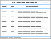
Table 1
Clinical data of 5 cases of gastrointestinal CFT
| Case | Age (Year) | Gender | Location | Clinical manifestations | Gastroscopic findings | Imaging findings | Treatment methods |
|---|---|---|---|---|---|---|---|
| 1 | 49 | Female | Subserosa of gastric body, subserosa of small intestinal wall, abdominopelvic wall and mesentery | Nausea and discomfort | Not performed | CT: Oval soft tissue density mass under the serosa of the gastric fundus, with clear and smooth boundaries. On plain scan, the parenchymal density of the tumor was higher than that of the normal gastric wall, with atypical calcification in the tumor, and mild progressive enhancement on enhanced scan | Laparoscopic resection of gastric lesion, resection of ileal lesion and resection of intra-abdominal mass |
| 2 | 54 | Female | Gastric fundus | Discovered during physical examination | Submucosal protrusion with smooth surface mucosa, accompanied by hyperemia and focal erosion | CT: No abnormality reported | Endoscopic submucosal dissection |
| 3 | 68 | Male | Gastric body | Discovered during physical examination | Submucosal protrusion with smooth surface mucosa | CT: Uniform enhancement after enhanced scan | Endoscopic submucosal dissection |
| 4 | 53 | Male | Gastric body | Discovered during physical examination | Submucosal protrusion with hyperemia, macular appearance and focal erosion | CT: No abnormality reported | Endoscopic submucosal dissection |
| 5 | 68 | Female | Gastric body | Upper abdominal pain accompanied by nausea and vomiting | Submucosal protrusion with smooth surface mucosa | CT: Mild enhancement after enhanced scan | Endoscopic full-thickness resection |

Figure 1
The elevated lesion showed a smooth surface mucosa, accompanied by mild hyperemia and a mottled appearance under gastroscopy


Figure 2
CT scan showed uniform enhancement after contrast enhancement


Figure 3
Pathological picture (Digital scanning slice) A: Subserosal calcifying fibrous tumor (×1); B: Submucosal calcifying fibrous tumor (×4); C: Scattered spindle cells, lymphocyles, and plasma cells were observed against a background of abundant dense collagen fibers(×10); D: The spindle cells exhibited a mild morphology, lymphocytes and plasma cells were distributed around blood vessels (×10); E: Lymphocytes were distributed in a "cull-like" patter around the periphery of the tumor and form lymphoid follicles (×4); F: Scallered psammoma bodies were observed(×4).


Figure 4
CD34 was partially positive(Digital scanning slice, Envision method, ×10)

Table 2
Clinicopathological features of 5 cases of gastrointestinal CFT
| Case | Disease location | Solitary / Multiple | Size(cm) | Gross | Pathological morphology | Psammoma bodies | "Cuff-like" structure | Immunophenotype |
|---|---|---|---|---|---|---|---|---|
| 1 | Subserosa of gastric body, subserosa of small intestinal wall, abdominopelvic wall and mesentery | Multiple | 1.1-2.0 | Well-circumscribed, grayish-white cut surface, firm or hard consistency | Hyaline degeneration of collagen fibers with a small amount of non-atypical fibroblasts | Present | Present | CD34 (partially+),CD117(partially+),DOG-1(-),S-100(-),SMA(partially+),Desmin(-) |
| 2 | Submucosa of gastric fundus | Solitary | 0.8 | Well-circumscribed, grayish-white cut surface, hard consistency | Hyaline degeneration of collagen fibers with a small amount of non-atypical fibroblasts | Absent | Absent | CD34(-),CD117(-),DOG-1(-),S-100 (-),SMA(-),Desmin(-) |
| 3 | Submucosa of gastric body | Solitary | 1.0 | Well-circumscribed, grayish-white cut surface, hard consistency | Hyaline degeneration of collagen fibers with a small amount of non-atypical fibroblasts | Absent | Absent | CD117(-),CD34(-),Desmin(-),DOG-1(-), SMA(-),S-100(-) |
| 4 | Submucosa of gastric body | Solitary | 1.7 | Well-circumscribed, grayish-white cut surface, moderate consistency | Hyaline degeneration of collagen fibers with a small amount of non-atypical fibroblasts | Present | Present | CD117(-),DOG-1(-),CD34(-),Desmin(-), SMA(-),S-100(-) |
| 5 | Submucosa of gastric body | Solitary | 1.7 | Well-circumscribed, grayish-white cut surface, moderate consistency | Hyaline degeneration of collagen fibers with a small amount of non-atypical fibroblasts | Present | Present | CD34(-),CD117(scattered+),DOG-1(-),Desmin(-),S-100(-),SMA(+) |
| [1] |
ROSENTHAL N S, ABDUL-KARIM F W. Childhood fibrous tumor with psammoma bodies. Clinicopathologic features in two cases[J]. Arch Pathol Lab Med, 1988, 112(8):798-800.
pmid: 3395217 |
| [2] | FLETCHER C D M, UNNI K, MERTENS F, et al. World Health Organization Classification of tumours. Pathology and genetics of tumours of soft tissue and bone[M]. Lyon France,IARC Press, 2002. |
| [3] |
CHORTI A, PAPAVRAMIDIS T S, MICHALOPOULOS A. Calcifying fibrous tumor: Review of 157 patients reported in international literature[J]. Medicine (Baltimore), 2016, 95(20):e3690.
doi: 10.1097/MD.0000000000003690 URL |
| [4] | 张雷, 魏建国, 方三高, 等. 钙化性纤维性肿瘤32例临床病理学分析[J]. 中华病理学杂志, 2020, 49(2):129-133. |
| ZHANG L, WEI J G, FANG S G, et al. Calcifying fibrous tumor: A clinicopathological analysis of 32 cases[J]. Chin J Pathol, 2020, 49(2):129-133. | |
| [5] |
FETSCH J F, MONTGOMERY E A, MEIS J M. Calcifyi-ng fibrous pseudotumor[J]. Am J Surg Pathol, 1993, 17(5):502-508.
doi: 10.1097/00000478-199305000-00010 URL |
| [6] |
NASCIMENTO A F, RUIZ R, HORNICK J L, et al. Calcifying fibrous 'pseudotumor': Clinicopathologic study of 15 cases and analysis of its relationship to inflammatory myofibroblastic tumor[J]. Int J Surg Pathol, 2002, 10(3):189-196.
doi: 10.1177/106689690201000304 pmid: 12232572 |
| [7] |
LARSON B K, BALZER B, GOLDWASSER J, et al. Calcifying fibrous tumor: an unrecognized IgG4---related disease?[J]. APMIS, 2015, 123(1):72-76.
doi: 10.1111/apm.2014.123.issue-1 URL |
| [8] |
VAN DORPE J, ECTORS N, GEBOES K, et al. Is calcif-ying fibrous pseudotumor a late sclerosing stage of inflammatory myofibroblastic tumor?[J]. Am J Surg Pathol, 1999, 23(3):329-335.
doi: 10.1097/00000478-199903000-00013 URL |
| [9] |
LEE D, SUH Y L, LEE S K. Calcifying fibrous pseudotumour arising in a gastric inflammatory myofibroblastic tumour[J]. Pathology, 2006, 38(6):588-591.
doi: 10.1080/00313020601023997 URL |
| [10] | PEZHOUH M K, REZAEI M K, SHABIHKHANI M, et al. Clinicopathologic study of calcifying fibrous tumor of the gastrointestinal tract: a case series[J]. Hum Pathol, 2017,62:199-205. |
| [11] |
AGAIMY A, BIHL M P, TORNILLO L, et al. Calcifying fibrous tumor of the stomach: Clinicopathologic and molecular study of seven cases with literature review and reappraisal of histogenesis[J]. Am J Surg Pathol, 2010, 34(2):271-278.
doi: 10.1097/PAS.0b013e3181ccb172 pmid: 20090503 |
| [12] | 李璐, 王淑妍, 邓茜, 等. 胃钙化性纤维性肿瘤24例临床病理分析[J]. 临床与实验病理学杂志, 2022, 38(2):211-213. |
| LI L, WANG S Y, DENG Q, et al. Clinicopathological analysis of 24 cases of calcific fibrous tumor of stomach[J]. J Clin Exp pathol, 2022, 38(2):211-213. | |
| [13] | 李红娜, 毛昕, 胡倩岚, 等. 回肠钙化性纤维性肿瘤1例[J]. 诊断病理学杂志, 2022, 29(8):782-783,封3. |
| LI H N, MAO X, HU Q L, et al. Calcific fibrous tumor of ileum: a case report[J]. J Diag Pathol, 2022, 29(8):782-783,i3. | |
| [14] |
WANG J, AO W, MAO G, et al. Gastric calcifying fibrous tumors: Computed tomography findings and clinical manifestations[J]. Medicine (Baltimore), 2021, 100(5):e23334.
doi: 10.1097/MD.0000000000023334 URL |
| [15] | 杨朝武, 王娟. 腹部钙化性纤维性肿瘤CT及MRI诊断价值[J]. 影像研究与医学应用, 2023, 7(9):174-176,180. |
| YANG C W, WANG J. Diagnostic value of CT and MRI in calcifying fibrous tumor of abdomen[J]. J Imaging Res Med Appl, 2023, 7(9):174-176,180. | |
| [16] | 唐晓婷, 刘振民, 房太勇. 胃钙化性纤维性肿瘤2例并文献复习[J]. 中华灾害救援医学, 2024, 11(11):1258-1262. |
| TANG X T, LIU Z M, FANG T Y. Two cases of gastric calcified fibrous tumors with literature review[J]. Chin J Disaster Med, 2024, 11(11):1258-1262. | |
| [17] | 杜志华, 余嘉惠, 林晓丰, 等. 胃肠道钙化性纤维性肿瘤17例临床病理分析[J]. 诊断病理学杂志, 2024, 31(3):208-212. |
| DU Z H, YU J H, LIN X F, et al. Calcifying fibrous tumor of the gastrointestinal tract:a clinicopathological analysis of 17 cases[J]. J Diag Pathol, 2024, 31(3):208-212. | |
| [18] | 高珊, 易慕华, 刘涛, 等. 胃钙化性纤维性肿瘤的诊断过程[J]. 巴楚医学, 2024, 7(2):27-29. |
| GAO S, YI M H, LIU T, et al. Diagnosis of gastric calcif-ying fibrous tumor[J]. Bachu Med J, 2024, 7(2):27-29. | |
| [19] | 彭金榜, 林佳佳, 叶丽萍, 等. 胃钙化性纤维性假瘤2例并文献复习[J]. 胃肠病学, 2014(11):703-704. |
| PENG J B, LIN J J, YE L P, et al. Gastric calcifying fibrous pseudotumor: Report of two cases and review of literature[J]. Chin J Gastroenterol, 2014(11):703-704. | |
| [20] |
TURBIVILLE D, ZHANG X. Calcifying fibrous tumor of the gastrointestinal tract: A clinicopathologic review and update[J]. World J Gastroenterol, 2020, 26(37):5597-5605.
doi: 10.3748/wjg.v26.i37.5597 URL |
| [21] | 朱常琰, 王修远, 胡玉秋, 等. 胃大弯侧外生性浆膜层下钙化性纤维性肿瘤1例[J]. 消化肿瘤杂志(电子版), 2024, 16(3):385-388. |
| ZHU C Y, WANG X Y, HU Y Q, et al. Exogenous subserosal calcifying fibrous tumor in gastric greater curvature:a case report[J]. J Dig Oncol(Electronic Version), 2024, 16(3):385-388. | |
| [22] | 周影, 陈百胜, 杨丹琪, 等. 胃钙化性纤维性肿瘤的内镜表现及病理特征[J]. 中国医药指南, 2020, 18(28):4-6. |
| ZHOU Y, CHEN B S, YANG D Q, et al. Endoscopic manifestations and pathological features of gastric calcif-ying fibrous tumors[J]. Guide Chin Med, 2020, 18(28):4-6. |
| [1] | GU Yijin, LI Anqi, DONG Lei, XU Haimin, SHEN Xia, XIE Jialing, YUAN Fei, WANG Chaofu. Correlation of KIT and PDGFRA mutation status with clinicopathologic features in primary and recurrent or metastatic gastrointestinal stromal tumors [J]. Journal of Diagnostics Concepts & Practice, 2021, 20(03): 257-264. |
| [2] | CHEN Xiaoyan, WANG Chaofu, DONG Lei, XU Haimin, JIN Xiaolong. Primary non-myxomatous cardiac tumor: clinicopathologic analysis of 19 cases [J]. Journal of Diagnostics Concepts & Practice, 2017, 16(01): 66-72. |
| [3] | . [J]. Journal of Diagnostics Concepts & Practice, 2012, 11(04): 382-386. |
| [4] | . [J]. Journal of Diagnostics Concepts & Practice, 2012, 11(02): 145-148. |
| [5] | . [J]. Journal of Diagnostics Concepts & Practice, 2010, 9(05): 453-456. |
| [6] | . [J]. Journal of Diagnostics Concepts & Practice, 2008, 7(01): 47-51. |
| [7] | . [J]. Journal of Diagnostics Concepts & Practice, 2005, 4(04): 296-299. |
| [8] | . [J]. Journal of Diagnostics Concepts & Practice, 2005, 4(03): 222-224. |
| Viewed | ||||||
|
Full text |
|
|||||
|
Abstract |
|
|||||
