

Journal of Diagnostics Concepts & Practice ›› 2026, Vol. 25 ›› Issue (03): 308-314.doi: 10.16150/j.1671-2870.2026.03.006
• Original articles • Previous Articles Next Articles
WANG Lili1, GUO Jian2( )
)
Received:2026-03-06
Revised:2026-03-31
Accepted:2026-05-11
Online:2026-06-25
Published:2026-06-27
Contact:
GUO Jian
E-mail:guojian1110@126.com
CLC Number:
WANG Lili, GUO Jian. Epidemiological status and drug resistance survey of protothecosis in China from 2015 to 2025[J]. Journal of Diagnostics Concepts & Practice, 2026, 25(03): 308-314.
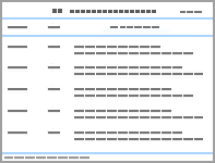
Table 1
Epidemiological characteristics of clinically isolated Prototheca strains
| Category | Subcategory | Number (%) |
|---|---|---|
| Age, years | 0-20 | 3 (3.6%) |
| 21-40 | 5 (6.0%) | |
| 41-60 | 29 (34.5%) | |
| 61-80 | 40 (47.6%) | |
| >81 | 7 (8.3%) | |
| Gender | Male | 43 (51.2%) |
| Female | 41 (48.8%) | |
| Species | Prototheca wickerhamii | 67 (79.8%) |
| Prototheca bovis | 10 (11.9%) | |
| Prototheca zopfii | 5 (6.0%) | |
| Prototheca ciferrii | 1 (1.2%) | |
| Prototheca miyajii | 1 (1.2%) | |
| Site of isolation | Skin tissue | 50 (59.5%) |
| Feces | 9 (10.7%) | |
| Ascitic fluid | 4 (4.8%) | |
| Cerebrospinal fluid | 3 (3.6%) | |
| Blood | 5 (6.0%) | |
| Sputum | 3 (3.6%) | |
| Bronchoalveolar lavage fluid | 3 (3.6%) | |
| Wound secretion | 3 (3.6%) | |
| Pus | 3 (3.6%) | |
| Joint fluid | 1 (1.2%) |
Table 2
The antimicrobial susceptibility profiles of all Prototheca strains
| Antibiotics | All (n=84) | Skin tissue (n=50) | Non-skin tissue (n=34) | ||||||
|---|---|---|---|---|---|---|---|---|---|
| MIC range (µg/mL) | MIC50 (µg/mL) | MIC90 (µg/mL) | MIC range (µg/mL) | MIC50 (µg/mL) | MIC90 (µg/mL) | MIC range (µg/mL) | MIC50 (µg/mL) | MIC90 (µg/mL) | |
| Isavuconazole | 0.015-1.000 | 0.060 | 0.250 | 0.015-0.500 | 0.060 | 0.120 | 0.015-1.00 | 0.120 | 0.500 |
| Itraconazole | 0.250-16.000 | 1.000 | 2.000 | 0.500-16.000 | 1.000 | 2.000 | 0.250-16.000 | 1.000 | 16.000 |
| Posaconazole | 0.500-8.000 | 1.000 | 2.000 | 0.500-4.000 | 1.000 | 2.000 | 0.500-8.000 | 1.000 | 2.000 |
| Voriconazole | 0.500-8.000 | 1.000 | 4.000 | 0.500-4.000 | 1.000 | 2.000 | 0.500-8.000 | 2.000 | 4.000 |
| Amphotericin B | 0.250-1.000 | 0.500 | 0.500 | 0.500-1.000 | 0.500 | 0.500 | 0.250-1.000 | 0.500 | 1.000 |
| Anidulafungin | 8.000-8.000 | 8.000 | 8.000 | 8.000-8.000 | 8.000 | 8.000 | 8.000-8.000 | 8.000 | 8.000 |
| Caspofungin | 4.000-4.000 | 4.000 | 4.000 | 4.000-4.000 | 4.000 | 4.000 | 4.000-4.000 | 4.000 | 4.000 |
| Micafungin | 8.00-8.000 | 8.000 | 8.000 | 8.000-8.000 | 8.000 | 8.000 | 8.000-8.000 | 8.000 | 8.000 |
| Fluconazole | 128.000-256.000 | 256.000 | 256.000 | 128.000-256.000 | 256.000 | 256.000 | 256.000-256.000 | 256.000 | 256.000 |
| 5-Fluorocytosine | 32.000-32.000 | 32.000 | 32.000 | 32.000-32.000 | 32.000 | 32.000 | 32.000-32.000 | 32.000 | 32.000 |

Figure 1
In vitro antimicrobial susceptibility of Prototheca strains

Table 3
In vitro antimicrobial susceptibility of different Prototheca species.
| Species | Minimum inhibitory concentration range (µg/mL) | |||||||||
|---|---|---|---|---|---|---|---|---|---|---|
| ISA | ITR | POS | VOR | AMB | ANI | CAS | MIC | FLU | 5-FC | |
| P. wickerhamii(n=67) | 0.015-0.500 | 0.500-16.000 | 0.500-4.000 | 0.500-4.000 | 0.250-1.000 | 8.000 | 4.000 | 8.000 | 128.000-256.000 | 32.000 |
| P. bovis(n=10) | 0.030-1.000 | 0.250-16.000 | 0.500-4.000 | 0.500-8.000 | 0.500-1.000 | 8.000 | 4.000 | 8.000 | 256.000 | 32.000 |
| P. zopfii(n=5) | 0.250-0.500 | 0.500-1.000 | 1.000-2.000 | 2.000-4.000 | 0.500-1.000 | 8.000 | 4.000 | 8.000 | 256.000 | 32.000 |
| P. ciferrii(n=1) | 0.015 | 0.250 | 0.500 | 0.500 | 0.500 | 8.000 | 4.000 | 8.000 | 256.000 | 32.000 |
| P. miyajii(n=1) | 0.120 | 16.000 | 8.00 | 1.000 | 0.250 | 8.000 | 4.000 | 8.000 | 256.000 | 32.000 |

Figure 2
Phylogenetic analysis of Prototheca strains based on the CYTB gene

| [1] |
THIELE D, BERGMANN A. Protothecosis in human medicine[J]. Int J Hyg Environ Health, 2002, 204(5/6):297-302.
doi: 10.1078/1438-4639-00105 URL |
| [2] |
LASS-FLÖRL C, MAYR A. Human protothecosis[J]. Clin Microbiol Rev, 2007, 20(2):230-242.
doi: 10.1128/CMR.00032-06 URL |
| [3] |
TODD J R, KING J W, OBERLE A, et al. Protothecosis: Report of a case with 20-year follow-up, and review of previously published cases[J]. Med Mycol, 2012, 50(7):673-689.
doi: 10.3109/13693786.2012.677862 pmid: 22571772 |
| [4] |
TSENG I L, LIU W T. Cutaneous protothecosis[J]. Acad Dermatol Venereol, 2026, 40(5):908-909.
doi: 10.1111/jdv.v40.5 URL |
| [5] |
INOUE M, MIYASHITA A, NOGUCHI H, et al. Case report of cutaneous protothecosis caused by Prototheca wickerhamii designated as genotype 2 and current status of human protothecosis in Japan[J]. J Dermatol, 2018, 45(1):67-71.
doi: 10.1111/jde.2018.45.issue-1 URL |
| [6] |
MATHEW L G, PULIMOOD S, THOMAS M, et al. Disseminated protothecosis[J]. Indian J Pediatr, 2010, 77(2):198-199.
doi: 10.1007/s12098-009-0251-6 pmid: 19936664 |
| [7] | JAGIELSKI T, GAWOR J, BAKUŁA Z, et al. cytbas a new genetic marker for differentiation of Prototheca Species[J]. J Clin Microbiol, 2018, 56(10):e00584-e00518. |
| [8] | WANG X, RAN Y, JIA S, et al. Human disseminated protothecosis: The skin is the “window”?[J]. Front Immunol, 2022,13:880196. |
| [9] |
SETHURAMAN N, RAO P, RAMANATHAN Y, et al. Disseminated protothecosis caused by Prototheca zopfii in a liver transplant recipient[J]. J Global Infect Dis, 2018, 10(4):228.
doi: 10.4103/jgid.jgid_55_17 URL |
| [10] | JAGIELSKI T, ISKRA M, BAKUŁA Z, et al. Occurrence ofProtothecaMicroalgae in aquatic ecosystems with a description of three new species, Prototheca fontanea, Prototheca lentecrescens, and Prototheca vistulensis[J]. Appl Environ Microbiol, 2022, 88(22):e01092-e01022. |
| [11] |
MAYORGA J, BARBA-GÓMEZ J F, VERDUZCO-MARTÍNEZ A P, et al. Protothecosis[J]. Clin Dermatol, 2012, 30(4):432-436.
doi: 10.1016/j.clindermatol.2011.09.016 pmid: 22682193 |
| [12] | PROSKURNICKA A, ŻUPNIK K, BAKUŁA Z, et al. Drug susceptibility profiling of Prototheca Species isolated from cases of human protothecosis[J]. Antimicrob Agents Chemother, 2023, 67(4):e01627-e01622. |
| [13] |
JAGIELSKI T, BAKUŁA Z, DI MAURO S, et al. A comparative study of the in vitro activity of iodopropynyl butylcarbamate and amphotericin B against Prototheca spp. isolates from European dairy herds[J]. J Dairy Sci, 2017, 100(9):7435-7445.
doi: 10.3168/jds.2017-12597 URL |
| [14] |
TORTORANO A M, PRIGITANO A, DHO G, et al. In vitro activity of conventional antifungal drugs and natural essences against the yeast-like Alga Prototheca[J]. J Antimicrob Chemother, 2008, 61(6):1312-1314.
doi: 10.1093/jac/dkn107 pmid: 18339634 |
| [15] |
MACESIC N, FLEMING S, KIDD S, et al. Protothecosis in hematopoietic stem cell transplantation: Case report and review of previous cases[J]. Transpl Infect Dis, 2014, 16(3):490-495.
doi: 10.1111/tid.12223 pmid: 24797402 |
| [16] |
TUOHUTI P, CHEN Y, ZHAO A, et al. Isavuconazole for the treatment of invasive fungal disease in hematology patients: A real-world retrospective study on efficacy and safety[J]. Microorganisms, 2025, 13(12):2677.
doi: 10.3390/microorganisms13122677 URL |
| [17] |
ÁLVAREZ-PÉREZ S, ANEGA B, DÍAZ-DE-TUESTA J A, et al. Susceptibility testing of Prototheca bovis isolates from cases of bovine mastitis using the CLSI reference broth microdilution method and the Sensititre YeastOne colorimetric panel[J]. Med Mycol, 2021, 59(12):1257-1261.
doi: 10.1093/mmy/myab063 URL |
| [18] |
MASUDA M, JAGIELSKI T, DANESI P, et al. Protothecosis in dogs and cats: New research directions[J]. Mycopathologia, 2021, 186(1):143-152.
doi: 10.1007/s11046-020-00508-y |
| [1] | LI Xinxin, XIAO Shuzhen, TAN Xijing, NI Yuxing, HAN Lizhong. Evaluation of TDR NH-AST kit for testing antimicrobial susceptibility of Haemophilus influenza [J]. Journal of Diagnostics Concepts & Practice, 2019, 18(05): 526-531. |
| [2] | WANG Su, ZHU Dedong, SUN Jingyong, HAN Lizhong. Identification and antimicrobial susceptibility test of clinical oxidase-negative gram-negative bacilli with TDR-300B Plus automatic microbial analysis system [J]. Journal of Diagnostics Concepts & Practice, 2017, 16(01): 48-54. |
| [3] | . [J]. Journal of Diagnostics Concepts & Practice, 2016, 15(01): 18-24. |
| [4] | . [J]. Journal of Diagnostics Concepts & Practice, 2013, 12(02): 199-204. |
| [5] | . [J]. Journal of Diagnostics Concepts & Practice, 2009, 8(02): 152-154. |
| [6] | . [J]. Journal of Diagnostics Concepts & Practice, 2006, 5(03): 243-246. |
| Viewed | ||||||
|
Full text |
|
|||||
|
Abstract |
|
|||||
