外科理论与实践 ›› 2022, Vol. 27 ›› Issue (03): 249-252.doi: 10.16139/j.1007-9610.2022.03.013
姚君良, 俞建平, 姜笑明, 顾超, 孙荣勋( )
)
收稿日期:2021-09-08
出版日期:2022-06-25
发布日期:2022-08-03
通讯作者:
孙荣勋
E-mail:srongxun@hotmail.com
YAO Junliang, YU Jianping, JIANG Xiaoming, GU Chao, SUN Rongxun()
Received:2021-09-08
Online:2022-06-25
Published:2022-08-03
Contact:
SUN Rongxun
E-mail:srongxun@hotmail.com
摘要:
目的:采用CT三维成像技术重建肌耻骨孔,研究腹股沟疝病人解剖数据。方法:采集2019年3月至2019年12月我院90例腹股沟疝病人的术前CT图像。其中斜疝79例,直疝9例,股疝2例。CT图像导入软件行三维重建。测量重建后肌耻骨孔的解剖数据,比较年龄、性别和体质量指数分组后的差异。结果:肌耻骨孔的平均总宽度为(7.67±0.75) cm,长度(7.34±0.38) cm,上缘长度(5.79±0.79) cm,下缘长度(6.57±0.50) cm,上下两区夹角(120.10±9.36)°。男女分组间的总宽度、长度和上缘长度差异有统计学意义(P<0.05)。年龄分组的上缘长度差异有统计学意义(P<0.05)。体质量指数分组的上下两区夹角差异有统计学意义(P<0.05)。结论:采用CT三维成像技术测量肌耻骨孔可行。采集的肌耻骨孔数据可指导不同年龄、性别以及体质量指数腹股沟疝手术病人的补片制作。
中图分类号:
姚君良, 俞建平, 姜笑明, 顾超, 孙荣勋. CT三维成像技术研究肌耻骨孔解剖[J]. 外科理论与实践, 2022, 27(03): 249-252.
YAO Junliang, YU Jianping, JIANG Xiaoming, GU Chao, SUN Rongxun. Study on myopectineal orifice anatomy using CT three-dimensional imaging[J]. Journal of Surgery Concepts & Practice, 2022, 27(03): 249-252.

图1
肌耻骨孔 A:耻骨结节;B:联合腱弓状缘与腹直肌外侧缘的交汇点;C:联合腱和腹股沟韧带的交汇点;D:髂外血管和腹股沟韧带交汇点;M:髂外血管和腹股沟韧带交汇点。
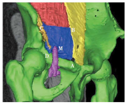

图2
肌耻骨孔上区与下区成一定角度 蓝色区域为肌耻骨孔。
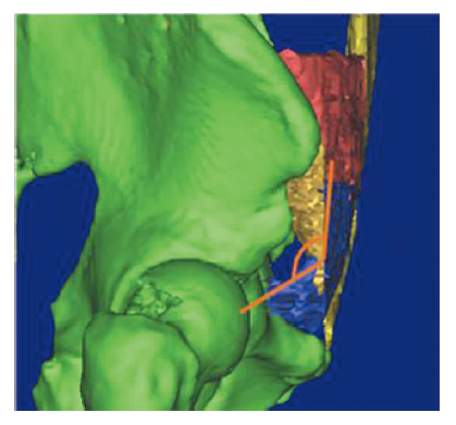
表1
性别对肌耻骨孔大小、形状的影响($\bar{x}\pm s$)
| 项目 | 男性组(76例) | 女性组(14例) | t值 | P值 | 总平均值 |
|---|---|---|---|---|---|
| 总宽度(cm) | 7.78±0.08 | 7.09±0.24 | 3.339 | 0.001a) | 7.67±0.75 |
| 长度(cm) | 7.38±0.04 | 7.13±0.10 | 2.301 | 0.024a) | 7.34±0.38 |
| 上、下两区夹角(°) | 120.66±1.05 | 117.07±2.86 | 1.317 | 0.191 | 120.10±9.36 |
| 上缘长度(cm) | 5.89±0.09 | 5.29±0.17 | 2.653 | 0.009a) | 5.79±0.79 |
| 下缘长度(cm) | 6.60±0.06 | 6.35±0.12 | 1.694 | 0.094 | 6.57±0.50 |
表2
年龄对肌耻骨孔大小、形状的影响($\bar{x}\pm s$)
| 项目 | 总平均值 | 成年组(21例) | 老年组(69例) | t值 | P值 |
|---|---|---|---|---|---|
| 总宽度(cm) | 7.67±0.75 | 7.81±0.13 | 7.63±0.09 | 0.963 | 0.338 |
| 长度(cm) | 7.34±0.38 | 7.44±0.07 | 7.31±0.05 | 1.413 | 0.161 |
| 上、下两区夹角(°) | 120.10±9.36 | 118.26±1.18 | 120.66±1.24 | -1.404 | 0.165 |
| 上缘长度(cm) | 5.79±0.79 | 5.36±0.13 | 5.93±0.10 | -3.004 | 0.003a) |
| 下缘长度(cm) | 6.57±0.50 | 6.63±0.95 | 6.55±0.06 | 0.648 | 0.518 |
表3
BMI对肌耻骨孔大小、形状的影响($\bar{x}\pm s$)
| 项目 | 总平均值 | 较瘦组(45例) | 较胖组(45例) | t值 | P值 |
|---|---|---|---|---|---|
| 总宽度(cm) | 7.67±0.75 | 7.66±0.12 | 7.68±0.11 | -0.155 | 0.877 |
| 长度(cm) | 7.34±0.38 | 7.32±0.06 | 7.36±0.06 | -0.603 | 0.548 |
| 上、下两区夹角(°) | 120.10±9.36 | 117.75±1.36 | 122.45±1.37 | -2.432 | 0.017a) |
| 上缘(cm) | 5.79±0.79 | 5.75±0.12 | 5.84±0.12 | -0.568 | 0.572 |
| 下缘(cm) | 6.57±0.50 | 6.56±0.07 | 6.57±0.78 | -0.145 | 0.885 |
| [1] |
Primatesta P, Goldacre MJ. Inguinal hernia repair: incidence of elective and emergency surgery, readmission and mortality[J]. Int J Epidemiol, 1996, 25(4):835-839.
pmid: 8921464 |
| [2] |
Mine Y, Fujita F, Kinoe H, et al. High incidence of recess formation at myopectineal orifice during laparosco-pic surgery[J]. Asian J Surg, 2018, 41(5):427-430.
doi: 10.1016/j.asjsur.2017.06.001 URL |
| [3] | 中华医学会外科学分会疝与腹壁外科学组. 成人腹股沟疝诊断和治疗指南(2018年版)[J]. 中国实用外科杂志, 2018, 38(7):704-706. |
| [4] |
Kingsnorth A, LeBlanc K. Hernias: inguinal and inci-sional[J]. Lancet, 2003, 362(9395):1561-1571.
pmid: 14615114 |
| [5] | 李建文, 乐飞. 腹股沟疝无张力修补术术式演变与合理选择[J]. 中国实用外科杂志, 2017, 37(11):1202-1205. |
| [6] |
Bracale U, Melillo P, Pignata G, et al. Which is the best laparoscopic approach for inguinal hernia repair: TEP or TAPP? a systematic review of the literature with a network meta-analysis[J]. Surg Endosc, 2012, 26(12):3355-3366.
doi: 10.1007/s00464-012-2382-5 URL |
| [7] |
Hiratsuka T, Shigemistu Y, Etoh T, et al. Appropriate mesh size in the totally extraperitoneal repair of groin hernias based on the intraoperative measurement of the myopectineal orifice[J]. Surg Endosc, 2021, 35(5):2126-2133.
doi: 10.1007/s00464-020-07616-2 URL |
| [8] |
Wolloscheck T, Konerding MA. Dimensions of the myopectineal orifice: a human cadaver study[J]. Hernia, 2009, 13(6):639-642.
doi: 10.1007/s10029-009-0559-1 pmid: 19763741 |
| [9] | 张继峰, 周学鲁, 周上军, 等. 中国人耻骨肌孔大小测量及其临床意义[J]. 中华疝和腹壁外科杂志(电子版), 2012, 6(3):835-839. |
| [10] | 耿兴隆, 戴勇, 赵亮, 等. 应用3D成像技术进行耻骨肌孔大小及角度的测量[J]. 中国内镜杂志, 2019, 25(9):48-52. |
| [11] |
Kang JS, Qiao F, Nie L, et al. Preperitoneal femoral hernioplasty: an “umbrella” technique[J]. Hernia, 2015, 19(5):805-808.
doi: 10.1007/s10029-014-1273-1 pmid: 24927966 |
| [12] | 钱鼎烽, 艾克拜, 沈文来, 等. 个体化裁剪补片配合开放腹膜前间隙疝修补术的临床应用[J]. 中华疝和腹壁外科杂志(电子版), 2016, 10(4):300-302. |
| [13] | 赵婉妮, 周鑫, 刘国勤, 等. 腹股沟区解剖在腹股沟疝修补术中的应用[J]. 中国现代普通外科进展, 2012, 15(4):310-312. |
| [14] | Guerron AD, Hui-Jie L, Jin Y, et al. Laparoscopic single-site inguinal hernia repair using a self-fixating mesh[J]. JSLS, 2017, 21(1):e2016.00103. |
| [15] | Dabić D, Cerović S, Azanjaç B, et al. Prolene hernia system,ultrapro hernia system and 3D patch devices in the treatment of inguinal, femoral, umbilical and small incisional hernias in outpatient surgery[J]. Acta ChirIugosl, 2010, 57(2):49-54. |
| [16] |
Song Z, Yang D, Wang Y, et al. Three-dimensional visualization and measurement of myopectineal orifice in non-inguinal hernia patients[J]. Surg Radiol Anat, 2020, 42(11):1315-1322.
doi: 10.1007/s00276-020-02543-2 URL |
| [17] |
Wang F, Yang XF. Application of computer tomography-based 3D reconstruction technique in hernia repair surgery[J]. World J Clin Cases, 2020, 8(23):5944-5951.
doi: 10.12998/wjcc.v8.i23.5944 URL |
| [1] | 杨良根, 朱俊强, 胡星辰. 腹腔镜经腹腹膜前疝修补术治疗嵌顿性腹股沟疝[J]. 外科理论与实践, 2022, 27(06): 551-554. |
| [2] | 于凡, 伍波, 康杰. 腹股沟疝Lichtenstein手术后的切口感染[J]. 外科理论与实践, 2022, 27(04): 357-358. |
| [3] | 蒋维荣, 俞永江. 预防性应用抗生素对无张力疝修补术后疗效的荟萃分析[J]. 外科理论与实践, 2022, 27(02): 173-179. |
| [4] | 黄磊. Lichtenstein手术在当今腹股沟疝治疗中的地位和再认识[J]. 外科理论与实践, 2021, 26(05): 386-389. |
| [5] | 李航宇, 魏士博. 成人腹股沟疝质量控制——以辽宁省基层医院实践经验为例[J]. 外科理论与实践, 2021, 26(05): 411-415. |
| [6] | 陈涛, 徐煜, 付学良, 袁志青, 花荣. 持续性非卧床腹膜透析病人并发腹股沟疝的外科治疗[J]. 外科理论与实践, 2021, 26(05): 425-429. |
| [7] | 方丽莉, 陆建平, 李绍杰, 唐健雄. 完全腹膜外腹腔镜腹股沟疝修补术中电外科器械无电输出的特殊病例及实验(附1例报告)*[J]. 外科理论与实践, 2021, 26(05): 449-451. |
| [8] | 林天龙, 唐坚. 腹腔镜腹股沟疝修补术合并引流治疗巨大滑疝及其对浆液肿的预防(附1例报告)[J]. 外科理论与实践, 2021, 26(05): 452-455. |
| [9] | 王丹, 倪燕婷, 吴德俊, 崔鹏, 徐明, 闵志钧, 王廷峰. 微创疝修补术后应用酮咯酸氨丁三醇与左旋布比卡因切口浸润镇痛的对比研究[J]. 外科理论与实践, 2020, 25(03): 259-262. |
| [10] | 李金东, 王晨星, 李健文, 郝晓晖, 冯波, 乐飞, 何子锐, 薛佩. 女性腹股沟疝合并子宫圆韧带囊肿的临床特点和腹腔镜治疗策略[J]. 外科理论与实践, 2020, 25(01): 69-73. |
| [11] | 李健, 武彪. 腹股沟疝术后下肢深静脉血栓形成的治疗体会[J]. 外科理论与实践, 2018, 23(05): 437-439. |
| [12] | 唐健雄, 李绍杰, 黄磊. 建立疝病诊治质量控制体系的设想[J]. 外科理论与实践, 2018, 23(04): 293-294. |
| [13] | 周红仙, 张潞英, 王毅. 频域低相干光干涉的折射率三维重建系统[J]. 实验室研究与探索, 2017, 36(5): 61-63. |
| [14] | 唐健雄, 郑民华, 陈杰, 陈双, 田文, 李健文, 王明刚,. 腹腔镜腹股沟疝手术操作指南(2017版)[J]. 外科理论与实践, 2017, 22(06): 483-488. |
| [15] | 吴向嵩, 靳云鹏, 穆嘉盛, 董谦,. 改良Kugel与Lichtenstein修补术治疗腹股沟复发疝的疗效比较[J]. 外科理论与实践, 2016, 21(06): 521-524. |
| 阅读次数 | ||||||
|
全文 |
|
|||||
|
摘要 |
|
|||||