外科理论与实践 ›› 2025, Vol. 30 ›› Issue (01): 21-26.doi: 10.16139/j.1007-9610.2025.01.05
苏一轩, 应涛( )
)
收稿日期:2024-12-03
出版日期:2025-01-25
发布日期:2025-04-25
通讯作者:
应涛,E-mail:yingtaomail@yeah.net
SU Yixuan, YING Tao()
Received:2024-12-03
Online:2025-01-25
Published:2025-04-25
摘要:
常规超声影像对于甲状腺滤泡性肿瘤的良、恶性鉴别较为困难,主要依赖于术后病理诊断。超声新技术和人工智能的进步,在提高诊断准确性、减少不必要手术和降低误诊率方面展现了较大潜力。超声超微血流成像、超声造影及超声弹性成像等新技术,为甲状腺滤泡性肿瘤术前良、恶性鉴别诊断提供了新的途径。本文总结并探讨了上述超声新技术及基于人工智能的多种建模方法在甲状腺滤泡性肿瘤术前诊断中的应用价值,以期为临床决策提供科学依据。
中图分类号:
苏一轩, 应涛. 甲状腺滤泡性肿瘤超声诊断新进展[J]. 外科理论与实践, 2025, 30(01): 21-26.
SU Yixuan, YING Tao. Recent advances in ultrasound diagnosis of thyroid follicular neoplasms[J]. Journal of Surgery Concepts & Practice, 2025, 30(01): 21-26.
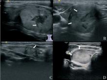
图1
FTC超声灰阶图像
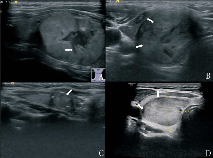

图2
FTC彩色多普勒血流成像
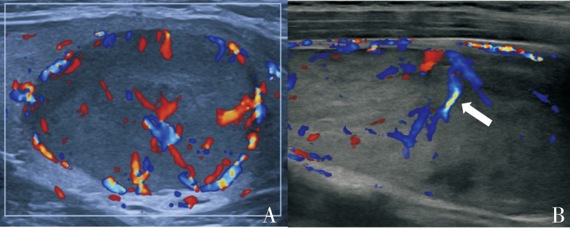

图3
甲状腺滤泡性肿瘤灰阶图像及超声造影表现[28]
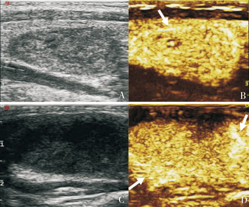

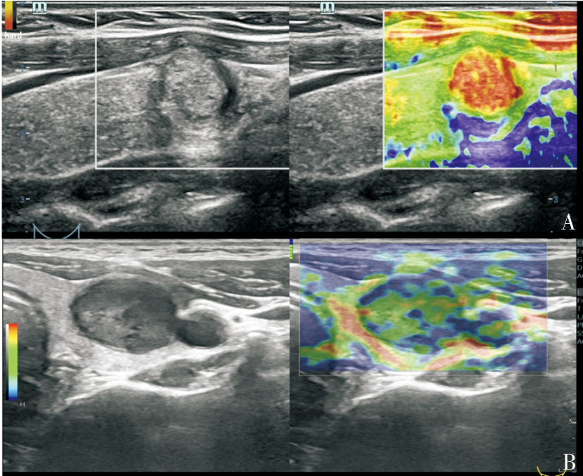
图4
FTC弹性成像
| [1] | HADDAD R I, NASR C, BISCHOFF L, et al. NCCN guidelines insights: thyroid carcinoma, version 2.2018[J]. J Natl Compr Canc Netw, 2018, 16(12):1429-1440. |
| [2] | 中华医学会超声医学分会浅表器官和血管学组, 中国甲状腺与乳腺超声人工智能联. 2020甲状腺结节超声恶性危险分层中国指南:C-TIRADS[J]. 中华超声影像学杂志, 2021, 30(3):185-200. |
| Superficial Organ and Vascular Ultrasound Group, Society of Ultrasound in Medicine, Chinese Medical Association; Chinese Artificial Intelligence Alliance for Thyroid and Breast Ultrasound. 2020 Chinese guidelines for ultrasound malignancy risk stratification of thyroid nodules:the C-TIRADS[J]. Chin J Ultrasonogr, 2021, 30(3):185-200. | |
| [3] | 李婷, 陈露, 李媛, 等. 甲状腺滤泡性腺瘤与滤泡癌的超声检查特征分析[J]. 医学影像学杂志, 2024, 34(8):30-32. |
| LI T, CHEN L, LI Y, et al. Analysis of ultrasound characteristics of thyroid follicular adenoma and follicular carcinoma[J]. J Med Imaging, 2024, 34(8):30-32. | |
| [4] |
HAYASHI C Y, JAUNE D T A, OLIVEIRA C C, et al. Indeterminate thyroid cytology: detecting malignancy using analysis of nuclear images[J]. Endocr Connect, 2021, 10(7):707-714.
doi: 10.1530/EC-20-0648 pmid: 34077391 |
| [5] | GRANI G, LAMARTINA L, DURANTE C, et al. Follicular thyroid cancer and Hürthle cell carcinoma: challenges in diagnosis, treatment, and clinical management[J]. Lancet Diabetes Endocrinol, 2018, 6(6):500-514. |
| [6] | WILHELM A, CONROY P C, CALTHORPE L, et al. Disease-specific survival trends for patients presenting with differentiated thyroid cancer and distant metastases in the United States, 1992-2018[J]. Thyroid, 2023, 33(1):63-73. |
| [7] | 唐婉晴, 张晓娟, 姜丹妮, 等. 甲状腺滤泡癌与滤泡腺瘤的超声特征分析[J]. 临床医学进展, 2022, 12(4):3724-3732. |
| TANG W Q, ZHANG X J, JIANG D N, et al. Sonographic characteristics and differentiation of follicular thyroid carcinoma and follicular adenoma[J]. Adv Clin Med, 2022, 12(4):3724-3732. | |
| [8] | KOIKE E, NOGUCHI S, YAMASHITA H, et al. Ultrasonographic characteristics of thyroid nodules: prediction of malignancy[J]. Arch Surg, 2001, 136(3):334-337. |
| [9] | 中华人民共和国国家卫生健康委员会医政医管局. 甲状腺癌诊疗指南(2022年版)[J]. 中国实用外科杂志, 2022, 42(12):1343-1357,1363. |
| National Health Commission of the People’s Republic of China Medical Administration and Hospital Administration. Guidelines for the diagnosis and treatment of thyroid carcinoma[J]. Chin J Pract Surg, 2022, 42(12):1343-1357,1363. | |
| [10] | 谢文婷, 陈轶洁, 黄伟钦, 等. 甲状腺滤泡癌与腺瘤的超声声像比较研究[J]. 肿瘤, 2020, 40(5):348-354. |
| XIE W T, CHEN Y J, HUANG W Q, et al. Comparative study of ultrasound imaging of thyroid follicular carcinoma and adenoma[J]. Tumor, 2020, 40(5):348-354. | |
| [11] | KUO T C, WU M H, CHEN K Y, et al. Ultrasonographic features for differentiating follicular thyroid carcinoma and follicular adenoma[J]. Asian J Surg, 2020, 43(1):339-346. |
| [12] | XU R, WEN W, ZHANG Y, et al. Diagnostic significance of ultrasound characteristics in discriminating follicular thyroid carcinoma from adenoma[J]. BMC Med Imaging, 2024, 24(1):299. |
| [13] |
OU D, YAO J, JIN J, et al. Ultrasonic identification and regression analysis of 294 thyroid follicular tumors[J]. J Cancer Res Ther, 2020, 16(5):1056-1062.
doi: 10.4103/jcrt.JCRT_913_19 pmid: 33004747 |
| [14] | ZHANG F, MEI F, CHEN W, et al. Role of ultrasound and ultrasound‐based prediction model in differentiating follicular thyroid carcinoma from follicular thyroid adenoma[J]. J Ultrasound Med, 2024, 43(8):1389-1399. |
| [15] | LIU B J, ZHANG Y F, ZHAO C K, et al. Conventional ultrasound characteristics, TI-RADS category and shear wave speed measurement between follicular adenoma and follicular thyroid carcinoma[J]. Clin Hemorheol Microcirc, 2020, 75(3):291-301. |
| [16] | LI H J, YANG Y P, LIANG X, et al. Comparison of the diagnostic performance of three ultrasound thyroid no-dule risk stratification systems for follicular thyroid neoplasm: K-TIRADS, ACR-TIRADS and C-TIRADS[J]. Clin Hemorheol Microcirc, 2023, 85(4):395-406. |
| [17] | LI J, LI C, ZHOU H, et al. US risk stratification system for follicular thyroid neoplasms[J]. Radiology, 2023, 309(2):e230949. |
| [18] |
KONG J, LI J C, WANG H Y, et al. Role of superb micro-vascular imaging in the preoperative evaluation of thyroid nodules: comparison with power Doppler flow imaging[J]. J Ultrasound Med, 2017, 36(7):1329-1337.
doi: 10.7863/ultra.16.07004 pmid: 28463412 |
| [19] | 王莹, 张岱, 杨凡, 等. 超微血流显像与超声造影对甲状腺实性结节的诊断价值[J]. 中国肿瘤临床, 2021, 48(14):711-715. |
| WANG Y, ZHANG D, YANG F, et al. The diagnostic value of superb micro-vacular imaging and contrast-enhanced ultrasound in solid thyroid nodules[J]. Chin J Clin Oncol, 2021, 48(14):711-715. | |
| [20] | 江琼, 林友国. 甲状腺滤泡状腺癌与良性结节的超声鉴别诊断[J]. 实用医学影像杂志, 2023, 24(3):221-225. |
| JIANG Q, LIN Y G. Ultrasonographic differential diagnosis of follicular thyroid carcinoma and thyroid benign nodule[J]. J Pract Med Imaging, 2023, 24(3):221-225. | |
| [21] | 邬宏恂, 王隽. 甲状腺滤泡状癌声像图分析[J]. 临床超声医学杂志, 2007, 9(9):535-538. |
| WU H X, WANG J. Analysis of ultrasonographic imaging in thyroid follicular carcinoma[J]. J Ultrasound in Clin Med, 2007, 9(9):535-538. | |
| [22] | CANNELLA R, PILATO G, MAZZOLA M, et al. New microvascular ultrasound techniques: abdominal applications[J]. Radiol Med, 2023, 128(9):1023-1034. |
| [23] |
ZHAO W, LU R, YIN L, et al. The value of superb microvascular imaging (SMI) scoring assignment method in differentiating benign and malignant thyroid nodules by conventional ultrasound[J]. Clin Hemorheol Microcirc, 2021, 78(4):355-363.
doi: 10.3233/CH-211235 pmid: 34366330 |
| [24] | BOJUNGA J, TRIMBOLI P. Thyroid ultrasound and its ancillary techniques[J]. Rev Endocr Metab Disord, 2024, 25(1):161-173. |
| [25] | GAO W, CHEN Y, WU Q, et al. Significance of maximum intensity projection technique of multimodal ultrasound imaging in differentiating follicular thyroid carcinoma from benign lesions[J]. Front Oncol, 2024,14:1407611. |
| [26] |
WU Q, QU Y, LI Y, et al. Logistic regression analysis of contrast-enhanced ultrasound and conventional ultrasound of follicular thyroid carcinoma and follicular adenoma[J]. Gland Surg, 2021, 10(10):2890-2900.
doi: 10.21037/gs-21-535 pmid: 34804877 |
| [27] | DIAO X H, CHEN L, YU B, et al. Follicular thyroid neoplasm on conventional and contrast-enhanced ultrasound[J]. Adv Ultrasound Diagn Ther, 2022, 6(2):48-57. |
| [28] | 李诗骜, 陆鑫, 姜珏, 等. 超声造影鉴别甲状腺滤泡状肿瘤及其与年龄、性别的相关性分析[J]. 中国临床医学影像杂志, 2023, 34(1):15-18. |
| LI S A, LU X, JIANG J, et al. Contrast-enhanced ultrasound in the differential diagnosis of follicular thyroid tumor and its correlation with age and gender[J]. J Chin Clin Med Imaging, 2023, 34(1):15-18. | |
| [29] | YOO M H, KIM H J, CHOI I H, et al. Efficacy of differential diagnosis of thyroid nodules by shear wave elastography—the stiffness map[J]. J Endocr Soc, 2021, 5(11):bvab154. |
| [30] |
COSGROVE D, BARR R, BOJUNGA J, et al. WFUMB guidelines and recommendations on the clinical use of ultrasound elastography: part 4. thyroid[J]. Ultrasound Med Biol, 2017, 43(1):4-26.
doi: S0301-5629(16)30153-3 pmid: 27570210 |
| [31] | 李宁, 杨丽春, 王丽伟, 等. 声辐射力弹性成像联合超声造影对甲状腺滤泡型肿瘤的诊断价值[J]. 放射学实践, 2020, 35(5):663-667. |
| LI N, YANG C L, WANG L W, et al. The diagnostic value of acoustic radiation force elastography combined with contrast-enhanced ultrasound in thyroid follicular tumor[J]. Radiol Pract, 2020, 35(5):663-667. | |
| [32] | SOLOMON C, PETEA-BALEA D R, DUDEA S M, et al. Role of ultrasound elastography and contrast-enhanced ultrasound (CEUS) in diagnosis and management of malignant thyroid nodules—an update[J]. Diagnostics(Basel), 2025, 15(5):599. |
| [33] |
SHIN I, KIM Y J, HAN K, et al. Application of machine learning to ultrasound images to differentiate follicular neoplasms of the thyroid gland[J]. Ultrasonography, 2020, 39(3):257-265.
doi: 10.14366/usg.19069 pmid: 32299197 |
| [34] |
CHEN W, NI X J, QIAN C, et al. The value of a neural network based on multi-scale feature fusion to ultrasound images for the differentiation in thyroid follicular neoplasms[J]. BMC Med Imaging, 2024, 24(1):74.
doi: 10.1186/s12880-024-01244-1 pmid: 38539143 |
| [35] | ZHENG Y, ZHANG Y J, LU K F, et al. Diagnostic value of an interpretable machine learning model based on clinical ultrasound features for follicular thyroid carcinoma[J]. Quant Imaging Med Surg, 2024, 14(9):6311-6324. |
| [1] | . 基于局部电极的压电材料全矩阵常数单样品表征[J]. J Shanghai Jiaotong Univ Sci, 2025, 30(2): 262-269. |
| [2] | 李易, 欧树彦, 梁伟栋, 董佳宝, 庄至栋. 飞行器低空大动压整流罩旋抛分离数值模拟[J]. 空天防御, 2025, 8(1): 102-108. |
| [3] | 李昂, 韩肖骅, 殷晓星. 肝细胞肝癌甲状腺转移(附1例报告)[J]. 外科理论与实践, 2025, 30(01): 66-69. |
| [4] | 陈灵勰, 赵起悟, 邱伟华. 甲状腺微创手术发展与未来[J]. 外科理论与实践, 2025, 30(01): 7-12. |
| [5] | 曾耀星, 丁敏, 费健. 吲哚菁绿甲状旁腺荧光成像在甲状腺手术中的应用与研究进展[J]. 外科理论与实践, 2025, 30(01): 84-87. |
| [6] | 郭雅文, 郑传铭, 葛明华. 无充气腋窝入路腔镜甲状腺手术的应用、创新与质控[J]. 外科理论与实践, 2025, 30(01): 1-6. |
| [7] | 赵文新, 黄其健, 张立永, 蔡少俊. 优于标准的喉返及喉上神经监测技术在腔镜及机器人甲状腺手术中的拓展应用[J]. 外科理论与实践, 2025, 30(01): 13-16. |
| [8] | 陈朋, 史加宁, 贾文俊, 方静. 经口腔前庭入路腔镜甲状腺手术的研究进展及技术要点[J]. 外科理论与实践, 2025, 30(01): 17-20. |
| [9] | 鲁姗姗, 纪元. 第5版WHO内分泌和神经内分泌肿瘤分类解读:甲状腺滤泡细胞起源肿瘤的更新及进展[J]. 外科理论与实践, 2025, 30(01): 27-33. |
| [10] | 柯晶1, 朱俊超2, 杨鑫1, 张浩林3, 孙宇翔1, 王嘉怡1, 鲁亦舟4, 沈逸卿5, 刘晟6, 蒋伏松7, 黄琴8. TshFNA-Examiner:甲状腺细胞学图像的核分割和癌症评估框架[J]. J Shanghai Jiaotong Univ Sci, 2024, 29(6): 945-957. |
| [11] | 粟华, 施振兴, 丁轩鹤, 张亚云, 龚春林. 高超声速飞行器多物理场虚拟飞行建模方法研究[J]. 空天防御, 2024, 7(5): 45-53. |
| [12] | 何国强, 秦飞, 魏祥庚, 张铎, 玉选斐, 魏黎明. 面向宽域飞行平台的涡轮增强组合动力总体性能分析[J]. 空天防御, 2024, 7(4): 7-17. |
| [13] | 李勇, 张顺家, 朱玉虎, 孔晓俊, 杜泽弘. 2023年美俄典型机载导弹发展动态与趋势分析[J]. 空天防御, 2024, 7(4): 30-37. |
| [14] | 臧红岩, 谢晓龙, 徐亚周, 陶业, 高长生. 基于循环神经网络的高超声速机动目标状态估计算法[J]. 空天防御, 2024, 7(4): 88-98. |
| [15] | 刘子博, 王一凯, 郑毅, 等. 脂肪间充质干细胞经超声刺激后分泌的细胞外囊泡对脂肪间充质干细胞生物学特性的影响[J]. 组织工程与重建外科杂志, 2024, 20(4): 403-. |
| 阅读次数 | ||||||
|
全文 |
|
|||||
|
摘要 |
|
|||||