外科理论与实践 ›› 2025, Vol. 30 ›› Issue (04): 325-331.doi: 10.16139/j.1007-9610.2025.04.06
黄文欣1, 何启宁1, 戚德彬1, 曹梓超1, 姜艳芝2a, 王普森2b, 阙伟涛2b, 钟林1,2b( )
)
收稿日期:2024-10-12
出版日期:2025-07-25
发布日期:2025-10-23
通讯作者:
钟林,E-mail: zhonglin1@medmail.com.cn基金资助:
HUANG Wenxin1, HE Qining1, Qi Debin1, Cao Zichao1, JIANG Yanzhi2a, WANG Pusen2b, QUE Weitao2b, ZHONG Lin1,2b()
Received:2024-10-12
Online:2025-07-25
Published:2025-10-23
摘要:
目的:探讨吲哚菁绿(ICG)荧光影像技术对原发性肝癌肝切除术的指导价值与对病人近期预后的分析。方法:回顾性采集了上海交通大学医学院附属第一人民医院肝胆外科2018年6月至2021年6月行肝癌切除术的166例原发性肝癌病人病例的临床资料,并按照病人术中是否使用ICG荧光影像技术分为ICG组(72例)和非ICG组(94例)。比较两组术前、术中、术后资料,并对ICG组病人肿瘤病灶的ICG荧光图像进行分析。结果:ICG荧光强度与肿瘤病理组织学、分化程度、肝硬化与否相关。肝细胞癌多显示为部分荧光,而肝内胆管癌多显示为环状荧光。高分化肿瘤多为完全荧光(7/11),中分化肿瘤多为部分荧光(28/51),低分化肿瘤多为环形荧光(7/10)。肝硬化病人多为部分荧光(18/35)或完全荧光(13/35)。ICG组血白蛋白水平术后第1天(34.6 g/L比31.4 g/L)及第3天(32.4 g/L比31.2 g/L)高于非ICG组(P<0.001),而手术时间(170 min比210 min)、肝门阻断率(9.7%比33.0%)、术中出血量(400 mL比430 mL)、术后住院时间(10 d比14 d)、术后近期并发症发生率(4.2%比20.2%)明显低于非ICG组(P<0.05)。结论:ICG荧光强度与原发性肝癌肿瘤病理组织学、分化程度、肝硬化与否相关。ICG荧光影像技术合理应用减少手术时间,且有利于提高病人近期预后,加速病人术后的康复。
中图分类号:
黄文欣, 何启宁, 戚德彬, 曹梓超, 姜艳芝, 王普森, 阙伟涛, 钟林. 吲哚菁绿荧光影像技术对原发性肝癌手术治疗的指导价值以及近期预后分析[J]. 外科理论与实践, 2025, 30(04): 325-331.
HUANG Wenxin, HE Qining, Qi Debin, Cao Zichao, JIANG Yanzhi, WANG Pusen, QUE Weitao, ZHONG Lin. Assessment of indocyanine green fluorescence imaging in hepatectomy for primary liver carcinoma: short-term prognostic analysis[J]. Journal of Surgery Concepts & Practice, 2025, 30(04): 325-331.
表1
ICG组与非ICG组术前一般情况比较[($\bar{x} \pm s$)/n(%)/M(Q1, Q3)]
| Item | ICG group (n=72) | non-ICG group (n=94) | t/χ²/Z value | P value |
|---|---|---|---|---|
| Age(years) | 61.9±12.3 | 58.8±11.2 | 1.703 | 0.091 |
| Gender | ||||
| Female | 17(23.6) | 30(31.9) | 1.385 | 0.239 |
| Male | 55(76.4) | 64(68.1) | ||
| HBsAg | ||||
| Negative | 34(47.2) | 33(35.1) | 2.486 | 0.115 |
| Positive | 38(52.8) | 61(64.9) | ||
| Preoperative examination | ||||
| Leukocyte(×109) | 5.5(4.2,7.7) | 5.47(4.6,6.4) | -0.370 | 0.711 |
| ALT(U/L) | 30.0(19.4,44.4) | 33.3(26.0,39.3) | -0.870 | 0.384 |
| AST(U/L) | 33.6(22.4,50.5) | 35.7(26.8,41.7) | -0.298 | 0.766 |
| AFP(μg/L) | 120.3(31.6,333.8) | 98.1(25.4,359.1) | -0.355 | 0.722 |
| Cirrhosis | ||||
| Negative | 37(51.4) | 46(48.9) | 0.098 | 0.754 |
| Positive | 35(48.6) | 48(51.1) | ||
| Child Pugh | ||||
| A | 68(94.4) | 89(94.7) | 0.004 | 0.947 |
| B | 4(5.6) | 5(5.3) | ||
| Tumor size (≥5 cm) | ||||
| Negative | 41(56.9) | 56(59.6) | 0.116 | 0.733 |
| Positive | 31(43.1) | 38(40.4) | ||
| Number of tumors | 1(1,1) | 1(1,1) | -0.74 | 0.941 |
| Degree of differentiation | ||||
| High | 11(15.3) | 9(9.6) | 1.290 | 0.525 |
| Moderate | 51(70.8) | 70(74.5) | ||
| Low | 10(13.9) | 15(16.0) | ||
| Tumor thrombus | ||||
| Negative | 66(91.7) | 90(95.7) | 1.198 | 0.274 |
| Positive | 6(8.3) | 4(4.3) | ||
| Tumor type | ||||
| Hepatocellular carcinoma | 48(66.7) | 67(71.3) | 0.422 | 0.810 |
| Intrahepatic bile duct carcinoma | 19(26.4) | 21(22.3) | ||
| Mixed liver carcinoma | 5(6.9) | 6(6.4) | ||
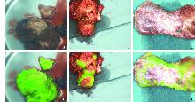
图1
肝脏肿瘤大体观与对应荧光模式图
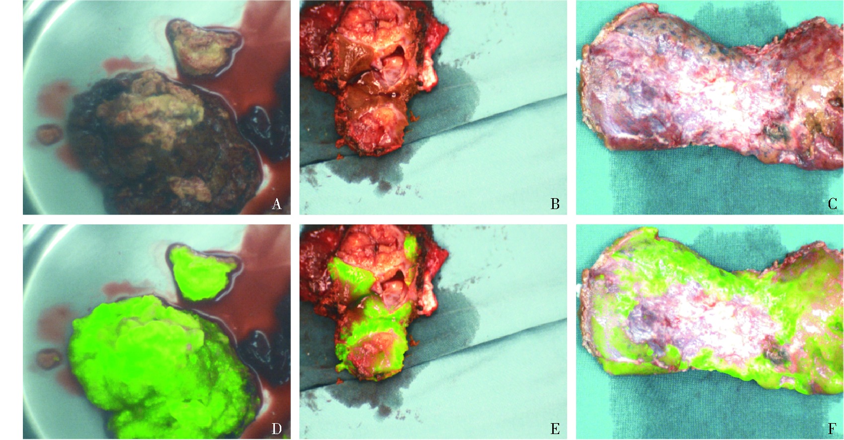
表2
ICG组不同荧光类型病例的特点[n(%)]
| Item | Non-fluorescent (n=3) | Partially-fluorescent (n=32) | Circular-fluorescence (n=18) | Completely-fluorescent (n=19) | χ² value | P value |
|---|---|---|---|---|---|---|
| HBs Ag+ | 1(33.3) | 15(46.9) | 11(61.1) | 11(57.9) | 1.604 | 0.659 |
| HCV Ag+ | 0 | 1(3.1) | 0 | 1(5.3) | 1.049 | 0.789 |
| Cirrhosis | 0 | 18(56.3) | 4(22.2) | 13(68.4) | 17.875 | <0.001 |
| Tumor type | ||||||
| Hepatocellular carcinoma | 0 | 25(78.1) | 8(44.4) | 15(78.9) | 18.112 | 0.006 |
| Intrahepatic bile duct carcinoma | 3(100) | 4(12.5) | 9(50.0) | 3(15.8) | ||
| Mixed liver carcinoma | 0 | 3(9.4) | 1(5.6) | 1(5.3) | ||
| Degree of differentiation | ||||||
| High | 1(33.3) | 2(6.3) | 1(5.6) | 7(36.8) | 24.746 | <0.001 |
| Moderate | 1(33.3) | 28(87.5) | 10(55.6) | 12(63.2) | ||
| Low | 1(33.3) | 2(6.3) | 7(38.9) | 0 |
表3
ICG组与非ICG组术中情况比较[n(%)/M(Q1, Q3)]
| Item | ICG group (n=72) | non-ICG group (n=94) | χ²/Z value | P value |
|---|---|---|---|---|
| Operation time | 170(140,200) | 210(166,348) | -4.416 | <0.001 |
| Portal blockade rate | 7(9.7) | 31(33.0) | 12.493 | <0.001 |
| Intraoperative blood loss | 400(150,587.5) | 430(325,752.5) | -2.053 | 0.040 |
| Intraoperative blood transfusion rate | 14(19.4) | 21(22.3) | 0.206 | 0.650 |
表4
ICG组与非ICG组术后情况比较[n(%)/M(Q1, Q3)]
| Item | ICG group(n=72) | non-ICG group (n=94) | χ²/Z value | P value |
|---|---|---|---|---|
| Surgical margin negative rate | 69(95.8%) | 86(91.5%) | 1.243 | 0.265 |
| Postoperative hospital stay | 10(8,12) | 14(10,21) | -4.317 | <0.001 |
| Number of painkillers used | 1(1,1) | 2(1,2) | -6.189 | <0.001 |
| Postoperative antibiotic use times | 1(1,1) | 2(1,2) | -5.738 | <0.001 |
| Drainage on the first day after surgery | 200(80,300) | 230(180,282.5) | -1.677 | 0.094 |
| The incidence of postoperative complications in the short term | 3(4.2) | 19(20.2) | 9.131 | 0.003 |
| Highest body temperature before discharge | 38(37.4,38.5) | 38.1(37.8,38.4) | -0.782 | 0.434 |
| Blood biochemistry on the first day after surgery | ||||
| Leukocyte(×109) | 11.6(8.5,16.0) | 14.3(12.5,16.3) | -3.680 | <0.001 |
| ALT(U/L) | 292.0(116.3,541.6) | 296.3(201.0,443.8) | -1.209 | 0.227 |
| AST(U/L) | 304.0(146.6,616.4) | 326.8(236.6,459.9) | -0.727 | 0.467 |
| TBil(μmol/L) | 23.5(18.1,29.7) | 27.0(18.2,33.6) | -1.060 | 0.289 |
| Albumin(g/L) | 34.6(30.4,37.0) | 31.4(29.3,33.6) | -3.611 | <0.001 |
| Blood biochemistry on the third day after surgery | ||||
| Leukocyte(×109) | 8.7(7.2,10.5) | 11.6(9.5,13.0) | -4.297 | <0.001 |
| ALT(U/L) | 180.8(100.5,504.5) | 193.4(143,265.3) | -1.267 | 0.205 |
| AST(U/L) | 94.9(55.8,270.2) | 124.5(84.2,168.5) | -2.042 | 0.041 |
| TBil(μmol/L) | 22.3(17.7,25.7) | 28.6(18.7,41.7) | -2.048 | 0.041 |
| Albumin(g/L) | 32.4(31.1,35.9) | 31.2(29.6,33.2) | -5.043 | <0.001 |
| Blood biochemistry on the seventh day after surgery | ||||
| Leukocyte(×109) | 8.8(6.2,10.0) | 7.7(6.4,9.4) | -1.069 | 0.285 |
| ALT(U/L) | 77.4(42.1,172.1) | 75.5(59.8,94.1) | -0.293 | 0.770 |
| AST(U/L) | 39.9(25.9,72.6) | 45.8(37.7,56.8) | -0.538 | 0.591 |
| TBil(μmol/L) | 19.6(15.8,23.9) | 22.3(14.9,32.9) | -1.396 | 0.163 |
| Albumin(g/L) | 34.4(31.0,36.5) | 32.8(30.9,34.9) | -1.785 | 0.074 |
| [1] |
RUMGAY H, ARNOLD M, FERLAY J, et al. Global burden of primary liver cancer in 2020 and predictions to 2040[J]. J Hepatol, 2022, 77(6):1598-1606.
doi: 10.1016/j.jhep.2022.08.021 pmid: 36208844 |
| [2] | SUNG H, FERLAY J, SIEGEL R L, et al. Global cancer statistics 2020:GLOBOCAN estimates of incidence and mortality worldwide for 36 cancers in 185 countries[J]. CA Cancer J Clin, 2021, 71(3):209-249. |
| [3] | CHEN W, ZHENG R, BAADE P D, et al. Cancer statistics in China, 2015[J]. CA Cancer J Clin, 2016, 66(2):115-132. |
| [4] | European Association for the Study of the Liver. EASL clinical practice guidelines:management of hepatocellular carcinoma[J]. J Hepatol, 2018, 69(1):182-236. |
| [5] |
LLOVET J M, DE BAERE T, KULIK L, et al. Locoregional therapies in the era of molecular and immune treatments for hepatocellular carcinoma[J]. Nat Rev Gastroenterol Hepatol, 2021, 18(5):293-313.
doi: 10.1038/s41575-020-00395-0 pmid: 33510460 |
| [6] |
MAČIANSKIENĖ R, ALMANAITYTĖ M, TREINYS R, et al. Spectral characteristics of voltage-sensitive indocyanine green fluorescence in the heart[J]. Sci Rep, 2017, 7(1):7983.
doi: 10.1038/s41598-017-08168-7 pmid: 28801595 |
| [7] | EGLOFF-JURAS C, BEZDETNAYA L, DOLIVET G, et al. NIR fluorescence-guided tumor surgery:new strategies for the use of indocyanine green[J]. Int J Nanomedicine, 2019,14:7823-7838. |
| [8] |
KOKUDO N. Indocyanine green fluorescence imaging as an indispensable tool for modern liver surgery[J]. Ann Surg, 2022, 275(6):1035-1036.
doi: 10.1097/SLA.0000000000005425 pmid: 35185123 |
| [9] | 骆洋, 俞旻皓, 叶光耀, 等. 术中吲哚菁绿荧光显像评估在降低腹腔镜直肠癌术后吻合口漏的应用价值[J]. 外科理论与实践, 2023, 28(3):249-253. |
| LUO Y, YU M H, YE G Y, et al. Application value of intraoperative indocyanine green fluorescence imaging in reducing anastomotic leakage after laparoscopic rectal cancer surgery[J]. J Surg Concepts Pract, 2019, 28(3):249-253. | |
| [10] | CHEN Q Y, XIE J W, ZHONG Q, et al. Safety and efficacy of indocyanine green tracer-guided lymph node dissection during laparoscopic radical gastrectomy in patients with gastric cancer:a randomized clinical trial[J]. JAMA surgery, 2020, 155(4):300-311. |
| [11] |
WANG X, TEH C S C, ISHIZAWA T, et al. Consensus guidelines for the use of fluorescence imaging in hepatobiliary surgery[J]. Ann Surg, 2021, 274(1):97-106.
doi: 10.1097/SLA.0000000000004718 pmid: 33351457 |
| [12] | HSU A, MU S Z, JAMES A, et al. Indocyanine green in bariatric surgery:a systematic review[J]. Obes Surg, 2023, 33(11):3539-3544. |
| [13] | HAN H W, SHI N, ZOU Y P, et al. Functional anatomical hepatectomy guided by indocyanine green fluorescence imaging in patients with localized cholestasis:report of four cases[J]. World J Gastrointest Surg, 2021, 13(3):323-329. |
| [14] | ALFANO M S, MOLFINO S, BENEDICENTI S, et al. Intraoperative ICG-based imaging of liver neoplasms:a simple yet powerful tool. Preliminary results[J]. Surg Endosc, 2019, 33(1):126-134. |
| [15] | WAKABAYASHI T, CACCIAGUERRA A B, ABE Y, et al. Indocyanine green fluorescence navigation in liver surgery:a systematic review on dose and timing of administration[J]. Ann Surg, 2022, 275(6):1025-1034. |
| [16] |
SPERBER A D, BANGDIWALA S I, DROSSMAN D A, et al. Worldwide prevalence and burden of functional gastrointestinal disorders, results of rome foundation global study[J]. Gastroenterology, 2021, 160(1):99-114.e3.
doi: 10.1053/j.gastro.2020.04.014 pmid: 32294476 |
| [17] | LU C, RONG D, ZHANG B, et al. Current perspectives on the immunosuppressive tumor microenvironment in hepatocellular carcinoma:challenges and opportunities[J]. Mol Cancer, 2019, 18(1):130. |
| [18] |
DAI Y, QIANG W, LIN K, et al. An immune-related gene signature for predicting survival and immunotherapy efficacy in hepatocellular carcinoma[J]. Cancer Immunol Immunother, 2021, 70(4):967-979.
doi: 10.1007/s00262-020-02743-0 pmid: 33089373 |
| [19] | WATANABE J, TAKEMASA I, KOTAKE M, et al. Blood perfusion assessment by indocyanine green fluorescence imaging for minimally invasive rectal cancer surgery (EssentiAL trial):a randomized clinical trial[J]. Ann Surg, 2023, 278(4):e688-e694. |
| [20] |
DUROT I, WILSON S R, WILLMANN J K. Contrast-enhanced ultrasound of malignant liver lesions[J]. Abdom Radiol (NY), 2018, 43(4):819-847.
doi: 10.1007/s00261-017-1360-8 pmid: 29094174 |
| [21] |
NAKASEKO Y, ISHIZAWA T, SAIURA A. Fluorescence-guided surgery for liver tumors[J]. J Surg Oncol, 2018, 118(2):324-331.
doi: 10.1002/jso.25128 pmid: 30098296 |
| [22] |
CASSINOTTI E, AL-TAHER M, ANTONIOU S A, et al. European association for endoscopic surgery (EAES) consensus on indocyanine green (ICG) fluorescence-guided surgery[J]. Surg Endosc, 2023, 37(3):1629-1648.
doi: 10.1007/s00464-023-09928-5 pmid: 36781468 |
| [23] | TANG Y, HUANG Z, ZHANG X, et al. Effect of visceral obesity on outcomes of fluorescence-guided lymphadenectomy during laparoscopic gastrectomy for gastric cancer:Post hoc analysis of a randomized phase 3 trial[J]. Chin J Cancer Res, 2024, 36(5):503-516. |
| [24] | CHEN Q Y, ZHONG Q, LIU Z Y, et al. Indocyanine green fluorescence imaging-guided versus conventional laparoscopic lymphadenectomy for gastric cancer:long-term outcomes of a phase 3 randomised clinical trial[J]. Nat Commun, 2023, 14(1):7413. |
| [25] | 周煦川, 刘宾, 王文飞, 等. 多点注射吲哚菁绿红外显影在下肢淋巴管-静脉吻合术中的应用[J]. 组织工程与重建外科杂志, 2023, 19(5):459-463 |
| ZHOU X C, LIU B, WANG W F, et al. Application of multi-point injection of indocyanine green infrared imaging in lymphangio-venous anastomosis of lower extremities[J]. J Tissue Eng Reconstr Surg, 2019, 19(5):459-463. | |
| [26] | RUZZENENTE A, CONCI S, ISA G, et al. The LIver SEntinel LYmph-node (LISELY) study:a prospective intraoperative real time evaluation of liver lymphatic drainage and sentinel lymph-node using near-infrared (NIR) imaging with indocyanine green (ICG)[J]. Eur J Surg Oncol, 2022, 48(12):2455-2459. |
| [27] | WU M R, HUANG Y Y, HSIAO J K. Use of indocyanine green (ICG), a medical near infrared dye, for enhanced fluorescent imaging-comparison of organic anion transporting polypeptide 1B3 (OATP1B3) and sodium-taurocholate cotransporting polypeptide (NTCP) reporter genes[J]. Molecules, 2019, 24(12):2295. |
| [28] | ANZAI K, TSURUYA K, MORIMACHI M, et al. The impact of a heterozygous SLCO1B3 null variant on the indocyanine green retention test[J]. J Pharm Sci, 2020, 109(10):3206-3209. |
| [29] | ISHIZAWA T, SAIURA A, KOKUDO N. Clinical application of indocyanine green-fluorescence imaging during hepatectomy[J]. Hepatobiliary Surg Nutr, 2016, 5(4):322-328. |
| [30] | FENG H L, LI Q, WANG L, et al. Indocyanine green clearance test combined with MELD score in predicting the short-term prognosis of patients with acute liver fai-lure[J]. Hepatobiliary Pancreat Dis Int, 2014, 13(3):271-275. |
| [1] | 曾耀星, 丁敏, 费健. 吲哚菁绿甲状旁腺荧光成像在甲状腺手术中的应用与研究进展[J]. 外科理论与实践, 2025, 30(01): 84-87. |
| [2] | 路志宇, 孙骥, 杜加录, 蒙轩, 罗漫, 刘玥, 王宏光. 腹腔镜解剖性半肝切除中肝蒂处理与吲哚菁绿剂量影响荧光染色效果的研究[J]. 外科理论与实践, 2024, 29(02): 138-142. |
| [3] | 周煦川, 刘宾, 王文飞, 等.
多点注射吲哚菁绿红外显影在下肢淋巴管 - 静脉吻合术中的应用
[J]. 组织工程与重建外科杂志, 2023, 19(5): 459-. |
| [4] | 叶枫, 龚笑勇, 任家俊, 蔡强, 陈胜. ERCP在原发性肝癌围术期胆道并发症诊治中的应用[J]. 外科理论与实践, 2023, 28(04): 355-360. |
| [5] | 骆洋, 俞旻皓, 叶光耀, 林海萍, 贡婷月, 李浩, 钟鸣. 术中吲哚菁绿荧光显像评估在降低腹腔镜直肠癌术后吻合口漏的应用价值[J]. 外科理论与实践, 2023, 28(03): 249-253. |
| [6] | 廖晓明 蒋奕 唐玮 杨华伟 姬逸男 韦莉颖. 薄层血管化腹股沟淋巴结皮瓣移植联合反向淋巴显影在继发性上肢淋巴水肿手术中的应用[J]. 组织工程与重建外科杂志, 2022, 18(1): 8-. |
| [7] | 任家俊, 陈拥军. 肝脾联合切除治疗原发性肝癌合并门静脉高压及脾功能亢进[J]. 外科理论与实践, 2022, 27(02): 139-144. |
| [8] | 朱鹏, 廖威, 张必翔, 陈孝平. 机器人肝癌肝切除应用现状与前景[J]. 外科理论与实践, 2022, 27(02): 95-99. |
| [9] | 苏梅茹, 冯艳平, 赵英杰. 地佐辛联合丙泊酚在腹腔镜肝切除术的应用效果[J]. 外科理论与实践, 2020, 25(01): 74-76. |
| [10] | 喜雯婧,李科,冯少清,章一新. 吲哚菁绿荧光造影在穿支皮瓣微循环检测中的应用[J]. 组织工程与重建外科杂志, 2018, 14(3): 139-142. |
| [11] | 侯振宇, 孔银龙, 张勇强, 朱科云, 杨雪娇, 陈平, 李慧锴, 崔云龙, 宋天强, 李强, 张倜. 术前白细胞计数预测肝切除治疗超“米兰标准”肝细胞癌病人的预后[J]. 外科理论与实践, 2018, 23(04): 358-362. |
| [12] | 张勇强, 张倜, 孔银龙, 侯振宇, 李慧锴, 崔云龙, 宋天强, 李强. 术中出血对早期肝细胞癌病人围术期及预后的影响[J]. 外科理论与实践, 2018, 23(04): 342-345. |
| [13] | 高志慧, 柏斗胜. 精准医学时代肝细胞癌破裂出血的诊治[J]. 外科理论与实践, 2018, 23(03): 217-220. |
| [14] | 金圣杰, 范逸群, 柏斗胜, 蒋国庆, 钱建军, 姚捷, 王小东, 高志慧, 张弛. 门静脉栓塞术在二期精准肝切除中的应用[J]. 外科理论与实践, 2018, 23(03): 247-251. |
| [15] | 李闻达, 陈亚进,. 腹腔镜肝切除在原发性肝癌治疗中的应用[J]. 外科理论与实践, 2017, 22(06): 471-474. |
| 阅读次数 | ||||||
|
全文 |
|
|||||
|
摘要 |
|
|||||