外科理论与实践 ›› 2024, Vol. 29 ›› Issue (05): 389-395.doi: 10.16139/j.1007-9610.2024.05.04
笪倩, 阮淼,*, 费晓春, 王朝夫( )
)
收稿日期:2024-08-12
出版日期:2024-09-25
发布日期:2025-01-23
通讯作者:
王朝夫,E-mail: wcf11956@rjh.com.cn作者简介:*共同第一作者
DA Qian, RUAN Miao,*, FEI Xiaochun, WANG Chaofu()
Received:2024-08-12
Online:2024-09-25
Published:2025-01-23
摘要:
乳腺癌是全球女性常见的癌症之一。病理数字切片扫描仪的诞生及深度学习算法的不断迭代推动了人工智能(AI)技术在乳腺癌诊疗领域的创新。本文对当前AI在乳腺癌病理诊断中的研究及应用现状作介绍,并总结该领域遇到的挑战及未来发展方向。
中图分类号:
笪倩, 阮淼, 费晓春, 王朝夫. 人工智能在乳腺癌病理诊断中的应用及研究展望[J]. 外科理论与实践, 2024, 29(05): 389-395.
DA Qian, RUAN Miao, FEI Xiaochun, WANG Chaofu. Application and research prospects of artificial intelligence in breast cancer pathological diagnosis[J]. Journal of Surgery Concepts & Practice, 2024, 29(05): 389-395.
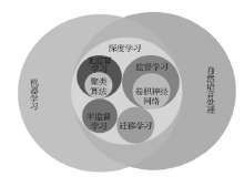
图1
乳腺癌病理诊断中常用的AI技术

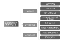
图2
乳腺癌病理诊断AI研究和应用现状

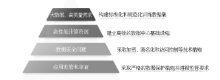
图3
乳腺癌病理诊断AI研究的挑战

| [7] |
BERA K, SCHALPER K A, RIMM D L, et al. Artificial intelligence in digital pathology - new tools for diagnosis and precision oncology[J]. Nat Rev Clin Oncol, 2019, 16(11):703-715.
doi: 10.1038/s41571-019-0252-y pmid: 31399699 |
| [8] | CHENG J, REN C, LIU G, et al. Development of high-resolution dedicated PET-based radiomics machine learning model to predict axillary lymph node status in early-stage breast cancer[J]. Cancers (Basel), 2022, 14(4):950. |
| [9] |
CAMPANELLA G, HANNA M G, GENESLAW L, et al. Clinical-grade computational pathology using weakly supervised deep learning on whole slide images[J]. Nat Med, 2019, 25(8):1301-1309.
doi: 10.1038/s41591-019-0508-1 pmid: 31308507 |
| [10] | JACKSON H W, FISCHER J R, ZANOTELLI V R T, et al. The single-cell pathology landscape of breast cancer[J]. Nature, 2020, 578(7796):615-620. |
| [11] | LIU M, HU L, TANG Y, et al. A deep learning method for breast cancer classification in the pathology images[J]. IEEE J Biomed Health Inform, 2022, 26(10):5025-5032. |
| [12] | ZHANG T, TAN T, WANG X, et al. RadioLOGIC, a healthcare model for processing electronic health records and decision-making in breast disease[J]. Cell Rep Med, 2023, 4(8):101131. |
| [13] |
NAM S, CHONG Y, JUNG CK, et al. Introduction to digital pathology and computer-aided pathology[J]. J Pathol Transl Med, 2020, 54(2):125-134.
doi: 10.4132/jptm.2019.12.31 pmid: 32045965 |
| [14] | SANDERS M E, SCHUYLER P A, DUPONT W D, et al. The natural history of low-grade ductal carcinoma in situ of the breast in women treated by biopsy only revealed over 30 years of long-term follow-up[J]. Cancer, 2005, 103(12):2481-2484. |
| [15] | YAMAMOTO Y, SAITO A, TATEISHI A, et al. Quantitative diagnosis of breast tumors by morphometric classifcation of microenvironmental myoepithelial cells using a machine learning approach[J]. Sci Rep, 2017,7:46732. |
| [16] | FONDÓN I, SARMIENTO A, GARCÍA A I, et al. Automatic classifcation of tissue malignancy for breast carcinoma diagnosis[J]. Comput Biol Med, 2018,96:41-51. |
| [17] | CRUZ-ROA A, GILMORE H, BASAVANHALLY A, et al. High-throughput adaptive sampling for whole-slide histopathology image analysis (HASHI) via convolutional neural networks: application to invasive breast cancer detection[J]. PLoS One, 2018, 13(5):e0196828. |
| [18] |
HAN Z, WEI B, ZHENG Y, et al. Breast cancer multi-classification from histopathological images with structured deep learning model[J]. Sci Rep, 2017, 7(1):4172.
doi: 10.1038/s41598-017-04075-z pmid: 28646155 |
| [19] |
VETA M, VAN DIEST P J, WILLEMS S M, et al. Assessment of algorithms for mitosis detection in breast cancer histopathology images[J]. Med Image Anal, 2015, 20(1):237-248.
doi: 10.1016/j.media.2014.11.010 pmid: 25547073 |
| [20] | WANG Y, ACS B, ROBERTSON S, et al. Improved breast cancer histological grading using deep learning[J]. Ann Oncol, 2022, 33(1):89-98. |
| [21] | ROMO-BUCHELI D, JANOWCZYK A, GILMORE H, et al. Automated tubule nuclei quantification and correlation with Oncotype DX risk categories in ER+ breast cancer whole slide images[J]. Sci Rep, 2016,6:32706. |
| [22] | KATAYAMA A, TOSS M S, PARKIN M, et al. Atypia in breast pathology: what pathologists need to know[J]. Pathology, 2022, 54(1):20-31. |
| [23] | NATEGHI R, DANYALI H, HELFROUSH M S. A deep learning approach for mitosis detection: application in tumor proliferation prediction from whole slide images[J]. Artif Intell Med, 2021,114:102048. |
| [24] |
ELSHARAWY K A, GERDS T A, RAKHA E A, et al. Artificial intelligence grading of breast cancer: a promising method to refine prognostic classification for management precision[J]. Histopathology, 2021, 79(2):187-199.
doi: 10.1111/his.14354 pmid: 33590486 |
| [25] | CHALLA B, TAHIR M, HU Y, et al. Artifcial intelligence-aided diagnosis of breast cancer lymph node metastasis on histologic slides in a digital workfow[J]. Mod Pathol, 2023, 36(8):100216. |
| [26] |
STEINER D F, MACDONALD R, LIU Y, et al. Impact of deep learning assistance on the histopathologic review of lymph nodes for metastatic breast cancer[J]. Am J Surg Pathol, 2018, 42(12):1636-1646.
doi: 10.1097/PAS.0000000000001151 pmid: 30312179 |
| [27] |
HAMMOND M E, HAYES D F, DOWSETT M, et al. American society of clinical oncology/college of American pathologists guideline recommendations for immunohistochemical testing of estrogen and progesterone receptors in breast cancer[J]. J Clin Oncol, 2010, 28(16):2784-2795.
doi: 10.1200/JCO.2009.25.6529 pmid: 20404251 |
| [28] |
VIALE G, REGAN M M, MAIORANO E, et al. Prognostic and predictive value of centrally reviewed expression of estrogen and progesterone receptors in a randomized trial comparing letrozole and tamoxifen adjuvant therapy for postmenopausal early breast cancer: BIG 1-98[J]. J Clin Oncol, 2007, 25(25):3846-3852.
doi: 10.1200/JCO.2007.11.9453 pmid: 17679725 |
| [29] |
AHERN T P, BECK A H, ROSNER B A, et al. Continuous measurement of breast tumour hormone receptor expression: a comparison of two computational pathology platforms[J]. J Clin Pathol, 2017, 70(5):428-434.
doi: 10.1136/jclinpath-2016-204107 pmid: 27729430 |
| [30] | KOOPMAN T, BUIKEMA H J, HOLLEMA H, et al. What is the added value of digital image analysis of HER2 immunohistochemistry in breast cancer in clinical practice? A study with multiple platforms[J]. Histopatho-logy, 2019, 74(6):917-924. |
| [31] | SERNA G, SIMONETTI S, FASANI R, et al. Sequential immunohistochemistry and virtual image reconstruction using a single slide for quantitative Ki67 measurement in breast cancer[J]. Breast, 2020,53:102-110. |
| [32] | HUMPHRIES M P, HYNES S, BINGHAM V, et al. Automated tumour recognition and digital pathology scoring unravels new role for PD-L1 in predicting good outcome in ER-/HER2+ breast cancer[J]. J Oncol, 2018,2018,2937012. |
| [33] | PILIPOW K, DARWICH A, LOSURDO A. T-cell-based breast cancer immunotherapy[J]. Semin Cancer Biol, 2021,72:90-101. |
| [34] | SAVAS P, TEO Z L, LEFEVRE C, et al. The subclonal architecture of metastatic breast cancer: results from a prospective community-based rapid autopsy program “CASCADE”[J]. PLoS Med, 2016, 13(12):e1002204. |
| [35] | 邢辉, 徐梓航, 董培, 等. 基于人工智能辅助乳腺癌新辅助治疗后肿瘤浸润淋巴细胞评估及可重复性分析[J]. 临床与实验病理学杂志, 2023, 39(7):776-781. |
| XING F, XU Z H, DONG P, et al. Evaluation and reproducibility of artificial intelligence-assisted TILs after neoadjuvant therapy for breast cancer[J]. J Clin Exp Pathol, 2023, 39(7):776-781. | |
| [36] |
RASMUSSON A, ZILENAITE D, NESTARENKAITE A, et al. Immunogradient indicators for antitumor response assessment by automated tumor-stroma interface zone detection[J]. Am J Pathol, 2020, 190(6):1309-1322.
doi: S0002-9440(20)30126-7 pmid: 32194048 |
| [37] | 李凤玲, 卫亚妮, 步宏. 人工智能在新辅助治疗后乳腺癌疗效及预后预测中的研究现状[J]. 临床与实验病理学杂志, 2023, 39(7):833-837. |
| LI F L, WEI Y N, BU H. Current research status of artificial intelligence in breast cancer efficacy and prognosis prediction after neoadjuvant therapy[J]. J Clin Exp Pathol, 2023, 39(7):833-837. | |
| [38] | DODINGTON D W, LAGREE A, TABBARAH S, et al. Analysis of tumor nuclear features using artificial intelligence to predict response to neoadjuvant chemotherapy in high-risk breast cancer patients[J]. Breast Cancer Res Treat, 2021, 186(2):379-389. |
| [39] |
SAEDNIA K, LAGREE A, ALERA M A, et al. Quantitative digital histopathology and machine learning to predict pathological complete response to chemotherapy in breast cancer patients using pre-treatment tumor biopsies[J]. Sci Rep, 2022, 12(1):9690.
doi: 10.1038/s41598-022-13917-4 pmid: 35690630 |
| [40] |
LI F, YANG Y, WEI Y, et al. Predicting neoadjuvant chemotherapy benefit using deep learning from stromal histology in breast cancer[J]. NPJ Breast Cancer, 2022, 8(1):124.
doi: 10.1038/s41523-022-00491-1 pmid: 36418332 |
| [41] | SAMMUT S J, CRISPIN-ORTUZAR M, CHIN S F, et al. Multi-omic machine learning predictor of breast cancer therapy response[J]. Nature, 2022, 601(7894):623-629. |
| [42] | 邓仕杰, 刘蘅安, 杨春雪, 等. 数字化智慧病理科建设中数字病理扫描仪的选择[J]. 临床与实验病理学杂志, 2023, 39(7):847-851. |
| DENG S J, LIU H A, YANG C X, et al. Selection of digital pathology scanners in the construction of digital smart pathology departments[J]. J Clin Exp Pathol, 2023, 39(7):847-851. | |
| [43] | 卞修武, 张培培, 平轶芳, 等. 下一代诊断病理学[J]. 中华病理学杂志, 2022, 51(1):3-6. |
| BIAN X W, ZHANG P P, PING Y F, et al. Next-generation diagnostic pathology[J]. Chin J Pathol, 2022, 51(1):3-6. | |
| [1] | HAN B, ZHENG R, ZENG H, et al. Cancer incidence and mortality in China,2022[J]. J Natl Cancer Cent, 2024, 4(1):47-53. |
| [2] |
CALDERARO J, KATHER J N. Artificial intelligence-based pathology for gastrointestinal and hepatobiliary cancers[J]. Gut, 2021, 70(6):1183-1193.
doi: 10.1136/gutjnl-2020-322880 pmid: 33214163 |
| [3] | GIULIANO A E, CONNOLLY J L, EDGE S B, et al. Breast Cancer-Major changes in the American joint committee on cancer eighth edition cancer staging manual[J]. CA Cancer J Clin, 2017, 67(4):290-303. |
| [4] | NIAZI M K K, PARWANI A V, GURCAN M N. Digital pathology and artificial intelligence[J]. Lancet Oncol, 2019, 20(5):e253-e261. |
| [5] | YU K H, HEALEY E, LEONG T Y, et al. Medical artificial intelligence and human values[J]. N Engl J Med, 2024, 390(20):1895-1904. |
| [6] | RAJKOMAR A, DEAN J, KOHANE I. Machine learning in medicine[J]. N Engl J Med, 2019, 380(14):1347-1358. |
| [1] | 詹何庆1,韩贵来1,魏传安1,李治群2. 人工智能结合磁共振成像和计算建模在心脏电生理与临床诊断中的应用[J]. J Shanghai Jiaotong Univ Sci, 2025, 30(1): 53-65. |
| [2] | 李晶洁, 陈秋燕. 人机协同智能写作发展演进分析与启示[J]. 当代外语研究, 2025, 25(1): 73-83. |
| [3] | 许悦婷, 古玥. 人工智能时代的外语教学与外语教师专业发展:挑战、身份认同危机与出路[J]. 当代外语研究, 2025, 25(1): 60-72. |
| [4] | 胡壮麟. 与时俱进,永无止境——有关多模态和人工智能研究的若干认识[J]. 当代外语研究, 2025, 25(1): 53-59. |
| [5] | 徐旺旺1,2,许良凤1,2,刘宁徽3,律娜3. 基于多注意力卷积神经网络的乳腺癌组织学图像诊断[J]. J Shanghai Jiaotong Univ Sci, 2025, 30(1): 91-106. |
| [6] | 潘幸怡, 黎子骏, 甄永环, 等.
人工智能在鼻整形手术中的应用
[J]. 组织工程与重建外科杂志, 2024, 20(6): 659-. |
| [7] | 袁国昊 曾立 汪海滨.
数字化技术在假体隆乳术中的应用与进展
[J]. 组织工程与重建外科杂志, 2024, 20(6): 653-. |
| [8] | 周毅, 周良才, 史迪, 赵小英, 闪鑫. 基于安全深度强化学习的电网有功频率协同优化控制[J]. 上海交通大学学报, 2024, 58(5): 682-692. |
| [9] | 卓玲. 外语教育学视域下人工智能对外语教学生态位的影响[J]. 当代外语研究, 2024, 24(5): 103-115. |
| [10] | 唐胜景, 王太岩, 赵刚练, 郭杰, 李佳丽, 尹航. 面向目标跟踪的多传感器数据融合研究综述[J]. 空天防御, 2024, 7(4): 18-29. |
| [11] | 宋达疆, 张天怡, 王志远, 等. 分叶游离下腹部皮瓣移植乳房再造的血管吻合方式选择 [J]. 组织工程与重建外科杂志, 2024, 20(4): 428-. |
| [12] | 梅冰. 师范生对人工智能赋能外语教学接受度探析[J]. 当代外语研究, 2024, 24(2): 89-99. |
| [13] | 郝昆, 孙宇光, 王仁贵, 等. 抽吸减容术治疗乳腺癌术后上肢淋巴水肿[J]. 组织工程与重建外科杂志, 2024, 20(1): 69-. |
| [14] | 徐雨凡, 胡煜奇, 陈泓宇, 等. 深度学习系统辅助成人发育性髋关节发育不良的分型培训[J]. 组织工程与重建外科杂志, 2024, 20(1): 114-. |
| [15] | 赵鑫, 高鹏, 陈洁. 机器人辅助手术系统在乳腺癌治疗中的应用及前景[J]. 外科理论与实践, 2024, 29(05): 376-381. |
| 阅读次数 | ||||||
|
全文 |
|
|||||
|
摘要 |
|
|||||