诊断学理论与实践 ›› 2019, Vol. 18 ›› Issue (03): 271-277.doi: 10.16150/j.1671-2870.2019.03.006
张淼a, 黄鹏b, 占世坤b, 孟宏平a, 黄新韵a, 林晓珠a, 张一帆a, 曹春燕b, 孙伯民b, 李彪a( ), 刘伟b()
收稿日期:2019-02-19
出版日期:2019-06-25
发布日期:2019-06-25
通讯作者:
李彪,刘伟
E-mail:lb10363@rjh.com.cn;doctorliuwei@163.com
基金资助:
ZHANG Miaoa, HUANG Pengb, ZHAN Shikunb, MENG Hongpinga, HUANG Xinyuna, LIN Xiaozhua, ZHANG Yifana, CAO Chunyanb, SUN Bominb, LI Biaoa(), LIU Weib()
Received:2019-02-19
Online:2019-06-25
Published:2019-06-25
Contact:
LI Biao,LIU Wei
E-mail:lb10363@rjh.com.cn;doctorliuwei@163.com
摘要:
目的: 探讨一体化18F-FDG PET/MRI(以下简化为PET/MRI)检查在癫痫精准定位中的应用价值。方法: 入组25例药物难治型癫痫患者,在术前发作间期行颅脑一体化PET/MRI检查,并在检查完成1个月内通过立体定向脑电图(stereot-actic electroencephalography, SEEG)或手术病理检查明确致癫痫灶。以SEEG及手术病理方法为金标准,对单一MRI形态学、PET形态分析法、PET/MRI融合成像3种方法检出、定位致痫灶的灵敏度和特异度进行对照研究;通过MI Neurology软件对致癫痫灶与正常PET/MRI脑代谢数据库进行配准比对,获得各脑区的标准差(standard deviation, SD)值,对比病变部位与对侧正常对照脑区SD值、平均标准化摄取值(mean standardized uptake value, SUVmean)值的差异。结果: 单一MRI形态学方法定位致癫痫灶的灵敏度为37.5%(9/24),特异度为100%(4/4),有15处致痫灶单一MRI形态学无法显示,其中14例病灶无结构异常,因而MRI无法显示。单一PET形态学方法确诊16处癫痫灶。结合PET(包括半定量PET)进行PET/MRI融合成像,可以检出12处MRI阴性病例,PET/MRI检查定位致癲痫灶的灵敏度为91.6%(22/24),明显高于单一MRI形态学(P<0.05)。PET/MRI融合成像减少了1例PET假阳性诊断,其成像特异度为100%,同时MRI可以清晰显示解剖结构,为手术提供精准定位。致癫痫灶SD值为-6.16±2.26,健侧SD值为-0.72±0.89,致癫痫灶的SD值明显低于健侧对照区(P<0.01)。结论: 一体化PET/MRI检查充分融合了2种显像方法的优势,对于无结构异常的致痫灶,结合PET(包括定量PET)可以发现MRI检查阴性的病灶,而MRI分辨率高,可清晰显示解剖结构,PET/MRI同机可对病灶的部位和范围进行精准定位,并可以进行功能融合成像,为手术方案制定提供了有力信息,在癫痫个体化精准医疗中显示出巨大应用潜力。
中图分类号:
张淼, 黄鹏, 占世坤, 孟宏平, 黄新韵, 林晓珠, 张一帆, 曹春燕, 孙伯民, 李彪, 刘伟. 一体化18F-FDG PET/MRI多模态分子影像在癫痫精准定位中的应用价值[J]. 诊断学理论与实践, 2019, 18(03): 271-277.
ZHANG Miao, HUANG Peng, ZHAN Shikun, MENG Hongping, HUANG Xinyun, LIN Xiaozhu, ZHANG Yifan, CAO Chunyan, SUN Bomin, LI Biao, LIU Wei. Clinical value of simultaneous 18F-FDG PET/MR molecular imaging in localizing seizure foci in epilepsy patients[J]. Journal of Diagnostics Concepts & Practice, 2019, 18(03): 271-277.
表1
不同成像模态诊断癫痫灶的效能[%(n)]
| 方法分组 | 灵敏度 | 特异度 | 阳性预测值 | 阴性预测值 |
|---|---|---|---|---|
| 单一MRI形态学 | 37.5%(9/24)* | 100%(4/4) | 81.8%(9/11) | 21.0% (4/19) |
| 单一PET形态学 | 66.6%(16/24) | 75.0%(3/4) | 88.8%(16/18) | 20.0%(2/10) |
| PET/MRI融合 | 91.6%(22/24) | 100%(4/4) | 100%(22/22) | 66.6%(4/6) |
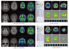
图1
36岁女性癫痫患者的PET及PET/MRI融合图像 患者为女性,36岁,其20余年前于睡眠中突发四肢抽搐,诊断为癫痫,当时未行系统治疗,后多次出现上述症状,发作频率为3~4年出现1次。患者曾在外院就诊,口服中成药(具体成分不详)治疗,过程中症状反复发作,近1年发作频率明显增加,约20 d出现1次。A:可见左侧扣带回局部信号异常,T2WI Flair呈高信号,T1WI局部灰白质信号欠清晰,PET形态分析较难显示,在结合MRI的情况下,需仔细观察才能发现左侧扣带回后部局部代谢较右侧低,B: PET半定量分析,病灶与标准脑库差值SD值为-5.5(蓝色区域),SUV平均值为4.4,健侧(右侧)对照区SD值为-0.6,SUV平均值为6.4;另外一处病灶位于左侧颞叶,C:在MRI形态学未发现结构异常,PET用形态分析方法难以分辨病灶(如图1C PET一列),D:定量PET可见左侧颞叶局灶性代谢减低(蓝色区域),与标准脑库差值SD值为-7.1,SUV平均值为5.3,健侧正常脑区SD值为-0.8,SUV平均值为5.7。该患者按照PET/MR提供的病灶信息进行了SEEG植入手术,结果SEEG电极记录发现了左侧扣带回后部、左侧颞叶部分异常癫痫信号, PET/MRI显像结果与SEEG完全一致。
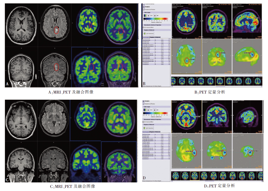

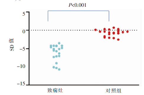
图2
致痫灶及健侧正常脑区与标准脑库差值SD值
| [1] |
Jost J, Raharivelo A, Ratsimbazafy V, et al. Availability and cost of major and first-line antiepileptic drugs: a comprehensive evaluation in the capital of Madagascar[J]. Springerplus, 2016, 5(1):1726.
doi: 10.1186/s40064-016-3409-5 URL |
| [2] | WHO. Atlas: epilepsy care in the world[R]. Geneva: World Health Organization, 2005. |
| [3] |
Jehi L, Yardi R, Chagin K, et al. Development and validation of nomograms to provide individualised predictions of seizure outcomes after epilepsy surgery: a retrospective analysis[J]. Lancet Neurol, 2015, 14(3):283-290.
doi: 10.1016/S1474-4422(14)70325-4 URL |
| [4] |
Najm I, Jehi L, Palmini A, et al. Temporal patterns and mechanisms of epilepsy surgery failure[J]. Epilepsia, 2013, 54(5):772-782.
doi: 10.1111/epi.12152 URL |
| [5] | 郝谦谦, 李迪彬, 李殿友, 等. PET/MRI异机融合图形对影像学阴性的难治性颞叶癫痫手术疗效的价值[J]. 中华神经外科杂志, 2014, 30(12):1262-1265. |
| [6] |
Rakheja R, DeMello L, Chandarana H, et al. Comparison of the accuracy of PET/CT and PET/MRI spatial registration of multiple metastatic lesions[J]. AJR Am J Roentgenol, 2013, 201(5):1120-1123.
doi: 10.2214/AJR.13.11305 URL |
| [7] |
Jadvar H, Colletti PM. Competitive advantage of PET/MRI[J]. Eur J Radiol, 2014, 83(1):84-94.
doi: 10.1016/j.ejrad.2013.05.028 URL |
| [8] |
Fisher RS, van Emde Boas W, Blume W, et al. Epileptic seizures and epilepsy: definitions proposed by the International League Against Epilepsy (ILAE) and the International Bureau for Epilepsy (IBE)[J]. Epilepsia, 2005, 46(4):470-472.
doi: 10.1111/j.0013-9580.2005.66104.x URL |
| [9] | Kumar A, Chugani HT. The role of radionuclide imaging in epilepsy, Part 1: Sporadic temporal and extratemporal lobe epilepsy[J]. J Nucl Med, 2013, 54(10):1775-1781. |
| [10] |
Gaillard WD, Kopylev L, Weinstein S, et al. Low incidence of abnormal (18)FDG-PET in children with new-o-nset partial epilepsy: a prospective study[J]. Neurology, 2002, 58(5):717-722.
pmid: 11889233 |
| [11] | 桑林, 张凯, 张建国, 等. PET-MRI影像融合技术在药物难治性癫痫术前评估中的价值[J]. 中华神经外科杂志, 2017, 33(6):559-563. |
| [12] |
Hu WH, Wang X, Liu LN, et al. Multimodality Image Post-processing in Detection of Extratemporal MRI-Nega-tive Cortical Dysplasia[J]. Front Neurol, 2018, 9:450.
doi: 10.3389/fneur.2018.00450 URL |
| [13] | Bisdas S, Lá Fougere C, Ernemann U. Hybrid MR-PET in Neuroimaging[J]. Clin Neuroradiol, 2015, 25(Suppl 2):275-281. |
| [14] | Wang K, Liu T, Zhao X, et al. Comparative Study of Voxel-Based Epileptic Foci Localization Accuracy between Statistical Parametric Mapping and Three-dimensional Stereotactic Surface Projection[J]. Front Neurol, 2016, 7:164. |
| [15] | 单艺, 卢洁, 李坤成. 一体化PET/MR评估脑血流量的研究进展[J]. 中国医学影像技术, 2017, 33(8):1269-1272. |
| [16] |
Shang K, Wang J, Fan X, et al. Clinical Value of Hybrid TOF-PET/MR Imaging-Based Multiparametric Imaging in Localizing Seizure Focus in Patients with MRI-Negative Temporal Lobe Epilepsy[J]. AJNR, 2018, 39(10):1791-1798.
doi: 10.3174/ajnr.A5814 URL |
| [17] | Garibotto V, Heinzer S, Vulliemoz S, et al. Clinical applications of hybrid PET/MRI in neuroimaging[J]. Clin Nucl Med, 2013, 38(1):e13-e18. |
| [18] | 臧玉峰, 冯逢, 霍力, 等. PET/fMRI对异常脑活动的精准定位:研究进展与展望[J]. 中华核医学与分子影像杂志, 2017, 37(12):802-808. |
| [1] | 马龙鑫, 汤杰, 林靖生, 曹青, 陈影, 陈尔真, 何萍. 大型方舱医院临床数据库建设和应用[J]. 诊断学理论与实践, 2022, 21(02): 205-211. |
| [2] | 沈文斌, 郭睿, 李彪. 18F-FDG PET/CT代谢参数在NK/T细胞淋巴瘤预后价值的评估[J]. 诊断学理论与实践, 2019, 18(03): 349-352. |
| [3] | . 诊断学理论与实践[J]. 诊断学理论与实践, 2016, 15(03): 337-. |
| [4] | . 诊断学理论与实践[J]. 诊断学理论与实践, 2015, 14(02): 86-. |
| [5] | . 诊断学理论与实践[J]. 诊断学理论与实践, 2014, 13(06): 645-. |
| [6] | . 诊断学理论与实践[J]. 诊断学理论与实践, 2014, 13(04): 449-. |
| [7] | 李彪,朱承谟. PET显像在中枢神经系统疾病诊断中的价值[J]. 诊断学理论与实践, 2005, 4(04): 269-272. |
| [8] | 李志刚,杨谦,张维,王林,周建平. 局部脑血流SPECT显像与其他影像对癫痫定位诊断比较的研究[J]. 诊断学理论与实践, 2005, 4(02): 110-112. |
| [9] | 王靖媛,赵阴环. 狼疮性癫痫发生的相关因素分析[J]. 诊断学理论与实践, 2004, 3(04): 29-30+36. |
| [10] | 王玉平. 脑电图检查在癫痫诊断中的应用[J]. 诊断学理论与实践, 2004, 3(02): 73-76. |
| 阅读次数 | ||||||
|
全文 |
|
|||||
|
摘要 |
|
|||||