诊断学理论与实践 ›› 2019, Vol. 18 ›› Issue (05): 548-554.doi: 10.16150/j.1671-2870.2019.05.012
杨志芳1, 方国平2, 詹维伟1( ), 吉日1
收稿日期:2019-01-23
出版日期:2019-10-25
发布日期:2019-10-25
通讯作者:
詹维伟
E-mail:shanghairuijin@126.com
YANG Zhifang1, FANG Guoping2, ZHAN Weiwei1(), JI Ri1
Received:2019-01-23
Online:2019-10-25
Published:2019-10-25
Contact:
ZHAN Weiwei
E-mail:shanghairuijin@126.com
摘要:
目的:分析1例罕见的去分化实体型甲状腺乳头状癌(solid variant papillary thyroid carcinoma, SVPTC)伴黏膜相关淋巴组织结外边缘区淋巴瘤(extranodal marginal zone lymphoma of mucosa-associated lymphoid tissue, MALT)患者的临床表现、超声及相关影像学特征,并对其病理特征进行探讨。方法:分析本例患者的临床资料、影像学特征及病理诊断特点,针对其SVPTC、去分化表现、MALT各相关特征进行分别论述。结果:甲状腺组织大体灰白质硬,光镜下可见肿瘤细胞排列呈实性、巢状,核分裂相易见;未见乳头状癌典型的细胞核特征;间质由形态各异的小淋巴细胞组成,可见较多Russell小体。免疫组织化学(immunohistochemistry, IHC)检测显示,CK19(+),Ki-67(80%+),TG(-),TTF-1(+),Bcl-6(+),CK5/6(-),CD117(-);周围淋巴细胞CD20(+)、CD79α(+)、CD3(+)、CD5(+)、CD43(+)、Bcl-2(+)。分子病理学检查结果示,BRAF基因未检测到突变,TERT启动子有突变。B淋巴瘤克隆基因重排结果为阳性。最终该患者经病理诊断为去分化SVPTC伴MALT。结论:去分化SVPTC伴MALT极其罕见,患者预后差,各病理特征都具有一定的临床价值,需仔细分析研究。
中图分类号:
杨志芳, 方国平, 詹维伟, 吉日. 去分化实体型甲状腺乳头状癌伴黏膜相关淋巴组织结外边缘区淋巴瘤1例病理特征并文献分析[J]. 诊断学理论与实践, 2019, 18(05): 548-554.
YANG Zhifang, FANG Guoping, ZHAN Weiwei, JI Ri. Dedifferentiated solid variant papillary thyroid carcinoma with extranodal marginal zone lymphoma of mucosa-associated lymphoid tissue: a case report with pathological characteristics and literature analysis[J]. Journal of Diagnostics Concepts & Practice, 2019, 18(05): 548-554.

图1
超声图像 A:右侧甲状腺形态肿大,回声弥漫性增粗,减低不均匀;B:右侧颈部Ⅲ区淋巴结肿大
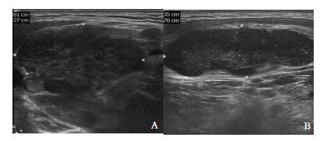
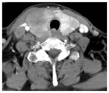
图2
双侧甲状腺低密度及钙化影
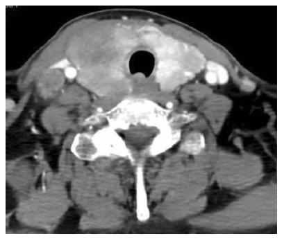

图3
PET-CT图像 双侧甲状腺肿大,代谢增高;颈部、锁骨上及纵隔内淋巴结肿大伴代谢增高
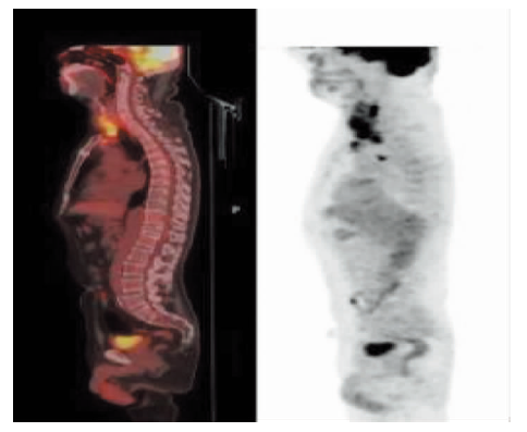
图4
病理组织图片(HE) A:肿瘤细胞排列呈实性、巢状,间质大量淋巴细胞浸润 (×100);B:脉管内见瘤栓(×100);C:肿瘤组织中央凝固性坏死,间质纤维组织增生(×100);D:肿瘤细胞核分裂相易见,未见乳头状癌典型的细胞核特征(×400);E:间质由形态各异的小淋巴细胞组成,可见Russell小体(×200)
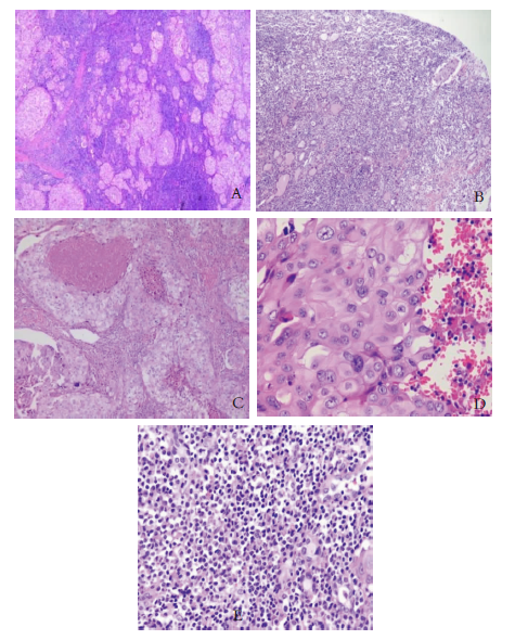
图5
免疫组化图片(Envision,×100) A、B、C、D分别为肿瘤细胞Tg失表达、TTF-1、CK19(+)、Ki-67(80%+)
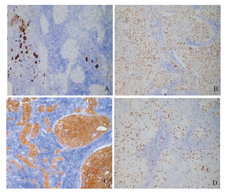
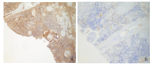
图6
免疫组化图片(Envision,×100) A、B λ∶κ>10∶1,提示轻链的限制性表达

| [1] |
Cabanillas ME, McFadden DG, Durante C. Thyroid cancer[J]. Lancet, 2016, 388(10061):2783-2795.
doi: S0140-6736(16)30172-6 pmid: 27240885 |
| [2] |
Haugen BR, Alexander EK, Bible KC, et al. 2015 Ame-rican Thyroid Association Management Guidelines for Adult Patients with Thyroid Nodules and Differentiated Thyroid Cancer: The American Thyroid Association Guidelines Task Force on Thyroid Nodules and Differentiated Thyroid Cancer[J]. Thyroid,2016, 26(1):1-133.
doi: 10.1089/thy.2015.0020 URL |
| [3] |
Roman S, Sosa JA. Aggressive variants of papillary thyroid cancer[J]. Curr Opin Oncol, 2013, 25(1):33-38.
doi: 10.1097/CCO.0b013e32835b7c6b URL |
| [4] |
Sakamoto A, Kasai N, Sugano H. Poorly differentiated carcinoma of the thyroid. A clinicopathologic entity for a high-risk group of papillary and follicular carcinomas[J]. Cancer, 1983, 52(10):1849-1855.
pmid: 6313176 |
| [5] |
Nikiforov YE, Erickson LA, Nikiforova MN, et al. Solid variant of papillary thyroid carcinoma: incidence, clinical-pathologic characteristics, molecular analysis, and biologic behavior[J]. Am J Surg Pathol, 2001, 25(12):1478-1484.
pmid: 11717536 |
| [6] |
Nikiforov Y, Gnepp DR. Pediatric thyroid cancer after the Chernobyl disaster. Pathomorphologic study of 84 cases (1991-1992) from the Republic of Belarus[J]. Cancer, 1994, 74(2):748-766.
pmid: 8033057 |
| [7] |
Vuong HG, Odate T, Duong UNP, et al. Prognostic importance of solid variant papillary thyroid carcinoma: A systematic review and meta-analysis[J]. Head Neck, 2018, 40(7):1588-1597.
doi: 10.1002/hed.25123 URL |
| [8] | 罗塞著, 郑杰译. 甲状腺[M]//罗塞. 外科病理学. 10版. 北京: 北京大学医学出版社, 2014:525. |
| [9] |
Galera-Davidson H, Bibbo M, Dytch HE, et al. Nuclear DNA in anaplastic thyroid carcinoma with a differentiated component[J]. Histopathology, 1987, 11(7):715-722.
pmid: 3623435 |
| [10] | Cardesa A, Slootweg PJ, Gale N, et al. thyroid and parathyroid glands[M]// Pathology of the Head And Neck.2 ed: Springer, 2016:648-649. |
| [11] | Cardesa A, Slootweg PJ, Gale N, et al. thyroid and parathyroid glands[M]// Pathology of the Head And Neck: Springer, 854:652-654. |
| [12] |
Santos L, Loo C, Chandraratnam E, et al. Anaplastic carcinoma dedifferentiation of solid variant of papillary thyroid carcinoma[J]. Pathology, 2004, 36(2):196-211.
doi: 10.1080/00313020410001672073 URL |
| [13] | Swerdlow SH, Campo E, Harris NL, et al. Mature B-cell neoplasms[M]// WHO Classification of Tumours of Haema-topoietic and Lymphoid Tissues. Revised 4th Editioned. Lyon:IARC, 2017:256-262. |
| [14] |
Isaacson P, Wright DH. Malignant lymphoma of mucosa-associated lymphoid tissue. A distinctive type of B-cell lymphoma[J]. Cancer, 1983, 52(8):1410-1416.
pmid: 6193858 |
| [15] |
Widder S, Pasieka JL. Primary thyroid lymphomas[J]. Curr Treat Options Oncol, 2004, 5(4):307-313.
doi: 10.1007/s11864-004-0021-7 URL |
| [16] | Peppa M, Nikolopoulos P, Korkolopoulou P, et al. Primary mucosa-associated lymphoid tissue thyroid lymphoma: a rare thyroid neoplasm of extrathyroid origin[J]. Rare Tumors, 2012, 4(1):e2. |
| [17] |
Watanabe N, Noh JY, Narimatsu H, et al. Clinicopathological features of 171 cases of primary thyroid lymphoma: a long-term study involving 24553 patients with Hashimoto's disease[J]. Br J Haematol, 2011, 153(2):236-243.
doi: 10.1111/j.1365-2141.2011.08606.x URL |
| [18] | 宋琳琳, 詹维伟, 周建桥, 等. 浅表器官结外淋巴瘤的超声表现[J]. 诊断学理论与实践, 2012, 11(5):471-475. |
| [19] | Shrestha P, Aderhold K, Swierczynski S, et al. Primary thyroid MALToma- a rare diagnosis of an unassuming thyroid nodule[J]. J Community Hosp Intern Med Perspect, 2018, 8(1):42-45. |
| [20] | Dündar HZ, Sarkut P, Kırdak T, et al. Primary thyroid lymphoma[J]. Ulus Cerrahi Derg, 2015, 32(1):75-77. |
| [21] | Peixoto R, Correia Pinto J, Soares V, et al. Primary thyroid lymphoma: A case report and review of the literature[J]. Ann Med Surg (Lond), 2016, 13:29-33. |
| [22] |
Pyke CM, Grant CS, Habermann TM, et al. Non-Hodgkin's lymphoma of the thyroid: is more than biopsy necessary?[J]. World J Surg, 1992, 16(4):604-609.
pmid: 1413831 |
| [23] |
Mack LA, Pasieka JL. An evidence-based approach to the treatment of thyroid lymphoma[J]. World J Surg, 2007, 31(5):978-986.
doi: 10.1007/s00268-005-0768-z URL |
| [24] |
Tsang RW, Gospodarowicz MK, Pintilie M, et al. Localized mucosa-associated lymphoid tissue lymphoma treated with radiation therapy has excellent clinical outcome[J]. J Clin Oncol, 2003, 21(22):4157-4164.
doi: 10.1200/JCO.2003.06.085 URL |
| [25] |
Watanabe N, Narimatsu H, Noh JY, et al. Long-Term Outcomes of 107 Cases of Primary Thyroid Mucosa-Associated Lymphoid Tissue Lymphoma at a Single Medical Institution in Japan[J]. J Clin Endocrinol Metab, 2018, 103(2):732-739.
doi: 10.1210/jc.2017-01478 pmid: 29165612 |
| [1] | 何亲羽, 王伟, 陈立芬, 张雪蕾, 董治亚. LHCGR基因突变致家族性男性性早熟2例报告及文献复习[J]. 诊断学理论与实践, 2022, 21(05): 598-605. |
| [2] | 陈志敏, 何浩岚. 艾滋病合并马尔尼菲篮状菌病的诊治现状[J]. 诊断学理论与实践, 2022, 21(04): 425-430. |
| [3] | 沈银忠. 《人类免疫缺陷病毒感染/艾滋病合并结核分枝杆菌感染诊治专家共识》解读[J]. 诊断学理论与实践, 2022, 21(04): 431-436. |
| [4] | 王文涵, 夏蜀珺, 詹维伟. 长链非编码RNA ENST00000489676在超声评估甲状腺乳头状癌颈部淋巴结转移中的应用[J]. 诊断学理论与实践, 2022, 21(04): 514-519. |
| [5] | 陈宏, 沈银忠. 人类免疫缺陷病毒感染/艾滋病合并结核病的诊治进展[J]. 诊断学理论与实践, 2022, 21(04): 530-534. |
| [6] | 何新, 陈慧, 冯炜炜. 机器学习算法在辅助超声诊断附件肿块良恶性中的应用研究进展[J]. 诊断学理论与实践, 2022, 21(04): 541-546. |
| [7] | 徐子真, 李擎天, 刘湘帆, 李莉, 李惠, 王也飞, 吴洁敏, 陈宁, 梁璆荔, 陈松立, 戴健敏, 宋珍, 丁磊. 实验诊断学在线课程的建立和实践[J]. 诊断学理论与实践, 2022, 21(04): 547-550. |
| [8] | 徐琛莹, 李嫣然, 倪晓枫, 徐上妍, 林青. 超声预测老年甲状腺乳头状癌患者颈部淋巴结转移的效能及相关超声征象分析[J]. 诊断学理论与实践, 2022, 21(03): 343-348. |
| [9] | 赵然, 詹维伟, 侯怡卿. 计算机辅助诊断系统辅助超声诊断甲状腺弥漫性病变合并结节良恶性的应用价值[J]. 诊断学理论与实践, 2022, 21(03): 390-394. |
| [10] | 郭业兵, 郑金峰. 阴道壁胃肠道外间质瘤一例报道并文献复习[J]. 诊断学理论与实践, 2022, 21(03): 405-407. |
| [11] | 王刚, 陈生弟. 神经病学的诊断:起源、发展及挑战[J]. 诊断学理论与实践, 2022, 21(01): 1-4. |
| [12] | 唐静仪, 余群, 刘军. 结合人工智能的结构影像分析对阿尔茨海默病的早期预测及精准诊断研究进展[J]. 诊断学理论与实践, 2022, 21(01): 12-17. |
| [13] | 魏文石. 直面我国阿尔茨海默病诊治的挑战——《中国阿尔茨海默病报告2021》解读[J]. 诊断学理论与实践, 2022, 21(01): 5-7. |
| [14] | 王蔚, 王小钦. 缺铁性贫血的病因诊断[J]. 诊断学理论与实践, 2021, 20(06): 529-532. |
| [15] | 岳婧婧, 宋琦, 江旭峰, 王黎, 赵维莅, 严福华. 磁共振全身扩散加权成像结合T2WI抑脂序列与FDG-PET/CT在初发淋巴瘤分期及病灶检出的对比研究[J]. 诊断学理论与实践, 2021, 20(06): 540-546. |
| 阅读次数 | ||||||
|
全文 |
|
|||||
|
摘要 |
|
|||||