诊断学理论与实践 ›› 2021, Vol. 20 ›› Issue (06): 540-546.doi: 10.16150/j.1671-2870.2021.06.005
岳婧婧1, 宋琦2a( ), 江旭峰2b, 王黎2c, 赵维莅2c, 严福华2a
收稿日期:2021-05-06
出版日期:2021-12-25
发布日期:2021-12-25
通讯作者:
宋琦
E-mail:sq10729@rjh.com.cn
YUE Jingjing1, SONG Qi2a(), JIANG Xufeng2b, WANG Li2c, ZHAO Weili2c, YAN Fuhua2a
Received:2021-05-06
Online:2021-12-25
Published:2021-12-25
Contact:
SONG Qi
E-mail:sq10729@rjh.com.cn
摘要:
目的: 比较磁共振全身扩散加权成像(whole body-diffusion weighted imaging,WB-DWI)结合T2WI抑脂序列(fat saturation T2-weighted imaging, FS-T2WI)及正电子发射计算机体层显像仪(positron emission tomography and computed tomography,PET/CT)在初发淋巴瘤分期诊断中的应用价值。方法: 回顾性分析2014年5月至2020年9月上海交通大学医学院附属瑞金医院血液科收治的初发、未经治疗且经病理证实的淋巴瘤患者25例,其中24例为非霍奇金淋巴瘤,1例为霍奇金淋巴瘤。患者分别行磁共振WB-DWI结合FS-T2WI序列及18F-氟代脱氧葡萄糖(18F-fluorodeoxyglucos,18F-FDG)PET/CT检查。以临床标准为金标准(由临床、病理、影像、骨髓穿刺及6个月随访结果综合分析,参照Ann-Arbor分期法),比较2种检查方法在病灶的检出及分期间的一致性。结果: 采用PET/CT、磁共振WB-DWI结合FS-T2WI序列检查评估了500处淋巴结区域,其中有466处检出结果相同,2种检查方法对结内病灶的诊断差异无统计学意义(P>0.05),具有较高的一致性(K=0.807)。采用PET/CT及磁共振WB-DWI结合FS-T2WI序列评估了450处结外脏器,其中有444处检出结果相同,2种检查方法对结外病灶的诊断差异无统计学意义(P>0.05),具有较高的一致性(K=0.857)。25例患者中20例采用2种检查方法获得的分期相同,具有良好的一致性(K=0.731)。与临床分期相比,PET/CT检查低估了3例患者的分期,而磁共振WB-DWI结合FS-T2WI检查诊断此3例分期正确。磁共振WB-DWI结合FS-T2WI检查低估了2例患者的分期,而PET/CT检查诊断此2例分期正确。结论: 磁共振WB-DWI结合FS-T2WI序列扫描与FDG-PET/CT检查对初发淋巴瘤病灶的检出及分期间具有较好的一致性,而WB-DWI结合FS-T2WI序列扫描无电离辐射、无需注射对比剂,在淋巴瘤分期中可与PET/CT检查相互补充。
中图分类号:
岳婧婧, 宋琦, 江旭峰, 王黎, 赵维莅, 严福华. 磁共振全身扩散加权成像结合T2WI抑脂序列与FDG-PET/CT在初发淋巴瘤分期及病灶检出的对比研究[J]. 诊断学理论与实践, 2021, 20(06): 540-546.
YUE Jingjing, SONG Qi, JIANG Xufeng, WANG Li, ZHAO Weili, YAN Fuhua. Comparison of magnetic resonance whole body diffusion weighted imaging with FS-T2WI and FDG-PET/CT for initial staging and detection of lesion in newly diagnosed lymphoma[J]. Journal of Diagnostics Concepts & Practice, 2021, 20(06): 540-546.
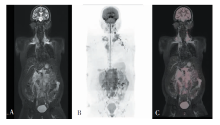
图1
FS-T2WI和WB-DWI重建及融合图像 男,38岁,弥漫大B细胞淋巴瘤,临床分期Ⅲ期。A~C分别为冠状位FS-T2WI、WB-DWI最大密度投影图像、冠状位FS-T2WI与WB-DWI融合图像。
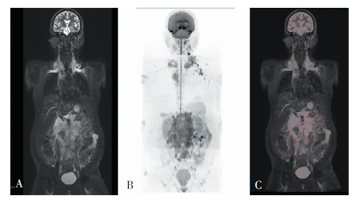
表1
磁共振WB-DWI结合FS-T2WI与PET/CT检查对结内病灶的评估
| WB-DWI结合FS-T2WI | PET/CT | ||
|---|---|---|---|
| 阳性 | 阴性 | 合计 | |
| 阳性 | 97 | 18 | 115 |
| 阴性 | 16 | 369 | 385 |
| 合计 | 113 | 387 | 500 |
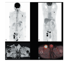
图2
WB-DWI及PET/CT检查评估结外脏器的示意图 患者为 75岁男性,弥漫大B细胞淋巴瘤,临床分期为Ⅲ期,淋巴瘤累及颈部、腋窝及腹股沟区淋巴结。A、B分别为WB-DWI、PET/CT图像,两者分期一致;C:右侧腹股沟淋巴结ADC值为0.61×10-3 mm2/s;D:SUV值为15。
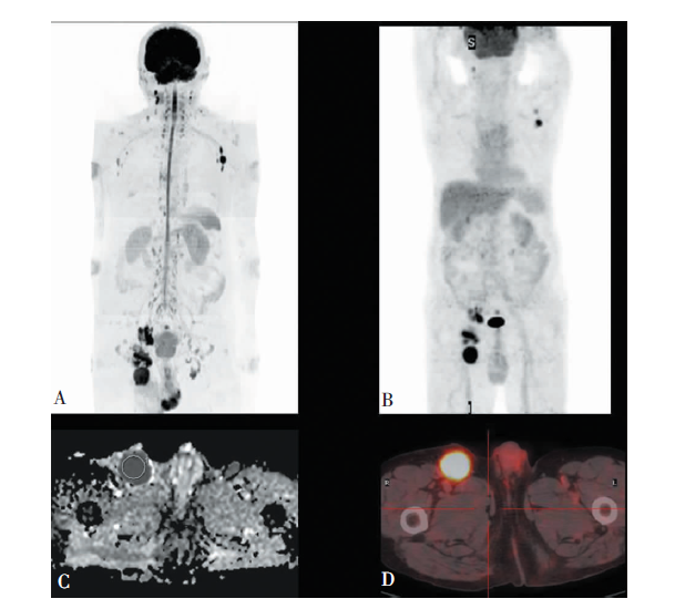
表2
磁共振WB-DWI结合FS-T2WI及PET/CT检查对结外病灶的评估
| WB-DWI结合FS-T2WI | PET/CT | ||
|---|---|---|---|
| 阳性 | 阴性 | 合计 | |
| 阳性 | 19 | 3 | 22 |
| 阴性 | 3 | 425 | 428 |
| 合计 | 22 | 428 | 450 |
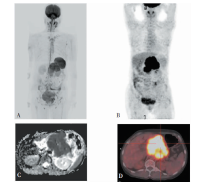
图3
WB-DWI及PET/CT检查评估结外脏器的示意图 患者为60岁女性,弥漫大B细胞淋巴瘤,临床分期为Ⅳ期,淋巴瘤弥漫浸润胰腺;A、B分别为WB-DWI、PET/CT图像,显示两者分期一致;C:ADC值为0.587×10-3 mm2/s;D:SUV值为37.5。

表3
磁共振WB-DWI结合FS-T2WI及PET/CT检查的淋巴瘤分期结果
| WB-DWI结合 FS-T2WI分期 | PET/CT分期 | ||||
|---|---|---|---|---|---|
| Ⅰ | Ⅱ | Ⅲ | Ⅳ | 合计 | |
| Ⅰ | 4 | 1 | 5 | ||
| Ⅱ | 4 | 4 | |||
| Ⅲ | 1 | 6 | 1 | 8 | |
| Ⅳ | 1 | 1 | 6 | 8 | |
| 合计 | 5 | 6 | 7 | 7 | 25 |
| [1] |
Sung H, Ferlay J, Siegel RL, et al. Global Cancer Statistics 2020: GLOBOCAN Estimates of Incidence and Mortality Worldwide for 36 Cancers in 185 Countries[J]. CA Cancer J Clin, 2021, 71(3):209-249.
doi: 10.3322/caac.21660 URL |
| [2] |
Zheng R, Zeng H, Zhang S, et al. Estimates of cancer incidence and mortality in China, 2013[J]. Chin J Cancer, 2017, 36(1):66.
doi: 10.1186/s40880-017-0234-3 URL |
| [3] |
McNamara C, Montoto S, Eyre TA, et al. The investigation and management of follicular lymphoma[J]. Br J Haematol, 2020, 191(3):363-381.
doi: 10.1111/bjh.16872 URL |
| [4] |
Ardeshna KM, Smith P, Norton A, et al. Long-term effect of a watch and wait policy versus immediate systemic treatment for asymptomatic advanced-stage non-Hodgkin lymphoma: a randomised controlled trial[J]. Lancet, 2003, 362(9383):516-522.
pmid: 12932382 |
| [5] | Pinilla I, Gómez-León N, Del Campo-Del Val L, et al. Diagnostic value of CT, PET and combined PET/CT performed with low-dose unenhanced CT and full-dose enhanced CT in the initial staging of lymphoma[J]. Q J Nucl Med Mol Imaging, 2011, 55(5):567-575. |
| [6] |
Robbins E. Radiation risks from imaging studies in children with cancer[J]. Pediatr Blood Cancer, 2008, 51(4):453-457.
doi: 10.1002/pbc.21599 URL |
| [7] |
Mayerhoefer ME, Karanikas G, Kletter K, et al. Evaluation of diffusion-weighted MRI for pretherapeutic assessment and staging of lymphoma: results of a prospective study in 140 patients[J]. Clin Cancer Res, 2014, 20(11):2984-2993.
doi: 10.1158/1078-0432.CCR-13-3355 pmid: 24696320 |
| [8] | 王燕, 宋琦. 磁共振全身弥散加权成像在淋巴瘤临床诊疗方面的应用价值及研究进展[J]. 诊断学理论与实践, 2019, 18(6):692-697. |
| [9] | 王根杰, 田颖. 不同影像学方法在淋巴瘤分期诊断中的应用[J]. 中国CT和MRI杂志, 2019, 17(4):150-152. |
| [10] | 沈俊杰, 张俊祥, 陈守康, 等. 3.0 T MRI类PET功能成像与PET/CT在淋巴瘤分期诊断中的对比研究[J]. 临床放射学杂志, 2017, 36(5):733-738. |
| [11] |
Cheson BD, Fisher RI, Barrington SF, et al. Recommendations for initial evaluation, staging, and response assessment of Hodgkin and non-Hodgkin lymphoma: the Lugano classification[J]. J Clin Oncol, 2014, 32(27):3059-3068.
pmid: 25113753 |
| [12] | 李莉, 强永乾, 任转琴, 等. 全身弥散加权成像在全身淋巴结病变中的应用价值[J]. 实用放射学杂志, 2013, 29(4):631-634. |
| [13] |
Spijkers S, Littooij AS, Humphries PD, et al. Imaging features of extranodal involvement in paediatric Hodgkin lymphoma[J]. Pediatr Radiol, 2019, 49(2):266-276.
doi: 10.1007/s00247-018-4280-z pmid: 30515533 |
| [14] |
Juweid ME, Stroobants S, Hoekstra OS, et al. Use of positron emission tomography for response assessment of lymphoma: consensus of the Imaging Subcommittee of International Harmonization Project in Lymphoma[J]. J Clin Oncol, 2007, 25(5):571-578.
pmid: 17242397 |
| [15] |
Klenk C, Gawande R, Uslu L, et al. Ionising radiation-free whole-body MRI versus (18)F-fluorodeoxyglucose PET/CT scans for children and young adults with cancer: a prospective, non-randomised, single-centre study[J]. Lancet Oncol, 2014, 15(3):275-285.
doi: 10.1016/S1470-2045(14)70021-X URL |
| [16] |
Regacini R, Puchnick A, Shigueoka DC, et al. Whole-body diffusion-weighted magnetic resonance imaging versus FDG-PET/CT for initial lymphoma staging: systematic review on diagnostic test accuracy studies[J]. Sao Paulo Med J, 2015, 133(2):141-150.
doi: 10.1590/1516-3180.2014.8312810 pmid: 25789779 |
| [17] |
Spijkers S, Littooij AS, Kwee TC, et al. Whole-body MRI versus an FDG-PET/CT-based reference standard for staging of paediatric Hodgkin lymphoma: a prospective multicentre study[J]. Eur Radiol, 2021, 31(3):1494-1504.
doi: 10.1007/s00330-020-07182-0 URL |
| [18] |
Kharuzhyk S, Zhavrid E, Dziuban A, et al. Comparison of whole-body MRI with diffusion-weighted imaging and PET/CT in lymphoma staging[J]. Eur Radiol, 2020, 30(7):3915-3923.
doi: 10.1007/s00330-020-06732-w pmid: 32103366 |
| [19] |
Stéphane V, Samuel B, Vincent D, et al. Comparison of PET-CT and magnetic resonance diffusion weighted imaging with body suppression(DWIBS) for initial stagi-ng of malignant lymphomas[J]. Eur J Radiol, 2013, 82(11):2011-2017.
doi: 10.1016/j.ejrad.2013.05.042 pmid: 23932096 |
| [20] | Ferrari C, Minoia C, Asabella AN, et al. Whole body magnetic resonance with diffusion weighted sequence with body signal suppression compared to (18)F-FDG PET/CT in newly diagnosed lymphoma[J]. Hell J Nucl Med, 2014, 17(Suppl 1):40-49. |
| [21] |
Tsuji K, Kishi S, Tsuchida T, et al. Evaluation of staging and early response to chemotherapy with whole-body diffusion-weighted MRI in malignant lymphoma patients: A comparison with FDG-PET/CT[J]. J Magn Reson Imaging, 2015, 41(6):1601-1607.
doi: 10.1002/jmri.24714 URL |
| [22] |
Tsushima Y, Takano A, Taketomi-Takahashi A, et al. Body diffusion-weighted MR imaging using high b-value for malignant tumor screening: usefulness and necessity of referring to T2-weighted images and creating fusion images[J]. Acad Radiol, 2007, 14(6):643-650.
pmid: 17502253 |
| [23] |
Spijkers S, Nievelstein RAJ, de Keizer B, et al. Fused high b-value diffusion weighted and T2-weighted MR ima-ges in staging of pediatric Hodgkin′s lymphoma: A pilot study[J]. Eur J Radiol, 2019, 121:108737.
doi: 10.1016/j.ejrad.2019.108737 URL |
| [24] | Baranska D, Matera K, Podgorski M, et al. Feasibility of diffusion-weighted imaging with DWIBS in staging Hodgkin lymphoma in pediatric patients: comparison with PET/CT[J]. MAGMA, 2019, 32(3):381-390. |
| [25] |
Wang D, Huo Y, Chen S, et al. Whole-body MRI versus 18F-FDG PET/CT for pretherapeutic assessment and sta-ging of lymphoma: a meta-analysis[J]. Onco Targets Ther, 2018, 11:3597-3608.
doi: 10.2147/OTT.S148189 URL |
| [26] |
Hong GS, Chae EJ, Ryu JS, et al. Assessment of naive indolent lymphoma using whole-body diffusion-weighted imaging and T2-weighted MRI: results of a prospective study in 30 patients[J]. Cancer Imaging, 2021, 21(1):5.
doi: 10.1186/s40644-020-00371-6 URL |
| [27] |
Asenbaum U, Nolz R, Karanikas G, et al. Bone marrow involvement in malignant lymphoma: evaluation of quantitative PET and MRI biomarkers[J]. Acad Radiol, 2018, 25(4):453-460.
doi: S1076-6332(17)30452-X pmid: 29199055 |
| [28] |
Wu LM, Chen FY, Jiang XX, et al. 18F-FDG PET, combined FDG-PET/CT and MRI for evaluation of bone marrow infiltration in staging of lymphoma: a systematic review and meta-analysis[J]. Eur J Radiol, 2012, 81(2):303-311.
doi: 10.1016/j.ejrad.2010.11.020 URL |
| [29] |
Barrington SF, Mikhaeel NG, Kostakoglu L, et al. Role of imaging in the staging and response assessment of lymphoma: consensus of the International Conference on Malignant Lymphomas Imaging Working Group[J]. J Clin Oncol, 2014, 32(27):3048-3058.
pmid: 25113771 |
| [30] |
Tomizawa M, Shinozaki F, Uchida Y, et al. Diffusion-weighted whole-body imaging with background body signal suppression/T2 image fusion and positron emission tomography/computed tomography of upper gastrointestinal cancers[J]. Abdom Imaging, 2015, 40(8):3012-3019.
doi: 10.1007/s00261-015-0545-2 pmid: 26350283 |
| [31] | Stecco A, Buemi F, Quagliozzi M, et al. Staging of primary abdominal lymphomas: comparison of whole-body MRI with diffusion-weighted imaging and (18)F-FDG-PET/CT[J]. Gastroenterol Res Pract, 2015, 2015:104794. |
| [1] | 冯国伟, 张晓娟, 郭睿, 关哲, 王越. 治疗前18F-FDG PET/CT显像对结外NK/T细胞淋巴瘤的预后判断价值[J]. 诊断学理论与实践, 2021, 20(06): 533-539. |
| [2] | 范春丽, 吴涛, 薛锋, 胡文雪, 王存邦, 白海. MUM1/IRF4阳性弥漫大B细胞淋巴瘤一例治疗报告并文献复习[J]. 诊断学理论与实践, 2021, 20(04): 399-400. |
| [3] | 赵茜, 赵肖庆, 刁立诚, 孙菲, 郑捷, 朱雪梅, 曹华. 伴皮肤紫癜的原发性干燥综合征患者的临床特征分析[J]. 诊断学理论与实践, 2021, 20(02): 155-160. |
| [4] | 沈小雁, 郑捷. 2005年至2018年皮肤淋巴瘤WHO-EORTC分类标准的发展与比较[J]. 诊断学理论与实践, 2021, 20(01): 15-20. |
| [5] | 李芹芹, 金晓龙, 袁菲. 儿童系统性EB病毒阳性T细胞淋巴瘤临床病理分析一例及文献复习[J]. 诊断学理论与实践, 2020, 19(1): 63-68. |
| [6] | 孟磊俊, 张晶, 王雪莉, 李治, 张泓, 曾乃燕. 儿童伯基特淋巴瘤中差异表达基因的鉴定及临床应用[J]. 诊断学理论与实践, 2020, 19(03): 248-257. |
| [7] | 丁燕飞, 陈平, 罗方秀, 吴云林. 以反复上消化道出血为首发表现的套细胞淋巴瘤一例[J]. 诊断学理论与实践, 2020, 19(02): 191-194. |
| [8] | 杨巧, 王佩, 王璐. 甲状腺细针穿刺细胞学诊断霍奇金淋巴瘤1例[J]. 诊断学理论与实践, 2019, 18(2): 215-217. |
| [9] | 王燕, 宋琦. 磁共振全身弥散加权成像在淋巴瘤临床诊疗方面的应用价值及研究进展[J]. 诊断学理论与实践, 2019, 18(06): 692-697. |
| [10] | 房莹, 吴东, 常春康. 癌基因与抑癌基因在伯基特淋巴瘤发生发展中的研究进展[J]. 诊断学理论与实践, 2019, 18(06): 630-633. |
| [11] | 杨志芳, 方国平, 詹维伟, 吉日. 去分化实体型甲状腺乳头状癌伴黏膜相关淋巴组织结外边缘区淋巴瘤1例病理特征并文献分析[J]. 诊断学理论与实践, 2019, 18(05): 548-554. |
| [12] | 沈文斌, 郭睿, 李彪. 18F-FDG PET/CT代谢参数在NK/T细胞淋巴瘤预后价值的评估[J]. 诊断学理论与实践, 2019, 18(03): 349-352. |
| [13] | 马媛媛, 王燕, 宋琦, 陈克敏, 赵夏, 王黎, 江旭峰, 赵维莅, 严福华. 肺原发性淋巴瘤的CT影像学特点及文献复习[J]. 诊断学理论与实践, 2018, 17(05): 533-537. |
| [14] | 武新洋, 张欢, 潘自来, 谭晶文, 杲霄源. 双源CT对原发性胃淋巴瘤和进展期胃癌的鉴别诊断价值[J]. 诊断学理论与实践, 2018, 17(01): 60-65. |
| [15] | 李佳明, 张苏江, 王莹, 严泽莹, 刘之茵, 孙海敏, 陈玉宝, 陈钰, 罗方秀, 孙静. 慢性粒-单核细胞白血病合并结外非霍奇金淋巴瘤的临床特征分析[J]. 诊断学理论与实践, 2018, 17(01): 76-81. |
| 阅读次数 | ||||||
|
全文 |
|
|||||
|
摘要 |
|
|||||