诊断学理论与实践 ›› 2020, Vol. 19 ›› Issue (1): 16-19.doi: 10.16150/j.1671-2870.2020.01.005

• 论著 • 上一篇    下一篇

新型冠状病毒(2019-nCoV)肺炎的CT征象分析

曹琪琪1a, 秦乐1a, 周慧娟1b, 杨之涛1c, 苏文婷2, 杨文洁1a, 程增辉1a, 陆勇1a, 严福华1a, 潘自来2()   

  1. 1.上海交通大学医学院附属瑞金医院 a. 放射科,b. 感染科,c. 急诊科,上海 200025
    2.上海交通大学医学院附属瑞金医院北院放射科,上海 201800
  • 收稿日期:2020-02-13 出版日期:2020-02-25 发布日期:2020-02-25
  • 通讯作者: 潘自来 E-mail:zilaipanlilly@163.com

CT features of 2019 novel coronavirus pneumonia

CAO Qiqi1a, QIN Le1a, ZHOU Huijuan1b, YANG Zhitao1c, SU Wenting2, YANG Wenjie1a, CHENG Zenghui1a, LU Yong1a, YAN Fuhua1a, PAN Zilai2()   

  1. 1a. Department of Radiology, Ruijin Hospital, Shanghai Jiao Tong University School of Medicine, 1b. Department of Infectious Diseases, 1c. Department of Emergency, Shanghai 200025, China
    2. Department of Radiology, Ruijin Hospital North, Shanghai Jiao Tong University School of Medicine, Shanghai 201800, China
  • Received:2020-02-13 Online:2020-02-25 Published:2020-02-25
  • Contact: PAN Zilai E-mail:zilaipanlilly@163.com

摘要:

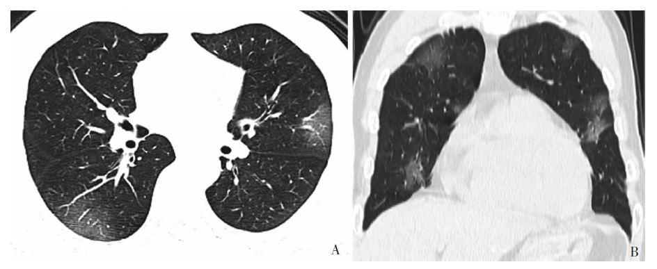
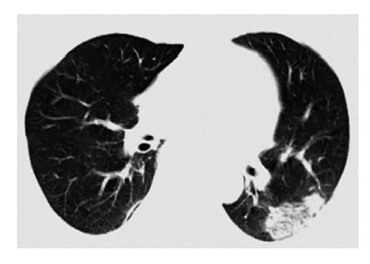
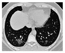
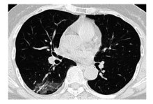
目的:观察分析新型冠状病毒(2019 novel coronavirus, 2019-nCoV)肺炎(新冠肺炎)患者的CT表现,探讨CT在诊断中的价值。方法:回顾性收集2020年1月20日至2月10日在上海交通大学医学院附属瑞金医院和瑞金北院发热门诊就诊者,患者依据实时荧光反转录聚合酶链反应(reverse transcription-polymerase chain reaction, RT-PCR)检测核酸结果阳性而确诊,分析所有患者的临床资料、首次实验室检查和CT图像。结果:确诊为新冠肺炎的病例共有12例,发病1周内CT检查结果显示,所有患者均有肺部浸润,累及多个肺叶和肺段。12例患者的病灶CT征像均表现为磨玻璃影(ground glass opacity,GGO),外周带多见,其中8例出现 GGO合并片状实变;10例患者的病灶内伴有局灶性网格影;9例病灶内出现空气支气管征;未见纵隔淋巴结肿大和胸腔积液。结论:新冠肺炎患者发病1周内,CT图像上多表现为多灶性、斑片状、外周分布为主、两肺下叶多见的GGO。疫情爆发期间,如果在CT影像上发现GGO多于实变时,应高度怀疑新型冠状病毒感染。CT检查结果对早期治疗和及时隔离至关重要。

关键词: 新型冠状病毒, 病毒性肺炎, 计算机断层扫描, 磨玻璃影

Abstract:

Objective: To analyze the CT features of patients with 2019 novel coronavirus (2019-nCoV) pneumonia, and explore the value of CT examination in the diagnosis. Methods: The clinical data of patients with fever and 2019-nCoV detected by real-time fluorescence RT-PCR from January 20 through February 10, 2020 were retrieved and imaging findings on CT were retrospectively reviewed. Results: Of all 12 patients with 2019-nCoV pneumonia, all had pulmonary infiltrations, involving multiple lung lobes and segments. Lesions on CT image showed GGO in all patients (12/12), predominantly located in the peripheral zone, with local reticular pattern within GGO and air bronchogram identified in 10 and 9 cases respectively. The patchy consolidation accompanying GGO presented in 8 cases. The lymphadenopathy and pleural effusion, however, were absent in all patients. Conclusion: For patients with novel coronavirus pneumonia within the first week of illness, the multifocal and patchy peripheral pure GGO, predominantly in the lower lung,is the most common findings on CT images. The extensive GGO with mild consolidation on CT scans is highly suspicious of 2019-nCoV infection and may prompt early treatment and prompt isolation in early outbreak.

Key words: Novel coronavirus, Viral pneumonia, Computed tomography, Ground glass opacity

中图分类号: