诊断学理论与实践 ›› 2020, Vol. 19 ›› Issue (1): 80-83.doi: 10.16150/j.1671-2870.2020.01.016
孙芙蓉, 陈克敏, 潘自来( ), 徐敬慈, 饶敏
收稿日期:2018-09-04
出版日期:2020-02-25
发布日期:2020-02-25
通讯作者:
潘自来
E-mail:zilaipanlilly@yahoo.com.cn
SUN Furong, CHEN Kemin, PAN Zilai(), XU Jingci, RAO Min
Received:2018-09-04
Online:2020-02-25
Published:2020-02-25
Contact:
PAN Zilai
E-mail:zilaipanlilly@yahoo.com.cn
摘要:
目的:探讨头颅多层螺旋CT血管造影(multi-slice spiral CT angiography, MSCTA)对枕动脉(occipital artery, OA)的显示能力及临床应用价值。方法:对80例头颅MSCTA原始图像数据分别进行遮盖容积重建(shade volume rendering,SVR)、最大密度投影(maximal intensity projection, MIP)、曲面重建(curved plannar reconstruction, CPR)处理,测量OA起始处及枕动脉沟中点处管径,并由2位医师分别评价3种重建图像的质量。结果:左侧OA起始处及枕动脉沟中点处管径分别为(2.33±0.26) mm、(2.32±0.24) mm;右侧OA起始处及枕动脉沟中点处管径分别为(2.28±0.24) mm、(2.30±0.25) mm。SVR与MIP间的图像质量评分差异无统计学意义(t=1.91,P>0.05),且均高于CPR(t=10.70、t=-10.03,P均<0.01)。结论:头颅MSCTA后处理SVR图像能清晰、准确地显示OA的解剖结构及走行,MIP图像能清晰显示血管本身情况,结合两者可以准确评估OA。
中图分类号:
孙芙蓉, 陈克敏, 潘自来, 徐敬慈, 饶敏. 枕动脉的多层螺旋CT血管造影及不同重建方法间的比较[J]. 诊断学理论与实践, 2020, 19(1): 80-83.
SUN Furong, CHEN Kemin, PAN Zilai, XU Jingci, RAO Min. Comparison of different image reconstruction methods in MSCT angiography of occipital artery[J]. Journal of Diagnostics Concepts & Practice, 2020, 19(1): 80-83.
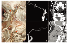
图1
3种后处理图像 A:右侧OA的SVR图像;B:右侧OA的MIP图像;C:右侧OA的CPR图像;D:左侧OA的SVR图像;E:左侧OA的MIP图像;F:左侧OA的CPR图像。A~F均为同一检查者,图A及图D箭头所指处为枕动脉沟中点处
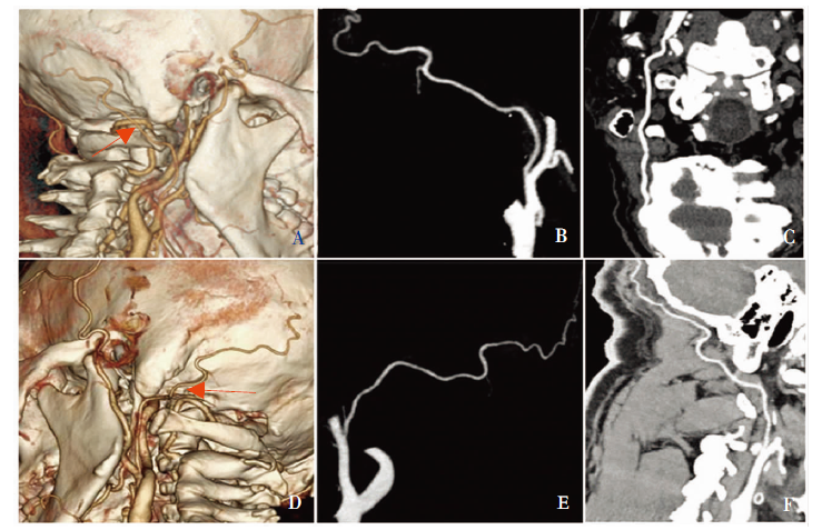
表1
SVR、MIP及CPR 重建方法对OA的显示情况
| 医师 | 重建方法 | 5分 | 4分 | 3分 | 2分 | 1分 | 合计 |
|---|---|---|---|---|---|---|---|
| 医师一 | SVR | 89 | 53 | 18 | 0 | 0 | 160 |
| 医师二 | SVR | 94 | 57 | 9 | 0 | 0 | 160 |
| 医师一 | MIP | 84 | 54 | 22 | 0 | 0 | 160 |
| 医师二 | MIP | 66 | 57 | 37 | 0 | 0 | 160 |
| 医师一 | CPR | 59 | 71 | 30 | 0 | 0 | 160 |
| 医师二 | CPR | 71 | 81 | 8 | 0 | 0 | 160 |
| [1] |
Iwai T, Izumi T, Inoue T, et al. Occipital artery arising from the anterior aspect of the internal carotid artery identified by three-dimensional computed tomography angiography[J]. Iran J Radiol, 2012, 9(2):103-105.
doi: 10.5812/iranjradiol.7809 URL |
| [2] |
Tubbs RS, Salter G, Oakes WJ. Continuation of the ascending cervical artery as the occipital artery in man[J]. Anat Sci Int, 2004, 79(1):43-45.
doi: 10.1111/j.1447-073x.2004.00062.x URL |
| [3] |
Aggarwal NR, Krishnamoorthy T, Devasia B, et al. Varia-nt origin of superior thyroid artery, occipital artery and ascending pharyngeal artery from a common trunk from the cervical segment of internal carotid artery[J]. Surg Radiol Anat, 2006, 28(6):650-653.
pmid: 17024310 |
| [4] |
Keser N, Avci E, Soylemez B, et al. Occipital artery and its segments in vertebral artery revascularization surgery: A microsurgical anatomic study[J]. World Neurosurg, 2018, 112:e534-e539.
doi: 10.1016/j.wneu.2018.01.073 URL |
| [5] | 胡继良, 罗伟坚, 王浩, 等. 远外侧入路中枕动脉的解剖特点及其在后循环搭桥手术中的意义[J]. 中华神经医学杂志, 2017, 16(1):51-54. |
| [6] | 党瑞山, 纪荣明, 张成, 等. 枕动脉与椎动脉寰椎部吻合术治疗椎动脉供血不全的应用解剖[J]. 解剖学杂志, 1990, 27(1):38-42. |
| [7] | Kazumata K, Kamiyama H, Saito H, et al. Direct anastomosis using occipital artery for additional revascularization in moyamoya disease after combined superficial temporal artery-middle cerebral artery and indirect bypass[J]. Oper Neurosurg(Hagerstown), 2017, 13(2):213-223. |
| [8] |
Hirano T, Mikami T, Suzuki H, et al. Occipital artery to middle cerebral artery bypass in cases of unavailable superficial temporal artery[J]. World Neurosurg, 2018, 112:101-108.
doi: 10.1016/j.wneu.2018.01.103 URL |
| [9] | Labauge R, Boukobza M, Pagès M, et al. Occlusion of the vertebral artery (100 personal cases)[J]. Rev Neurol (Paris), 1987, 143(6-7):490-509. |
| [10] |
Flossmann E, Rothwell PM. Prognosis of vertebrobasilar transient ischaemic attack and minor stroke[J]. Brain, 2003, 126(Pt 9):1940-1954.
doi: 10.1093/brain/awg197 pmid: 12847074 |
| [11] |
Katsuki M, Yamamoto Y, Wada N, et al. Occipital artery to extracranial vertebral artery anastomosis for bilateral vertebral artery stenosis at the origin: A case report[J]. Surg Neurol Int, 2018, 9:82.
doi: 10.4103/sni.sni_20_18 URL |
| [12] | 张永力, 石祥恩, 周忠清, 等. 枕动脉-小脑下后动脉吻合术治疗颅内段椎动脉梭形动脉瘤四例[J]. 中国脑血管病杂志, 2009, 6(11):596-598. |
| [13] | 李荷欢, 贺佳妮, 韩思源, 等. 应用枕动脉跨区供血头皮瓣修复头皮肿瘤术后大面积复杂头皮缺损[J]. 中国美容整形外科杂志, 2011, 22(3):142-144. |
| [1] | 何亲羽, 王伟, 陈立芬, 张雪蕾, 董治亚. LHCGR基因突变致家族性男性性早熟2例报告及文献复习[J]. 诊断学理论与实践, 2022, 21(05): 598-605. |
| [2] | 陈志敏, 何浩岚. 艾滋病合并马尔尼菲篮状菌病的诊治现状[J]. 诊断学理论与实践, 2022, 21(04): 425-430. |
| [3] | 沈银忠. 《人类免疫缺陷病毒感染/艾滋病合并结核分枝杆菌感染诊治专家共识》解读[J]. 诊断学理论与实践, 2022, 21(04): 431-436. |
| [4] | 陈宏, 沈银忠. 人类免疫缺陷病毒感染/艾滋病合并结核病的诊治进展[J]. 诊断学理论与实践, 2022, 21(04): 530-534. |
| [5] | 何新, 陈慧, 冯炜炜. 机器学习算法在辅助超声诊断附件肿块良恶性中的应用研究进展[J]. 诊断学理论与实践, 2022, 21(04): 541-546. |
| [6] | 徐子真, 李擎天, 刘湘帆, 李莉, 李惠, 王也飞, 吴洁敏, 陈宁, 梁璆荔, 陈松立, 戴健敏, 宋珍, 丁磊. 实验诊断学在线课程的建立和实践[J]. 诊断学理论与实践, 2022, 21(04): 547-550. |
| [7] | 赵然, 詹维伟, 侯怡卿. 计算机辅助诊断系统辅助超声诊断甲状腺弥漫性病变合并结节良恶性的应用价值[J]. 诊断学理论与实践, 2022, 21(03): 390-394. |
| [8] | 郭业兵, 郑金峰. 阴道壁胃肠道外间质瘤一例报道并文献复习[J]. 诊断学理论与实践, 2022, 21(03): 405-407. |
| [9] | 王刚, 陈生弟. 神经病学的诊断:起源、发展及挑战[J]. 诊断学理论与实践, 2022, 21(01): 1-4. |
| [10] | 唐静仪, 余群, 刘军. 结合人工智能的结构影像分析对阿尔茨海默病的早期预测及精准诊断研究进展[J]. 诊断学理论与实践, 2022, 21(01): 12-17. |
| [11] | 魏文石. 直面我国阿尔茨海默病诊治的挑战——《中国阿尔茨海默病报告2021》解读[J]. 诊断学理论与实践, 2022, 21(01): 5-7. |
| [12] | 王蔚, 王小钦. 缺铁性贫血的病因诊断[J]. 诊断学理论与实践, 2021, 20(06): 529-532. |
| [13] | 岳婧婧, 宋琦, 江旭峰, 王黎, 赵维莅, 严福华. 磁共振全身扩散加权成像结合T2WI抑脂序列与FDG-PET/CT在初发淋巴瘤分期及病灶检出的对比研究[J]. 诊断学理论与实践, 2021, 20(06): 540-546. |
| [14] | 王昭晖, 吴海波. 胃神经鞘瘤31例临床病理学分析[J]. 诊断学理论与实践, 2021, 20(06): 552-556. |
| [15] | 王广宇, 杨昕, 张立娟, 谭姣容. 住院新诊断2型糖尿病男性患者血浆总睾酮水平与骨钙素的相关性研究[J]. 诊断学理论与实践, 2021, 20(06): 573-578. |
| 阅读次数 | ||||||
|
全文 |
|
|||||
|
摘要 |
|
|||||