诊断学理论与实践 ›› 2021, Vol. 20 ›› Issue (06): 552-556.doi: 10.16150/j.1671-2870.2021.06.007
王昭晖, 吴海波( )
)
收稿日期:2021-04-06
出版日期:2021-12-25
发布日期:2021-12-25
通讯作者:
吴海波
E-mail:bbwuhaibo@sina.com
WANG Zhaohui, WU Haibo()
Received:2021-04-06
Online:2021-12-25
Published:2021-12-25
Contact:
WU Haibo
E-mail:bbwuhaibo@sina.com
摘要:
目的: 探讨胃神经鞘瘤(gastric schwannoma,GS)的临床病理学特征、诊断、鉴别诊断、治疗及预后。方法: 2014年1月至2020年3月间,中国科学技术大学附属第一医院经病理确诊为各种类型胃肿瘤的手术标本共有17 223例,其中GS有31例,约占0.18%,回顾性分析这31例GS患者的临床病理学特征和免疫表型,并复习相关文献,讨论GS的诊断、鉴别、治疗及预后。结果: 31例GS患者中,男性为12例,女性为19例,年龄为22~70岁,中位年龄为51岁。其中20例患者术前影像学检查提示为胃肠道间质瘤(gastrointestinal stromal tumor,GIST)。29例患者的肿瘤病灶位于胃黏膜层至浆膜层之间,2例病灶突破浆膜层。肉眼观察,GS的边界清晰,均无包膜。在光学显微镜下,可见肿瘤边界较清晰,周边常见淋巴细胞套;肿瘤主要由梭形细胞组成,呈束状、小簇状或杂乱排列,经典软组织神经鞘瘤常见的交错分布的Antoni A区和Antoni B区常不明显;细胞核可有一定异型性,但核分裂象罕见。免疫组织化学检测显示,肿瘤组织呈弥漫性S100、SOX10阳性。结论: GS是胃罕见的间叶源性肿瘤,为良性,术前易被误诊为GIST,诊断时需结合组织学特征及免疫表型,与GIST等肿瘤进行鉴别。
中图分类号:
王昭晖, 吴海波. 胃神经鞘瘤31例临床病理学分析[J]. 诊断学理论与实践, 2021, 20(06): 552-556.
WANG Zhaohui, WU Haibo. Clinicopathological analysis of 31 cases of gastric schwannoma[J]. Journal of Diagnostics Concepts & Practice, 2021, 20(06): 552-556.
表1
31例胃神经鞘瘤的临床病理学特点
| 序号 | 性别 | 年龄(岁) | 部位 | 肿瘤直径(cm) | 临床症状 | 术前诊断 | 治疗方式 |
|---|---|---|---|---|---|---|---|
| 1 | 女 | 43 | 胃体大弯 | 4.5 | 上腹部不适 | 胃间质瘤 | 手术 |
| 2 | 男 | 44 | 胃体大弯 | 6.0 | 上腹隐痛 | 恶性胃间质瘤 | 手术 |
| 3 | 男 | 48 | 胃体大弯 | 6.5 | 黑便 | 胃间质瘤 | 手术 |
| 4 | 女 | 70 | 胃体大弯 | 6.0 | 上腹部隐痛 | 胃间质瘤 | 手术 |
| 5 | 女 | 53 | 胃底 | 5.0 | 上腹部胀痛 | 胃间质瘤 | 手术 |
| 6 | 女 | 50 | 胃体大弯 | 3.8 | 体检 | 胃间质瘤 | 手术 |
| 7 | 女 | 51 | 胃体大弯 | 3.2 | 上腹部不适 | 胃间质瘤 | 手术 |
| 8 | 女 | 48 | 胃体大弯 | 3.0 | 体检 | 胃间质瘤 | 手术 |
| 9 | 女 | 60 | 贲门小弯 | 6.0 | 呕吐 | 胃占位 | 手术 |
| 10 | 女 | 50 | 胃角 | 4.5 | 上腹部胀痛 | 胃间质瘤 | 手术 |
| 11 | 女 | 66 | 胃体小弯 | 5.0 | 体检 | 胃间质瘤 | 手术 |
| 12 | 女 | 66 | 贲门小弯 | 1.7 | 上腹部隐痛 | 胃癌 | 手术 |
| 13 | 女 | 39 | 胃体前壁 | 2.0 | 上腹部阵痛 | 胃间质瘤 | 内镜 |
| 14 | 男 | 22 | 胃体大弯 | 2.0 | 腹胀 | 胃间质瘤(合并食管隆起病变) | 内镜 |
| 15 | 男 | 28 | 胃角前壁 | 1.5 | 急性胃肠炎 | 胃黏膜病变 | 内镜 |
| 16 | 女 | 51 | 胃体大弯 | 2.5 | 体检 | 胃间质瘤 | 手术 |
| 17 | 男 | 41 | 胃体大弯 | 7.0 | 呕吐 | 胃间质瘤 | 手术 |
| 18 | 男 | 48 | 胃体大弯 | 6.5 | 上腹部不适 | 恶性胃间质瘤 | 手术 |
| 19 | 男 | 50 | 胃体小弯 | 4.0 | 上腹部不适 | 胃间质瘤 | 手术 |
| 20 | 女 | 49 | 胃体前壁 | 6.0 | 嗳气 | 胃间质瘤 | 手术 |
| 21 | 女 | 50 | 胃体小弯 | 4.5 | 体检 | 胃间质瘤 | 手术 |
| 22 | 女 | 53 | 胃体大弯 | 3.5 | 上腹部不适 | 胃间质瘤 | 手术 |
| 23 | 女 | 60 | 胃体小弯 | 4.0 | 腹痛 | 胃间质瘤 | 手术 |
| 24 | 男 | 56 | 胃体大弯 | 4.0 | 体检 | 胃肿物 | 手术 |
| 25 | 女 | 54 | 胃体大弯 | 3.0 | 上腹部不适 | 胃占位 | 手术 |
| 26 | 男 | 60 | 胃体大弯 | 0.4 | 合并食管癌 | 食管癌 | 手术 |
| 27 | 女 | 58 | 胃体小弯 | 2.0 | 上腹部隐痛 | 胃间质瘤 | 内镜 |
| 28 | 女 | 64 | 胃体后壁 | 4.0 | 上腹部不适 | 胃占位 | 手术 |
| 29 | 男 | 28 | 胃角前壁 | 2.2 | 急性胃肠炎 | 胃黏膜病变 | 内镜 |
| 30 | 女 | 54 | 胃体小弯 | 4.0 | 体检 | 胃间质瘤 | 手术 |
| 31 | 男 | 70 | 胃体大弯 | 2.0 | 大便带血 | 直肠癌 | 手术 |
图1
GS病理图片(HE) A:肿瘤组织位于黏膜肌层,边界清晰,周边淋巴组织增生形成淋巴细胞套(×40);B:肿瘤细胞呈束状排列,散在核深染细胞(×100);C:肿瘤细胞呈栅栏状排列(×100);D:肿瘤细胞的细胞质呈多空泡状(×400);E:肿瘤间质局灶间黏液变性(×100);F:肿瘤内见大小不一的不规则厚壁血管,血管壁不同程度玻璃样变性(×100)
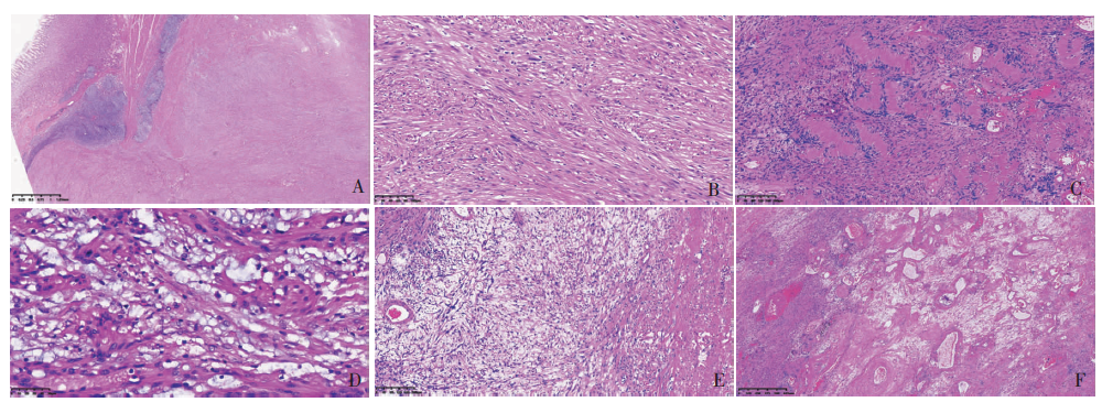
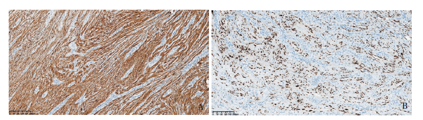
图2
GS的免疫组化图片(EnVision法,×100)
| [1] |
Rosario MS, Yamamoto N, Hayashi K, et al. A case of infected schwannoma mimicking malignant tumor[J]. World J Surg Oncol, 2016, 14(1):302.
pmid: 27923374 |
| [2] | Hu BG, Wu FJ, Zhu J, et al. Gastric schwannoma: a tumor must be included in differential diagnoses of gastric Submucosal tumors[J]. Case Rep Gastrointest Med, 2017, 2017:9615359. |
| [3] |
Yanagawa S, Kagemoto K, Tanji H, et al. A rare case of gastric schwannoma: a case report and literature review[J]. Case Rep Oncol, 2020, 13(1):330-335.
doi: 10.1159/000506450 URL |
| [4] | Yoon W, Paulson K, Mazzara P, et al. Gastric schwannoma: a rare but important differential diagnosis of a gastric submucosal mass[J]. Case Rep Surg, 2012, 2012:280982. |
| [5] |
Voltaggio L, Murray R, Lasota J, et al. Gastric schwannoma: a clinicopathologic study of 51 cases and critical review of the literature[J]. Hum Pathol, 2012, 43(5):650-659.
doi: 10.1016/j.humpath.2011.07.006 pmid: 22137423 |
| [6] |
Mekras A, Krenn V, Perrakis A, et al. Gastrointestinal schwannomas: a rare but important differential diagnosis of mesenchymal tumors of gastrointestinal tract[J]. BMC Surg, 2018, 18(1):47.
doi: 10.1186/s12893-018-0379-2 URL |
| [7] | Tao LP, Huang EJ, Li P, et al. Schwannoma of stomach: a clinicopathologic study of 12 cases[J]. Int J Clin Exp Pathol, 2018, 11(3):1679-1683. |
| [8] | Zheng L, Wu X, Kreis ME, et al. Clinicopathological and immunohistochemical characterisation of gastric schwannomas in 29 cases[J]. Gastroenterol Res Pract, 2014, 2014:202960. |
| [9] |
Cho H, Watanabe T, Aoyama T, et al. Small bud of pro-bable gastrointestinal stromal tumor within a laparoscopically-resected gastric schwannoma[J]. Int J Clin Oncol, 2012, 17(3):294-298.
doi: 10.1007/s10147-011-0296-1 URL |
| [10] |
Dalton BG, Thomas PG, Sharp NE, et al. Inflammatory myofibroblastic tumors in children[J]. J Pediatr Surg, 2016, 51(4):541-544.
doi: 10.1016/j.jpedsurg.2015.11.015 pmid: 26732283 |
| [11] |
Zhou Y, Zheng S, Ullah S, et al. Endoscopic Resection for gastric schwannoma: our clinical experience of 28 Cases[J]. J Gastrointest Surg, 2020, 24(9):2135-2136.
doi: 10.1007/s11605-020-04679-3 URL |
| [12] |
Lomdo M, Setti K, Oukabli M, et al. Gastric schwannoma: a diagnosis that should be known in 2019[J]. J Surg Case Rep, 2020, 2020(1):rjz382.
doi: 10.1093/jscr/rjz382 URL |
| [13] |
Hong X, Wu W, Wang M, et al. Benign gastric schwannoma: how long should we follow up to monitor the recurrence? A case report and comprehensive review of lite-rature of 137 cases[J]. Int Surg, 2015, 100(4):744-747.
doi: 10.9738/INTSURG-D-14-00106.1 URL |
| [1] | 陈平, 徐莹, 吴云林. 消化内镜在早期胃癌诊断中的应用进展[J]. 诊断学理论与实践, 2022, 21(05): 551-554. |
| [2] | 王亚雷. 重视胃癌高危人群的内镜精查[J]. 诊断学理论与实践, 2022, 21(05): 555-559. |
| [3] | 马乾宸, 张本炎, 芮炜玮, 王婷, 罗方秀, 王朝夫, 袁菲. 中国3 071例胃癌病理分型分析[J]. 诊断学理论与实践, 2022, 21(05): 560-566. |
| [4] | 杨蕊馨, 杜宇童, 燕然林, 朱正纲, 李琛, 于颖彦. 消化道肿瘤单细胞转录组测序研究中生物样本前处理改良的探索[J]. 诊断学理论与实践, 2022, 21(05): 567-574. |
| [5] | 鲍萍萍, 吴春晓, 顾凯, 庞怡, 王春芳, 施亮, 向詠梅, 龚杨明, 窦剑明, 吴梦吟, 付晨, 施燕. 上海市2016年胃癌发病特征及2002年至2016年胃癌发病趋势分析[J]. 诊断学理论与实践, 2022, 21(04): 462-469. |
| [6] | 李娜娜, 齐涛, 朱黎明. 血清胃蛋白酶原、胃泌素17和幽门螺杆菌IgG抗体在胃部疾病初筛中的临床价值[J]. 诊断学理论与实践, 2022, 21(04): 509-513. |
| [7] | 郭业兵, 郑金峰. 阴道壁胃肠道外间质瘤一例报道并文献复习[J]. 诊断学理论与实践, 2022, 21(03): 405-407. |
| [8] | 李娟, 刘劲松, 李梅, 李殿炜, 朱弘. 细支气管腺瘤10例临床病理分析及文献复习[J]. 诊断学理论与实践, 2021, 20(05): 466-470. |
| [9] | 谭英斌, 谢玲, 吴云林, 陈平. 自身免疫性胃炎关联疾病3例报道并文献复习[J]. 诊断学理论与实践, 2021, 20(05): 484-490. |
| [10] | 吴冬梅, 吴丽莉, 陈佳, 刘坤. 淋巴上皮样肝细胞肝癌一例报告附文献复习[J]. 诊断学理论与实践, 2021, 20(05): 498-501. |
| [11] | 崔芷萌, 任刚, 蔡嵘. EBV阳性胃淋巴上皮瘤样癌的诊治进展[J]. 诊断学理论与实践, 2021, 20(05): 502-506. |
| [12] | 王虹. 胃食管反流病的临床表现分类及精准治疗策略[J]. 诊断学理论与实践, 2021, 20(03): 251-256. |
| [13] | 顾怡瑾, 李安琪, 董磊, 许海敏, 申霞, 谢嘉玲, 袁菲, 王朝夫. 原发、复发及转移胃肠道间质瘤KIT及PDGFRA突变与临床病理特征间的关系[J]. 诊断学理论与实践, 2021, 20(03): 257-264. |
| [14] | 韦若蕖, 余红, 姚志荣. 儿童成纤维细胞结缔组织痣一例报道并文献复习[J]. 诊断学理论与实践, 2021, 20(02): 190-194. |
| [15] | 林雨轩, 赵延华, 王筱婧. 丙泊酚镇静下无痛胃镜术中低氧血症的发生率及危险因素分析[J]. 诊断学理论与实践, 2020, 19(06): 594-599. |
| 阅读次数 | ||||||
|
全文 |
|
|||||
|
摘要 |
|
|||||