诊断学理论与实践 ›› 2024, Vol. 23 ›› Issue (04): 385-391.doi: 10.16150/j.1671-2870.2024.04.006
伏秋燚, 展颖, 谭令, 朱宏, 朱乃懿, 孙琨, 柴丽, 柴维敏( )
)
收稿日期:2022-12-22
接受日期:2023-06-15
出版日期:2024-08-25
发布日期:2024-08-25
通讯作者:
柴维敏 E-mail:cwm11394@rjh.com.cn基金资助:
FU Qiuyi, ZHAN Ying, TAN Ling, ZHU Hong, ZHU Naiyi, SUN Kun, CHAI Li, CHAI Weimin()
Received:2022-12-22
Accepted:2023-06-15
Published:2024-08-25
Online:2024-08-25
摘要:
目的:分析全野数字乳腺X线摄影(full field digital mammography, FFDM)单独及联合数字乳腺断层合成X线摄影(digital breast tomosynthesis, DBT)对不同类型乳腺病灶的诊断能力差异。方法:前瞻性纳入2021年11月至2022年6月在上海交通大学医学院附属瑞金医院进行术前乳腺X线检查的患者共计389例,每例患者行Combo模式拍摄的同时获得FFDM图像和DBT图像,所有图像经具有乳腺影像诊断工作10年以上经验的放射科医师阅片。以病理学结果作为金标准,分析乳腺的影像学评价与病理学金标准的一致性,评价FFDM与FFDM+DBT数体诊断效能[灵敏度、特异度、准确率、阳性预测值(positive predictive value, PPV)、阴性预测值(negative predictive value, NPV)、受试者操作特征(receiver operating characteristic, ROC)曲线的线下面积(area under curve, AUC)诊断能力]的差异,并比较FFDM与FFDM+DBT在不同类型乳腺病灶中的诊断效能差异。结果:FFDM+DBT的整体诊断能力较FFDM显著提高(P<0.0001),FFDM+DBT的特异度、灵敏度、准确率、PPV、NPV显著提高(86.96%、89.11%、88.15%、86.54%、89.45%比80.19%、87.16%、84.05%、83.42%、84.53%),差异有统计学意义(P<0.05);FFDM+DBT诊断乳腺癌ROC的AUC较FFDM显著提高(0.906比0.869, P<0.01)。FFDM+DBT对肿块的检出率较FFDM显著提高(62.5%,46.55%,P<0.05),FFDM+DBT对结构扭曲的检出率较FFDM显著提高(11.42%比5.17%,P<0.05);对于肿块病灶良恶性,FFDM+DBT诊断的AUC较FFDM显著提高(0.918 6比0.8759,P=0.004)。结论:相比于FFDM单独检查,FFDM+DBT检查对乳腺肿块和相关结构扭曲的显示更有优势,对乳腺良恶性病灶的诊断效能更高。
中图分类号:
伏秋燚, 展颖, 谭令, 朱宏, 朱乃懿, 孙琨, 柴丽, 柴维敏. 全野数字乳腺X线摄影及联合数字乳腺断层合成X线摄影在乳腺癌诊断中效能评价[J]. 诊断学理论与实践, 2024, 23(04): 385-391.
FU Qiuyi, ZHAN Ying, TAN Ling, ZHU Hong, ZHU Naiyi, SUN Kun, CHAI Li, CHAI Weimin. Evaluation of clinical efficacy of full field digital mammography (FFDM) used alone and in combition with digital breast tomosynthesis in diagnosis of breast cancer[J]. Journal of Diagnostics Concepts & Practice, 2024, 23(04): 385-391.
表1
入组病例的年龄分布、病理分布
| Item | Groups | Numbers |
|---|---|---|
| Age distribution (case level) | 10-20 | 1 |
| 20-30 | 22 | |
| 30-40 | 92 | |
| 40-50 | 98 | |
| 50-60 | 65 | |
| 60-70 | 65 | |
| 70-80 | 41 | |
| 80-90 | 5 | |
| Total | 389 | |
| Pathological distribution (breast level) | Benign | 257 |
| Fibroadenoma | 94 | |
| Intraductal papilloma | 26 | |
| Benign/Borderline phyllodes tumor | 12 | |
| Granulomatous mastitis | 5 | |
| Complex sclerosing lesion | 5 | |
| Adenomyoepithelioma | 3 | |
| Others# | 112 | |
| Malignant | 207 | |
| Invasive ductal carcinoma | 103 | |
| Invasive breast carcinoma* | 55 | |
| Ductal carcinoma in situ | 24 | |
| Invasive lobular carcinoma | 5 | |
| Solid papillary carcinoma in situ | 6 | |
| Invasive solid papillary carcinoma | 4 | |
| Mucinous carcinoma | 5 | |
| Metaplastic carcinoma | 2 | |
| Invasive apocrine carcinoma | 1 | |
| Invasive micropapillary carcinoma | 1 | |
| Encapsulated papillary carcinoma | 1 | |
| Total | 464 |
表2
阅片评价标准
| Assessment | Likelihood of Cancer | |
|---|---|---|
| BI-RADS Categories | Category 1 | 0 |
| Category 2 | 0 | |
| Category 3 | 0-2% | |
| Category 4A | 2%-10% | |
| Category 4B | 10%-50% | |
| Category 4C | 50%-95% | |
| Category 5 | ≥95% | |
表3
FFDM与FFDM+DBT的诊断效能对比
| Item | FFDM | FFDM+DBT | Χ2 | P values |
|---|---|---|---|---|
| Sensitivity | 80.19% | 86.96% | 87.425 | <0.000 1 |
| Specificity | 87.16% | 89.11% | 152.44 | <0.000 1 |
| PPV | 83.42% | 86.54% | 101.45 | <0.000 1 |
| NPV | 84.53% | 89.45% | 154.62 | <0.000 1 |
| Accuracy | 84.05% | 88.15% | 4.7921 | 0.029 0 |
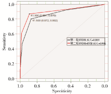
图1
FFDM与FFDM+DBT诊断乳腺病灶的ROC曲线
表4
FFDM与FFDM+DBT对不同类型病灶(BI-RADS类别最高)检出率的对比
| Item | FFDM | FFDM+DBT | Χ2 | P values |
|---|---|---|---|---|
| Masses | 46.55%(216/464) | 62.5%(290/464) | 4.5231 | 0.033 0 |
| Calcifications | 33.84%(157/464) | 23.92%(111/464) | 75.325 | <0.000 1 |
| Architectural distortion | 5.17%(24/464) | 11.42%(53/464) | 344.3 | <0.000 1 |
| Asymmetries | 20.91%(97/464) | 8.41%(39/464) | 206.1 | <0.000 1 |
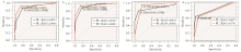
图2
FFDM与FFDM+DBT诊断不同病灶表现AUC对比 a:肿块;b:钙化;c:结构扭曲;d:不对称。
表5
FFDM与FFDM+DBT鉴别病灶表现的AUC分析
| Item | FFDM | FFDM+DBT | Χ2 | P values | Confidence interval |
|---|---|---|---|---|---|
| Masses | 0.875 9 | 0.918 6 | -2.878 1 | 0.004 | [-0.071 8,-0.013 6] |
| Calcifications | 0.918 7 | 0.949 3 | -1.504 5 | 0.132 | [-0.070 3,0.009 2] |
| Architectural distortion | 0.861 5 | 0.910 2 | -0.919 74 | 0.358 | [-0.152 7,0.055 1] |
| Asymmetries | 0.827 9 | 0.779 2 | 1.049 9 | 0.294 | [-0.042 2,0.139 6] |
| [1] | GINSBURG O, YIP C H, BROOKS A, et al. Breast cancer early detection: A phased approach to implementation[J]. Cancer, 2020, 126 Suppl 10(Suppl 10):2379-2393. |
| [2] | BUDH D P, SAPRA A. Breast Cancer Screening[M]. StatPearls. Treasure Island (FL), 2022. |
| [3] | MONTICCIOLO D L. Digital breast tomosynthesis: a decade of practice in review[J]. J Am Coll Radiol, 2023, 20(2):127-133. |
| [4] | 敬文波. 乳腺三维断层X线摄影联合磁共振成像在致密型乳腺中乳腺癌诊断的应用价值[D]. 新疆: 新疆医科大学, 2020. |
| JING W B. The value of digital breast tomosynthesis combined with magnetic resonance imaging for diagnosing breast cancers in dense breast[D]. Xinjiang Medical University, 2020. | |
| [5] | 程兰兰, 刘斌, 胡汉金, 等. 数字乳腺断层摄影与常规影像学检查对乳腺肿块型病变诊断的对比研究[J]. 临床放射学杂志, 2019, 38(9):1637-1641. |
| CHENG L L, LIU B, HU H J, et al. A comparative study of diagnostic of breast mass lesions betweendigital breast tomosynthesis and conventional imaging methods[J]. J of Clin Radiol, 2019, 38(9):1637-1641. | |
| [6] |
CONANT E F, BARLOW W E, HERSCHORN S D, et al. Association of digital breast tomosynthesis vs digital mammography with cancer detection and recall rates by age and breast density[J]. JAMA Oncol, 2019, 5(5):635-642.
doi: 10.1001/jamaoncol.2018.7078 pmid: 30816931 |
| [7] | ROGANOVIC D, DJILAS D, VUJNOVIC S, et al. Breast MRI, digital mammography and breast tomosynthesis: comparison of three methods for early detection of breast cancer[J]. Bosn J Basic Med Sci, 2015, 15(4):64-68. |
| [8] |
WIECHMANN L, FRIEDLANDER L C. Management of radiographic lesions of the breast[J]. Surg Clin North Am, 2022, 102(6):1031-1041.
doi: 10.1016/j.suc.2022.06.005 pmid: 36335923 |
| [9] | WANG L C, PHILIP M, BHOLE S, et al. Pathologic outcomes in single versus multiple areas of architectural distortion on digital breast tomosynthesis[J]. AJR Am J Roentgenol, 2023, 220(1):50-62. |
| [10] | GEORGIAN-SMITH D, OBUCHOWSKI N A, LO J Y, et al. Can digital breast tomosynthesis replace full-field digital mammography? A multireader, multicase study of wide-angle tomosynthesis[J]. Am J Roentgenol. 2019, 212(6):1393-1399. |
| [11] | CHAN H P, GOODSITT M M, HELVIE M A, et al. Digital breast tomosynthesis: observer performance of clustered microcalcification detection on breast phantom images acquired with an experimental system using variable scan angles, angular increments, and number of projection views[J]. Radiology, 2014, 273(3):675-685. |
| [12] | GOODSITT M M, CHAN H P, SCHMITZ A, et al. Digital breast tomosynthesis: studies of the effects of acquisition geometry on contrast-to-noise ratio and observer prefe-rence of low-contrast objects in breast phantom images[J]. Phys Med Biol, 2014, 59(19):5883-5902. |
| [13] | KHANANI S, HRUSKA C, LAZAR A, et al. Performance of wide-angle tomosynthesis with synthetic mammography in comparison to full field digital mammography[J]. Acad Radiol, 2023, 30(1):3-13. |
| [14] |
ALABOUSI M, WADERA A, KASHIF AL-GHITA M, et al. Performance of digital breast tomosynthesis, synthetic mammography, and digital mammography in breast cancer screening: a systematic review and meta-analysis[J]. J Natl Cancer Inst, 2021, 113(6):680-690.
doi: 10.1093/jnci/djaa205 pmid: 33372954 |
| [15] |
MALL S, NOAKES J, KOSSOFF M, et al. Can digital breast tomosynthesis perform better than standard digital mammography work-up in breast cancer assessment clinic?[J]. Eur Radiol, 2018, 28(12):5182-5194.
doi: 10.1007/s00330-018-5473-4 pmid: 29846804 |
| [16] |
COCHON L R, GIESS C S, KHORASANI R. Comparing diagnostic performance of digital breast tomosynthesis and Full-Field Digital Mammography[J]. J Am Coll Radiol, 2020, 17(8):999-1003.
doi: S1546-1440(20)30035-1 pmid: 32068009 |
| [17] | LUPARIA A, MARISCOTTI G, DURANDO M, et al. Accuracy of tumour size assessment in the preoperative sta-ging of breast cancer: comparison of digital mammography, tomosynthesis, ultrasound and MRI[J]. Radiol Med, 2013, 118(7):1119-1136. |
| [18] | GAO Y, REIG B, HEACOCK L, et al. Magnetic resonance imaging in screening of breast cancer[J]. Radiol Clin North Am, 2021, 59(1):85-98. |
| [19] |
BAN K, TSUNODA H, TOGASHI S, et al. Comparative study of the usefulness of adjunctive tomosynthesis in breast cancer screening by mammography and ultrasound in Japan[J]. Breast Cancer, 2022, 29(5):790-795.
doi: 10.1007/s12282-022-01358-w pmid: 35585469 |
| [20] |
KIM S A, CHANG J M, CHO N, et al. Characterization of breast lesions: comparison of digital breast tomosynthesis and ultrasonography[J]. Korean J Radiol, 2015, 16(2):229-238.
doi: 10.3348/kjr.2015.16.2.229 pmid: 25741187 |
| [21] | 杨骏宇, 沈吉, 沈思平, 等. 基于双模态超声的乳腺癌患者术前淋巴结转移负荷模型构建分析[J]. 中华全科医学, 2024, 22(4):646-650. |
| YANG J Y, SHEN J, SHEN S P, et al. Construction and analysis of preoperative lymph node metastasis load model of breast cancer patients based on dual-mode ultrasound[J]. Chin J Gen Pract, 2024, 22(4): 646-650. | |
| [22] | 邹明池, 冉海涛, 姚延峰. 人工智能在乳腺癌超声筛查中的应用进展[J]. 中国临床研究, 2024, 37(3):344-347,364. |
| ZOU M C, RAN H T, YAO Y F. Construction and analysis of preoperative lymph node metastasis load model of breast cancer patients based on dual-mode ultrasound[J]. Chin J Clin Res, 2024, 37(3):344-347,364. | |
| [23] | 谢文, 汪珺莉, 夏菲. 乳腺癌超声征象与p120ctn及CD133表达的相关性[J]. 安徽医学, 2023, 44(2):181-184. |
| XIE W, WANG J L, XIA F. Correlation between ultrasonographic signs and the expression of p120ctn and CD133 in breast cancer[J]. Anhui Med, 2023, 44(2):181-184. | |
| [24] | Murakami R, Tani H, Kumita S, et al. Diagnostic performance of digital breast tomosynthesis for predicting response to neoadjuvant systemic therapy in breast cancer patients: A comparison with magnetic resonance imaging, ultrasound, and full-field digital mammography[J]. Acta Radiol Open, 2021, 10(12):20584601211063746. |
| [1] | 何新, 陈慧, 冯炜炜. 机器学习算法在辅助超声诊断附件肿块良恶性中的应用研究进展[J]. 诊断学理论与实践, 2022, 21(04): 541-546. |
| [2] | 赵然, 詹维伟, 侯怡卿. 计算机辅助诊断系统辅助超声诊断甲状腺弥漫性病变合并结节良恶性的应用价值[J]. 诊断学理论与实践, 2022, 21(03): 390-394. |
| [3] | 李伟伟, 吴迎, 周伟, 詹维伟, 周庆华, 陶玲玲, 杨雁雯. 超微血管三维立体成像技术对BI-RADS 4类乳腺肿块血流显示及鉴别良恶性的价值研究[J]. 诊断学理论与实践, 2020, 19(06): 583-587. |
| [4] | 唐桢云, 詹维伟. 剪切波弹性成像在乳腺癌诊断中的应用现状[J]. 诊断学理论与实践, 2019, 18(2): 223-227. |
| [5] | 李伟伟, 詹维伟, 周伟, 陶玲玲, 王怡, 樊金芳, 费圆欣, 况李君, 徐文颖. 超微血管三维立体成像技术在乳腺癌血流分布模式中的应用[J]. 诊断学理论与实践, 2019, 18(2): 139-143. |
| [6] | 姚洁洁, 朱樱, 詹维伟, 陈小松, 费晓春. 非肿块型乳腺导管内癌超声特征及与临床、病理、免疫组化指标表达间的相关性[J]. 诊断学理论与实践, 2018, 17(06): 676-681. |
| [7] | 金正吉, 詹维伟, 贾晓红, 周建桥, 张晓晓, 朱樱,. 女性乳腺超声分型与乳腺肿块良恶性间的相关性[J]. 诊断学理论与实践, 2013, 12(05): 549-552. |
| [8] | 周俊宇, 詹维伟, 沈理, 董屹婕,. 超声引导下乳腺小肿块穿刺活检临床价值初步探讨[J]. 诊断学理论与实践, 2012, 11(01): 67-70. |
| [9] | 郭续胜,王丽,刘瑞林,张晓卉,范厚臻. 382例乳腺肿块细针吸取细胞学检查结果分析[J]. 诊断学理论与实践, 2005, 4(04): 326-327. |
| [10] | 陈岗,黄平. 肺内胸腺瘤一例报告[J]. 诊断学理论与实践, 2003, 2(01): 69-. |
| [11] | 丁晓毅,管永靖,江浩,何国祥,刘建军,陈克敏. 色素沉着绒毛结节性滑膜炎的MRI表现[J]. 诊断学理论与实践, 2002, 1(03): 59-61. |
| 阅读次数 | ||||||
|
全文 |
|
|||||
|
摘要 |
|
|||||