诊断学理论与实践 ›› 2019, Vol. 18 ›› Issue (2): 139-143.doi: 10.16150/j.1671-2870.2019.02.004
李伟伟1, 詹维伟2, 周伟2( ), 陶玲玲1, 王怡1, 樊金芳1, 费圆欣1, 况李君1, 徐文颖1
收稿日期:2019-01-01
出版日期:2019-04-25
发布日期:2019-04-25
通讯作者:
周伟
E-mail:zw11468@126.com
LI Weiwei1, ZHAN Weiwei2, ZHOU Wei2(), TAO Lingling1, WANG Yi1, FAN Jinfang1, FEI Yuanxin1, KUANG Lijun1, XU Wenying1
Received:2019-01-01
Online:2019-04-25
Published:2019-04-25
Contact:
ZHOU Wei
E-mail:zw11468@126.com
摘要:
目的:研究乳腺肿块的超微血管三维立体成像(smart three-dimensional superb microvascular imaging,Smart 3D SMI)血流分布特点,探讨乳腺癌血流分布模式的三维图像特征。方法:研究经病理证实的145例乳腺癌患者的Smart 3D SMI微血流声像图特征,并结合血流显示区域相应的超声造影(contrast-enhanced ultrasound, CEUS)图像特征,评估乳腺癌的血流分布模式特征。结果:145例乳腺癌患者的Smart 3D SMI检查血流分布模式中,血流最丰富的球形型血流分布模式最多见(58例,40%),其次是分枝型(32例,22%),点线型最少(11例,8%)。进一步比较CEUS与Smart 3D SMI图像上病变区血流分布模式特征,结果显示,CEUS图像均显示有超声对比剂灌注。Smart 3D SMI图像显示为点线型的患者中有约55%在CEUS图像中显示存在粗大血流,而在显示为分枝型、球形型、扩散型、混合型的患者中,CEUS图像显示存在粗大血流的比例分别为75%、72%、78%、82%。Smart 3D SMI各型(点线型、分枝型、球形型、扩散型、混合型)患者,在CEUS图像中显示为无法辨识清晰的分支血流的比例分别为82%、78%、67%、60%和58%。Smart 3D SMI分枝型和球形型的病灶在CEUS上较多表现为血流显示区高强度灌注,与点线型相比差异有统计学意义(P=0.018、0.006)。Smart 3D SMI点线型、分枝型及球形型病灶的灌注区多位于肿块内部,仅少数病例在病灶周边也可见到较强的放射状灌注。结论:乳腺癌的Smart 3D SMI血流分布评估中常见“球形型”和“分枝型”,从超声微血流三维检测的角度提供了乳腺癌微血流的血供分布区域及分布特征,表现出显著的三维形态学特征,为乳腺癌血流分布模式的深入研究提供了更多的信息。
中图分类号:
李伟伟, 詹维伟, 周伟, 陶玲玲, 王怡, 樊金芳, 费圆欣, 况李君, 徐文颖. 超微血管三维立体成像技术在乳腺癌血流分布模式中的应用[J]. 诊断学理论与实践, 2019, 18(2): 139-143.
LI Weiwei, ZHAN Weiwei, ZHOU Wei, TAO Lingling, WANG Yi, FAN Jinfang, FEI Yuanxin, KUANG Lijun, XU Wenying. The diagnostic value of smart three-dimensional superb microvascular imaging in detecting blood flow distribution pattern of breast lesion[J]. Journal of Diagnostics Concepts & Practice, 2019, 18(2): 139-143.
表1
Smart 3D SMI血流分布模式与该模式下CEUS灌注区表现(n)
| CEUS对比剂灌注 | Smart 3D SMI | ||||
|---|---|---|---|---|---|
| 点线型(n=11) | 分枝型(n=32) | 球形型(n=58) | 扩散型(n=20) | 混合型(n=24) | |
| 灌注与否 | |||||
| 有 | 11 | 32 | 58 | 20 | 24 |
| 无 | 0 | 0 | 0 | 0 | 0 |
| 粗大血流 | |||||
| 有 | 6 (55%) | 24 (75%) | 42(72%) | 16(80%) | 19(79%) |
| 无 | 5 | 8 | 16 | 4 | 5 |
| 分支血流 | |||||
| 有 | 2 | 7 | 19 | 8 | 10 |
| 无 | 9(82%) | 25 (78%) | 39 (67%) | 12 (60%) | 14(58%) |
| 强度 | |||||
| 强 | 5 (45%) | 27 (84%)(p1) | 50 (86%) (P2) (P3) | 15 (75%) | 17 (71%) |
| 中或弱 | 6 | 5 | 8 | 5 | 7 |
| 均匀与否 | |||||
| 是 | 4 (36%) | 15 (47%)(p4) | 36 (62%) (P5) (P6) | 6 (30%) | 8 (33%) |
| 否 | 7 | 17 | 22 | 14 | 16 |
| 位于肿块 | |||||
| 内部 | 11 | 29 | 54 | 0 | 0 |
| 周边扩散状 | 0 | 0 | 0 | 14 | 0 |
| 内及周边 | 0 | 3 | 4 | 6 | 24 |

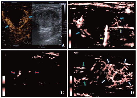
图1
乳腺浸润性导管癌 A:CEUS图像,蓝色箭头示肿块内及边缘环形灌注,中低强度灌注,不均匀;B:Smart 3D SMI图像,蓝色箭头示与A图同一肿块内及边缘环形血流,病灶内部局部呈分枝型血流,并可分辨分枝血流;黄色箭头示边缘粗大穿支血流(CEUS无法显示位于不同平面的两支穿支血流信息);C: Smart 3D SMI图像,红色箭头示病灶内点线型血流分布; D:Smart 3D SMI图像,绿色箭头示病灶局部扩散型血流,白色箭头示病灶局部球形型血流,该病例属于混合型血流分布
| [1] | 孙可欣, 李贺, 孙可欣, 等. 2014年中国恶性肿瘤发病和死亡分析[J]. 中华肿瘤杂志, 2018, 40(1):5. |
| [2] | Wang M, Feng HL, Liu YQ, et al. Angiogenesis Research in Mouse Mammary Cancer Based on Contrast-enhanced Ultrasonography: Exploratory Study[J]. Acad Radiol, 2018, 25(7):889-897. |
| [3] | Holmgren L, O'Reilly MS, Folkman J. Dormancy of micrometastases: balanced proliferation and apoptosis in the presence of angiogenesis suppression[J]. Nat Med, 1995, 1(2):149-153. |
| [4] | Yongfeng Z, Ping Z, Wengang L, et al. Application of a Novel Microvascular Imaging Technique in Breast Lesion Evaluation[J]. Ultrasound Med Biol, 2016, 42(9):2097-2105. |
| [5] | Park AY, Seo BK. Up-to-date Doppler techniques for breast tumor vascularity: superb microvascular imaging and contrast-enhanced ultrasound[J]. Ultrasonography, 2018, 37(2):98-106. |
| [6] | Adler DD, Carson PL, Rubin JM, et al. Doppler ultrasound color flow imaging in the study of breast cancer: preliminary findings[J]. Ultrasound Med Biol, 1990, 16(6):553-559. |
| [7] | Li Q, Hu M, Chen Z, et al. Meta-Analysis: Contrast-Enhanced Ultrasound Versus Conventional Ultrasound for Differentiation of Benign and Malignant Breast Lesions[J]. Ultrasound Med Biol, 2018, 44(5):919-929. |
| [8] | Wan CF, Liu XS, Wang L, et al. Quantitative contrast-enhanced ultrasound evaluation of pathological complete response in patients with locally advanced breast cancer receiving neoadjuvant chemotherapy[J]. Eur J Radiol, 2018, 103:118-123. |
| [9] | Wang XY, Kang LK, Lan CY. Contrast-enhanced ultrasonography in diagnosis of benign and malignant breast lesions[J]. Eur J Gynaecol Oncol, 2014, 35(4):415-420. |
| [10] | Machado P, Segal S, Lyshchik A, et al. A Novel Microvascular Flow Technique: Initial Results in Thyroids[J]. Ultrasound Q, 2016, 32(1):67-74. |
| [11] | Mao Y, Mu J, Zhao J, et al. The value of superb microvascular imaging in differentiating benign renal mass from malignant renal tumor: a retrospective study[J]. Br J Radiol, 2018, 91(1082):20170601. |
| [12] | Karaca L, Oral A, Kantarci M, et al. Comparison of the superb microvascular imaging technique and the color Doppler techniques for evaluating children's testicular blood flow[J]. Eur Rev Med Pharmacol Sci, 2016, 20(10):1947-1953. |
| [13] | He MN, Lv K, Jiang YX, et al. Application of superb microvascular imaging in focal liver lesions[J]. World J Gastroenterol, 2017, 23(43):7765-7775. |
| [14] | Ma Y, Li G, Li J, et al. The Diagnostic Value of Superb Microvascular Imaging (SMI) in Detecting Blood Flow Signals of Breast Lesions: A Preliminary Study Compari-ng SMI to Color Doppler Flow Imaging[J]. Medicine (Baltimore), 2015, 94(36):e1502. |
| [15] | 朱慧, 徐卫平, 陈红燕, 等. 超声造影联合超微血管显像对乳腺癌微血管的评价[J]. 中国临床医学影像杂志, 2016, 27(6):390-392. |
| [16] | 陈欣, 肖晓云, 吴欢, 等. 微细血流成像技术在乳腺肿瘤鉴别中的应用[J]. 中国超声医学杂志, 2016, 32(5):407-410. |
| [1] | 何亲羽, 王伟, 陈立芬, 张雪蕾, 董治亚. LHCGR基因突变致家族性男性性早熟2例报告及文献复习[J]. 诊断学理论与实践, 2022, 21(05): 598-605. |
| [2] | 陈志敏, 何浩岚. 艾滋病合并马尔尼菲篮状菌病的诊治现状[J]. 诊断学理论与实践, 2022, 21(04): 425-430. |
| [3] | 沈银忠. 《人类免疫缺陷病毒感染/艾滋病合并结核分枝杆菌感染诊治专家共识》解读[J]. 诊断学理论与实践, 2022, 21(04): 431-436. |
| [4] | 陈宏, 沈银忠. 人类免疫缺陷病毒感染/艾滋病合并结核病的诊治进展[J]. 诊断学理论与实践, 2022, 21(04): 530-534. |
| [5] | 何新, 陈慧, 冯炜炜. 机器学习算法在辅助超声诊断附件肿块良恶性中的应用研究进展[J]. 诊断学理论与实践, 2022, 21(04): 541-546. |
| [6] | 徐子真, 李擎天, 刘湘帆, 李莉, 李惠, 王也飞, 吴洁敏, 陈宁, 梁璆荔, 陈松立, 戴健敏, 宋珍, 丁磊. 实验诊断学在线课程的建立和实践[J]. 诊断学理论与实践, 2022, 21(04): 547-550. |
| [7] | 赵然, 詹维伟, 侯怡卿. 计算机辅助诊断系统辅助超声诊断甲状腺弥漫性病变合并结节良恶性的应用价值[J]. 诊断学理论与实践, 2022, 21(03): 390-394. |
| [8] | 郭业兵, 郑金峰. 阴道壁胃肠道外间质瘤一例报道并文献复习[J]. 诊断学理论与实践, 2022, 21(03): 405-407. |
| [9] | 王刚, 陈生弟. 神经病学的诊断:起源、发展及挑战[J]. 诊断学理论与实践, 2022, 21(01): 1-4. |
| [10] | 唐静仪, 余群, 刘军. 结合人工智能的结构影像分析对阿尔茨海默病的早期预测及精准诊断研究进展[J]. 诊断学理论与实践, 2022, 21(01): 12-17. |
| [11] | 魏文石. 直面我国阿尔茨海默病诊治的挑战——《中国阿尔茨海默病报告2021》解读[J]. 诊断学理论与实践, 2022, 21(01): 5-7. |
| [12] | 王蔚, 王小钦. 缺铁性贫血的病因诊断[J]. 诊断学理论与实践, 2021, 20(06): 529-532. |
| [13] | 岳婧婧, 宋琦, 江旭峰, 王黎, 赵维莅, 严福华. 磁共振全身扩散加权成像结合T2WI抑脂序列与FDG-PET/CT在初发淋巴瘤分期及病灶检出的对比研究[J]. 诊断学理论与实践, 2021, 20(06): 540-546. |
| [14] | 王昭晖, 吴海波. 胃神经鞘瘤31例临床病理学分析[J]. 诊断学理论与实践, 2021, 20(06): 552-556. |
| [15] | 王广宇, 杨昕, 张立娟, 谭姣容. 住院新诊断2型糖尿病男性患者血浆总睾酮水平与骨钙素的相关性研究[J]. 诊断学理论与实践, 2021, 20(06): 573-578. |
| 阅读次数 | ||||||
|
全文 |
|
|||||
|
摘要 |
|
|||||